Medical Radiology - Lazar A.P. 2008
Radiation examination of the maxillofacial region
Cysts and tumors
Cysts and Tumors are classified into odontogenic and non-odontogenic.
In dental practice, it is crucial to timely identify odontogenic cysts, specifically follicular and radicular ones.
A follicular (dentigerous) cyst develops due to the pathological formation of a normal dental follicle or a supernumerary follicle. It occurs predominantly in the Mandible. Radiographically, it presents as a dental arch defect accompanied by a well-defined round cavity within the jaw bone, containing a crown of a tooth while its ROOT lies outside the cavity (Fig. 308). Occasionally, multiple tooth germs at various Selection/3.html">Stages of development may be present within the cyst cavity.
A radicular (periapical) cyst develops on The basis of a periapical granuloma and grows slowly. Gradually, the Teeth become displaced, facial Asymmetry develops, and a pathological jaw fracture may occur. Radiographically, the cyst appears periapically as a round focus of bone destruction with clear, smooth, and sometimes sclerotic borders (Fig. 309).
Class="center">
Fig. 309. Radicular cyst at the root of tooth 34.

Fig. 310. Ameloblastoma in the area of the left mandibular ramus.
Infiltration and suppuration of a radicular cyst are possible. A radicular cyst that continues to grow after the extraction of the causative tooth is referred to as a residual cyst.
Odontogenic tumors are benign, whereas non-odontogenic tumors include both benign and malignant types. Benign odontogenic tumors include odontoma and ameloblastoma.
Benign non-odontogenic tumors include osteoma, hemangioma, osteoblastoclastoma, and several others.
Ameloblastoma arises from the epithelium of the enamel organ. In 80% of cases, it occurs in the mandible. It grows slowly and asymptomatically, typically being detected at the age of 40-50. Malignant transformation occurs in 2-4% of cases. Radiographically, it appears as a single or multilocular round cavity with distinct scalloped borders (Fig. 310), causing mandibular deformity.
An odontoma grows slowly and represents a conglomerate of multiple tooth-like structures and tooth germs. Radiographically, it is defined as a dense, heterogeneous, irregularly round or oval formation with well-defined borders, surrounded by a thin radiolucent rim—a capsule (Fig. 311). An odontoma causes displacement and destruction of the cortical plate, and its growth leads to facial asymmetry. A distinctive feature of an odontoma is its ability to erupt into the Oral Cavity.

Fig. 311. Odontoma.
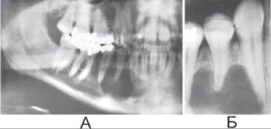
Fig. 312. Osteoblastoclastoma on panoramic (A) and periapical (B) radiographs.
Osteoma is a non-odontogenic benign tumor. Structurally, compact, cancellous, and mixed types are distinguished. Osteomas are classified into exophytic osteomas, growing on the jaw surface, and intraosseous osteomas, developing within the bone. An exophytic osteoma appears as an additional Bone Formation in the Oral Cavity and most commonly grows in the Paranasal Sinuses and on the inner surface of the mandible.
Hemangioma is a benign vascular tumor that is more frequently observed in women. Radiographically, it presents as a round focus of destruction with distinct borders, a fine-meshed Structure, and slight expansion. Hemangiomas are better visualized using CT.
Osteoblastoclastoma (giant Cell tumor) occurs in trabecular (multilocular) and lytic forms. The trabecular form of osteoblastoclastoma is radiographically defined as an expanded area with fairly distinct borders and thin septa that form locules of varying sizes (Fig. 312). The lytic form of the giant cell tumor is characterized by a radiolucency within the expanded area, presenting an irregular oval shape.
Malignant tumors of the jaws, both Primary and secondary, are rare. Primary malignant jaw tumors include osteogenic Sarcoma, chondrosarcoma, reticulosarcoma, and Ewing's sarcoma (Fig. 312). The variants and radiographic appearances of these neoplasms are described in the chapter "Malignant Bone tumors".

Fig. 313. Osteogenic sarcoma in the area of tooth 46.
Secondary infiltrating tumors cause destruction in various PARTS OF THE jaws corresponding to the site of direct extension from the oral mucosa. In the Maxilla, contact involvement results from mucosal Cancer of the maxillary sinus. The mandible is affected by direct extension from carcinomas of the floor of the Mouth, gingiva, buccal mucosa, or Lips. Oral mucosal cancer extending to the jaw is the most frequently observed.
Radiographic examination reveals osteolytic destruction with indistinct borders, lacking sclerotic demarcation and periosteal reaction. Diagnostic Imaging Modalities allow for determining the extent of the process in Bone tissue (radiography, orthopantomography, CT) and soft Tissues (CT, MRI, ultrasound), as well as performing Differential Diagnosis with inflammatory processes (Osteomyelitis). However, the definitive diagnosis is established through biopsy.
Last update: 08/08/2026
Editorial and Educational Adaptation: This material has been compiled based on the primary/original source text. The project team performed an editorial review, corrected technical inaccuracies, structured sections, and adapted the content for an educational format.
What was processed:
- elimination of formatting defects (OCR errors, structural breaks, corrupted characters);
- editorial organization of content;
- standardization of terminology in accordance with academic sources;
- verification of factual statements against the original source text.
All mentions of the author, publication year, and origin of the primary text have been preserved in accordance with the source.