Medical Radiology - Lazar A.P. 2008
Radiological Examination of the Maxillofacial Region
Caries and Inflammatory Diseases
Dental caries is a widespread condition characterized by the destruction of enamel, dentin, and cementum.
Radiological examination for caries is performed only when visual inspection is impossible due to an inaccessible Location (such as on the proximal contact surface of a tooth, the neck or ROOT, or under a filling or crown). An X-ray makes it possible to detect a carious defect and clarify its relationship to the pulp chamber. It is essential to determine whether the dentin layer remains intact or has been destroyed.
The lesion in dental caries is surrounded by demineralized dentin, which appears as a radiolucent band up to 1 mm wide, with bone density decreasing from the center toward the periphery.
Inflammatory conditions include pulpitis, periodontitis, Osteomyelitis, and periodontitis.
Pulpitis cannot be detected radiologically. Only the presence of a deep carious cavity can serve as an indirect sign of pulpitis. However, chronic lesions sometimes lead to The formation of intraradicular granulomas or cysts. As a result of dentin resorption in the tooth root, defects connected to the root canal develop, which appear on radiographs as rounded radiolucencies with well-defined margins. In addition, chronic pulp inflammation may cause the formation of calcified deposits and denticles—dense structures composed of dentin-like material. Radiologically, they appear within the pulp chamber as dense round or oval opacities.
Acute periodontitis arises as a complication of pulpitis. It is an inflammatory process surrounding the tooth that frequently spreads not only to the periodontium but also to the periodontium proper, affecting the alveolar wall and spongy bone. Acute periodontitis is not visible on radiographs.
Chronic periodontitis. There are three main forms of chronic periodontitis: granulomatous, granulating, and fibrous.
Granulating periodontitis is characterized by the intensive proliferation of granulation tissue with a tendency toward purulent breakdown of adjacent Tissues and the formation of fistulae. Radiographically, widening of the periodontal ligament space is observed around the apex of the affected tooth root, transitioning into a destructive lesion with blurred, irregular margins. The lamina dura of the alveolar socket is destroyed near the tooth apex. The root apex is also partially destroyed, acquiring irregular, jagged contours.
Granulomatous periodontitis (granuloma) is distinguished by a localized proliferation of granulation tissue near the apex of the affected tooth.
Class="center">
Fig. 305. Chronic apical periodontitis.
Radiological signs: progressive widening of the periodontal ligament space near the tooth apex, transitioning into a rounded destructive lesion with clear, even margins; the lamina dura of the alveolar socket near the tooth apex is partially destroyed, and the root apex projects into the granuloma and is partially resorbed (Fig. 305).
In children with caries, when the root is still developing, its growth zone is sometimes mistaken for a granuloma. It should be remembered that, unlike a granuloma, the cortical lamina dura of the alveolar socket remains intact during root formation.
Fibrous periodontitis is a benign form of chronic periodontal inflammation. Radiological signs: uneven widening of the periodontal ligament space in a localized area or along its entire length while the cortical plate of the socket remains intact.
Osteomyelitis of the jaws. Acute osteomyelitis presents with acute clinical symptoms in the absence of radiological signs. The first radiographic symptoms appear 8–14 days after the onset of the disease. They manifest as blurring of the bone trabeculae and cortical margins, fraying of the bone trabeculae, and the initial formation of osteolytic destruction foci (Fig. 306).

Fig. 306. Acute osteomyelitis following the extraction of tooth 46, with the formation of destruction foci (arrows).
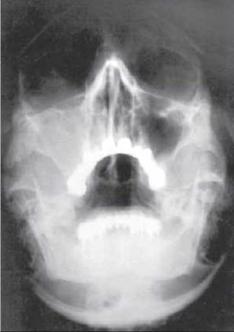
Fig. 307. Acute right-sided odontogenic maxillary sinusitis.
Subacute osteomyelitis presents with the following radiological features: multiple destructive lesions appearing as an irregular trabecular Structure, often accompanied by marginal defects on the alveolar process of the jaw containing residual tooth roots and the formation of sequestra. A small zone of sclerosis can be identified around the destructive foci.
Chronic osteomyelitis is characterized by an indolent course. Radiological signs: areas of destruction containing sequestra against a Background of sclerosis and hyperostosis. When osteomyelitis affects the Maxilla, the maxillary sinuses must be examined, as odontogenic osteomyelitis is frequently complicated by sinusitis (Fig. 307).
Periodontitis is a progressive inflammatory, degenerative, and dystrophic process affecting the periodontium, which can damage the alveolar processes of both jaws even in the presence of intact Teeth.
Radiological examination is used for early Diagnosis and subsequently serves as the primary METHOD FOR DETERMINING the disease's progression pattern and stage. The most informative imaging method is panoramic tomography, while conventional intraoral periapical radiography of the upper and lower jaws covering the incisor and premolar regions is also reliable.
Morphologically, the process is characterized by resorption of the alveolar crest with dental calculus deposits on the tooth roots. There are four stages of periodontitis.
The First stage begins with the deposition of dental calculus. Radiographically, bone resorption of the crests of the interdental and interradicular septa is observed up to 1/4 of the root length, along with regional Osteoporosis.
In the second stage, oral discomfort appears, accompanied by symptoms of gingivitis and heavy dental calculus accumulation. Radiological signs: bone resorption of the tooth sockets reaches 1/2 of the root length.

Fig. 308. Follicular cysts; teeth 18 and 48 are present in the follicles.
The Third Stage is characterized by purulent discharge from the gingival pockets and exposure of the tooth necks. Radiological signs: alveolar resorption reaches 2/3 of the root length.
In the Fourth Stage, purulent discharge from the gingival pockets is observed, and the teeth become loose and fall out. Radiological signs: complete resorption of the socket walls. The tooth roots are exposed and surrounded by granulation tissue.
Upon completion of the process after tooth loss, inflammation and suppuration cease. Radiologically, the formation of a new lamina dura is observed along the alveolar ridge of the edentulous, atrophied jaw.
Last update: 08/08/2026
Editorial and Educational Adaptation: This material has been compiled based on the primary/original source text. The project team performed an editorial review, corrected technical inaccuracies, structured sections, and adapted the content for an educational format.
What was processed:
- elimination of formatting defects (OCR errors, structural breaks, corrupted characters);
- editorial organization of content;
- standardization of terminology in accordance with academic sources;
- verification of factual statements against the original source text.
All mentions of the author, publication year, and origin of the primary text have been preserved in accordance with the source.