Vertebrate Zoology - V. M. Konstantinov 2011
Chordates
Terrestrial, or Tetrapod, Vertebrates — Vertebrates with Embryonic Membranes
Class Birds
Birds are an advanced, highly specialized branch of reptiles that have adapted for flight. The progressive organizational features that fundamentally distinguish birds from reptiles include: 1) a higher level of Nervous system development and, consequently, more diverse and sophisticated adaptive behavior; 2) a high and constant body Temperature associated with a significantly increased metabolic rate and more efficient thermoregulation; 3) the capacity for flight, which in the vast majority of cases does not preclude The ability to move along solid substrates or climb; 4) more advanced reproduction: laying and incubating eggs, brooding, protecting, and feeding the nestlings.
These traits have enabled birds, despite the relative evolutionary youth of the Class, to spread across the globe and inhabit A wide variety of environments. Due to their wide distribution and diverse living conditions, the class of birds exhibits greater species diversity and Abundance than reptiles. It comprises approximately 10,000 living species grouped into 30 — 35 orders.
Morphologically, birds are characterized by a body covered in feathers, forelimbs modified into wings, pneumatic bones, a Skull with a single occipital condyle, a four-chambered Heart with a single right aortic arch, and the complete absence of Teeth in modern birds, which are functionally replaced by a horny beak.
Morphophysiological Overview
Integument and its derivatives. The avian Skin is thin, lacks any bony structures, and is almost entirely devoid of glands. The only exception is the uropygial (preen) gland, located above the Base of the tail, the secretion of which is used to oil the feathers and render the plumage waterproof. The uropygial gland is highly developed in waterbirds, whereas in certain terrestrial species inhabiting arid climates, such as ostriches and bustards, it is absent.
Alongside the absence of bony structures in the skin, horny epidermal derivatives are abundant and diverse. For instance, the upper and lower jaws are covered in horny sheaths that form the beak. The tips of the digits bear claws, and the lower PARTS OF THE legs (toes, tarsometatarsus, and in some species the Tibia) are covered with horny scutes. The body is covered in feathers, which in the overwhelming majority of species are not distributed uniformly, but rather restricted to specific areas known as pterylae. Other regions, called apteria, are entirely or almost entirely devoid of feathers. This patchy feather arrangement, characteristic of flying birds, has adaptive significance because it facilitates Muscle contraction, skin mobility, and the shifting of body plumage associated with wing movements. Apteria play a similar role during the Movements of the hind limbs and neck (Fig. 126).

Fig. 126. Pterylae and apteria of a pigeon:
ventral (A) and dorsal (B) views; 1 — cervical pterylia; 2 — ventral pterylia; 3 — ventral apterium; 4 — cervical pterylia; 5 — humeral pterylia; 6 — alar pterylia; 7 — dorsal pterylia; 8 — femoral pterylia; 9 — crural pterylia; 10 — caudal pterylia; 11 — cervical apterium; 12 — lateral apterium
The total number of contour feathers is greater in large birds than in small ones. For example, the ruby-throated hummingbird has about 100, small passerines 1.5 — 2.5 thousand, gulls 5 — 6 thousand, ducks 10 — 12 thousand, and swans 25 thousand.
Bird feathers vary in Structure and function. The exterior of the body is covered with contour feathers, consisting of a hollow shaft to which two lateral plates, the vanes, are attached more or less symmetrically. The lower part of the shaft is embedded in the skin and is called the calamus; the large upper portion of the shaft bearing the vanes is called the rachis (Fig. 127). Each vane consists of numerous long barbs of the first order, which in turn bear barbs of the second order ( barbules). The latter are equipped with very tiny hooklets that interlock adjacent barbules. Consequently, the vane forms a resilient, elastic plate (Fig. 128).
Contour feathers form The basis of the plumage. They protect the bird's body from heat loss and mechanical impact, form the lifting surface of the wing and the steering plane of the tail. Depending on their position on the body, contour feathers are divided into groups. Thus, long feathers located along the trailing edge of the forelimbs and forming the wing blades are called remiges (flight feathers), long tail feathers are rectrices (tail feathers), feathers covering the upper and lower surfaces of the wings are wing coverts, the upper tail coverts cover the base of the tail, and so on. Beneath the contour feathers lie small down feathers. Their shafts are thin and lack second-order barbules, so the vanes do not form closed plates. Often the shaft of a down feather is so shortened that the barbs emerge from the tip in a single tuft; such a feather is termed true down. Down feathers and down are particularly well developed in waterfowl and species inhabiting cold regions. Their primary role is to reduce heat loss.
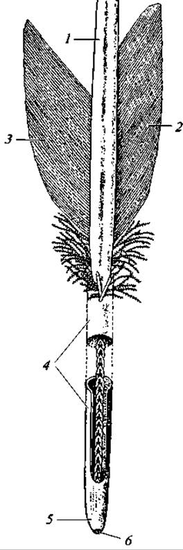
Fig. 127. Contour feather (calamus opened to show the inferior umbilicus):
1 — rachis; 2 — outer vane; 3 — inner vane; 4 — calamus; 5 — inferior umbilicus; 6 — inferior umbilicus margin (arch)
Sensory filoplumes are interspersed among the down, and many birds possess bristles at the corners of the Mouth. In insectivorous species that catch prey on the wing, these bristles form a funnel-like trap when the beak is open, increasing the efficiency of insect capture.
The initiation and Cytology/cytology/16.html">Early stages of feather development resemble reptilian scales. A feather germ, much like a horny scale primordium, is a dermal Connective Tissue papilla covered externally by the epidermis. As it grows, the bud tilts backward, and its base sinks deep into the skin, forming the follicle of the future feather and its Blood-rich papilla, through which the growing feather is nourished. The ectodermal portion of the primordium proliferates and differentiates into a longitudinal thickening—the future rachis—and two longitudinal keels that subsequently break down into the barbs of the vanes. Initially, the feather germ is covered on the outside by a thin horny sheath, which is later shed as the feather develops. Once this sheath is shed, the vanes are released, and their left and right halves spread apart (Fig. 129). Feathers are molted and replaced regularly. Many birds undergo not one, but two or three molts per year. In the latter case, usually not the entire plumage is replaced, but only specific areas. Multiple molts are associated with seasonal polymorphism and breeding plumage. The pattern of molting varies among birds. Birds of prey and aerial insectivores molt gradually and do not lose the ability to flight. Gallinaceous birds and inhabitants of forests, shrublands, and grassy thickets molt rapidly; during this time, they take flight with difficulty and remain in secluded spots, hiding in dense bushes or grass when danger approaches. Ducks, geese, swans, alcids, grebes, loons, and most rails exhibit a very distinct molting pattern. Their flight feathers are shed almost simultaneously, and the birds lose the ability to fly for an extended period. Geese, certain ducks, and swans gather during this time in remote, inaccessible areas along the shores of rivers, lakes, and seas, concentrating in massive numbers, sometimes reaching many thousands of individuals.
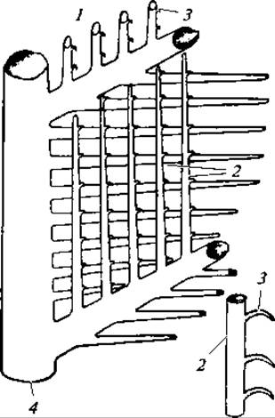
Fig. 128. Diagram of vane structure:
1 — barb; 2 — barbules; 3 — hooklets; 4 — rachis
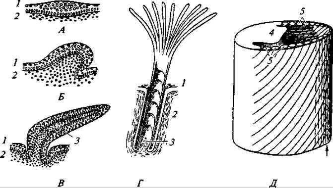
Fig. 129. Diagram of feather development:
A, B, and C — longitudinal sections through feather germs of different ages; D — sectional view of an embryonic feather; E — stereogram of a developing contour feather; 1 — epidermis; 2 — dermis (cutis); 3 — feather papilla; 4 — rachis primordium; 5 — barb primordia. After shedding the outer skin layer, the vane barbs are released and spread to both sides along the direction indicated by the arrow
Molting involves not only the replacement of plumage, but also changes in its structure (in A number of species). For instance, the summer plumage of the Eurasian siskin comprises approximately 1,500 feathers, whereas its winter plumage numbers 2,100 — 2,400; one species of tit has 1,100 feathers in summer and 1,700 in winter. In the willow ptarmigan, the length of dorsal contour feathers averages 5.4 cm in winter and 3.8 cm in summer; their downy sections measure 1.8 and 1.4 cm, respectively; and the aftershaft measures 3.7 — 2.5 cm (A. V. Mikheev, 1960).
Musculature. The avian musculature exhibits several distinctive features closely related to their primary life activities. First, The Muscular System is more differentiated than that of reptiles, driven by the complex movements required for flight, walking, climbing, and foraging. Second, the largest Muscles that drive the wings are located on the trunk, while tendons extend to the limbs themselves. Finally, third, to support the immense workload performed by the wings, the bulk of the musculature is situated not on the DORSAL SIDE OF the body, but on the chest, which houses the wing-stroke muscles.
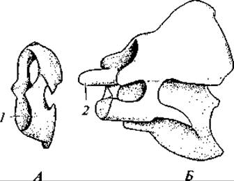
Fig. 130. Atlas (A) and axis (B) of a goose:
1 — articular facet for the occipital condyle; 2 — odontoid process
The limb muscles deserve special attention. The massive pectoral muscles, attached to the sternal keel and accounting for up to 20% of the bird's total body weight, serve to depress the wing. The subclavius muscles lying beneath them are somewhat smaller and serve to elevate the wings. The hind limbs feature a highly complex musculature (comprising up to 35 muscles). Of particular interest is the ambiens muscle found in some species. It originates on the pelvis, extends along the thigh, loops over the knee as a tendon, and then connects to the digital flexor. When a bird perches on a branch, it bends its knees, thereby pulling the ambiens muscle and consequently the digital flexor, which causes the toes to clamp down tightly around the branch. The lower the bird settles, the greater the tension on the ambiens muscle and the tighter the bird grips the branch with its toes. A sleeping bird is thus automatically held on its branch.
In other species (such as passerines), The Mechanism of automatic toe flexion is different. It is driven by a muscle known as the deep digital flexor. Its tendons, extending to the tips of the toes, have a heavily roughened lower surface and move within sheaths whose inner surfaces feature transverse ridges. When the bird lands on a branch and grasps it with its toes, the roughened surface of the tendons is pressed against the sheath under the bird's weight and locks onto its ridges. As a result, the toes are fixed in a flexed position and securely grip the branch without any muscular effort.
Skeleton. The avian skeleton possesses a number of specific features associated with adaptations for flight and terrestrial locomotion exclusively on the hind limbs. These structural peculiarities lie, first, in the unique modifications of the limbs and their girdles, and second, in the lightweight yet robust Nature of the entire skeleton. Its lightness is ensured by the pneumaticity of most bones, while its strength results from the mineralization and fusion of many bones at early Stages of Ontogeny; consequently, in adult birds, the sutures between individual bones are often completely indiscernible.
Vertebral Column. THE VERTEBRAL COLUMN comprises cervical, thoracic, lumbar, sacral, and caudal regions. The cervical region is notably long and highly mobile. This mobility is enabled by the distinctive shape of the vertebrae, which feature saddle-shaped articular surfaces. Such vertebrae are termed heterocoelous.
The number of cervical vertebrae varies among bird species, ranging from 11 to 25. The first two cervical vertebrae exhibit the typical amniote structure, i.e., the atlas and axis (Fig. 130). The cervical region participates in complex HEAD movements. The angle of head rotation typically reaches 180°, and in owls even 270°.
The thoracic vertebrae (ranging from 3 to 10) are fused to each other and to the synsacrum. They bear Ribs that articulate movably with the Sternum. Each rib consists of two parts—vertebral and sternal—which are movably joined to form an angle pointing backward. Due to this rib structure, the contraction of corresponding muscles allows the sternum to move either toward or away from the vertebral column. This alters the volume of the thoracic cavity and plays a vital role in the respiratory mechanism.
The sternum has a unique structure. It is a broad, slightly outward-curved bony plate to which the thoracic ribs articulate. In modern flying birds, the sternum bears a bony keel. The wing-stroke muscles attach symmetrically to the left and right of this keel. Only ratites (a few species that secondarily lost the ability to fly) lack a keel, their sternum being only slightly convex. In penguins, the keel is strongly developed because their forelimbs perform heavy work during swimming.
The lumbar vertebrae fuse with one another, with the iliac bones, and with the sacral vertebrae. A portion of the caudal vertebrae also fuses with the sacral vertebrae. Consequently, this forms the complex sacrum (synsacrum) characteristic of birds, consisting of A large number of vertebrae (from 10 to 22). However, birds retain only two true sacral vertebrae, just like reptiles. The synsacrum has important adaptive significance, providing support for the bird's body solely on the hind limbs during terrestrial locomotion. There are 6 to 9 free caudal vertebrae; the caudal region terminates in a vertical bony plate—the pygostyle—which represents several fused vertebrae. The pygostyle serves as a supporting base for the attachment of tail feathers.
Skull. The overall structural plan of the avian skull is close to that of reptiles. The occipital region is composed of the usual four occipital bones (basioccipital, two exoccipitals, and supraoccipital). There is a single occipital condyle, as in reptiles. The auditory capsule is formed, similarly to reptiles, by three otic bones, which fuse into a single bone in adult birds.

Fig. 131. SKULL OF A young pigeon:
lateral view (A); ventral view (B); dorsal view (C); bones: 1 — basioccipital; 2 — exoccipital; 3 — occipital condyle; 4 — foramen magnum; 5 — supraoccipital; 6 — otic; 7 — basisphenoid; 8 — presphenoid; 9 — alisphenoid; 10 — orbitosphenoid; 11 — interorbital septum; 12 — mesethmoid; 13 — parietal; 14 — frontal; 15 — nasal; 16 — lacrimal; 17 — premaxilla; 18 — Maxilla; 19 — jugal; 20 — quadratojugal; 21 — squamosal; 22 — quadrate; 23 — Vomer; 24 — palatine; 25 — articular; 26 — dentary; 27 — angular
The floor of the skull is formed by the basisphenoid and presphenoid bones, along with the palatines and pterygoids. The skull roof is formed by the paired nasals, frontals, parietals, and squamosals. The upper jaw is represented by the premaxillae and maxillae. The latter articulate posteriorly with rod-like jugal and quadratojugal bones, which in turn connect to the quadrate bones. This forms the lower temporal bar characteristic of birds, which separates the Orbit from the temporal fossa. The lower jaw consists of the articular bone (homologous to Meckel's Cartilage) and dermal bones: the dentary, splenial, angular, and surangular (supraangular) (Fig. 131).
The hyoid apparatus is bony, taking the form of an elongated plate with very long horns homologous to the first pair of gill arches, and lies in the laryngeal region.
There is a single auditory ossicle (the columella or stapes), just as in reptiles.
Along with similarities in the General structural plan, the avian skull differs significantly from the reptilian skull in several adaptive features. Notable are the relatively large volume of the braincase and the enormous orbital cavities, which are associated with the high Development of the Brain (primarily its hemispheres) and very large eyes, serving as a vital sensory organ in birds. The jaws are greatly elongated, and their bones fuse together, creating an exceptionally efficient and robust grasping apparatus. The BONES OF THE braincase are thin and fuse very early, making the braincase both light and strong. Additionally, a number of skull bones are pneumatic.
Forelimbs and their girdle. In connection with flight adaptations, the forelimbs and their girdle exhibit a series of distinctive features. The girdle consists of the scapula, coracoid, and clavicle, whose proximal ends form the socket for the Articulation of the humerus. The scapulae are long, saber-shaped, and lie upon the ribs, over which they can glide freely. The large, strongly developed coracoid abuts the sternum at one end, while the other end provides support for the shoulder articulation. The left and right clavicles fuse to form the furcula (or 'wishbone') characteristic of birds, imparting special elasticity to the girdle (Fig. 132).
The Skeleton of the wing itself comprises all the typical divisions of a pentadactyl limb. Here, the humerus and forearm remain essentially unmodified, whereas the manus (hand), conversely, displays several unique peculiarities. The carpus is heavily reduced; its proximal elements fuse into two small bones, while the distal elements fuse with the metacarpus. The metacarpus consists of two elongated bones fused at both the proximal and distal ends, ultimately forming a complex carpometacarpus (the 'buckle' or composite bone). The primary joint is the intercarpal joint. Of the digits, only three are retained—the second, third, and fourth—with only the third digit possessing two Phalanges, while the second and fourth have one each.
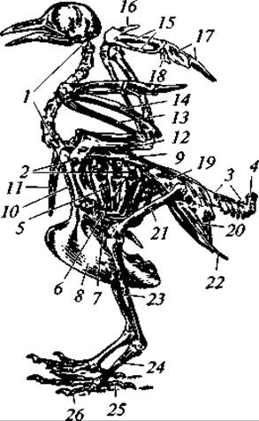
Fig. 132. Skeleton of a pigeon:
1 — cervical vertebrae; 2 — thoracic vertebrae; 3 — caudal vertebrae; 4 — pygostyle; 5 — vertebral rib with uncinate process; 6 — sternal rib; 7 — sternum; 8 — sternal keel; 9 — scapula; 10 — coracoid; 11 — clavicle; 12 — humerus; 13 — radius; 14 — ulna; 15 — carpometacarpus; 16 — digit II of the wing; 17 — digit III of the wing; 18 — digit IV of the wing; 19 — ilium; 20 — ischium; 21 — Femur; 22 — pubis; 23 — tibiotarsus (shank); 24 — tarsometatarsus (tarsus); 25 — digit I of the FOOT; 26 — digit IV of the foot
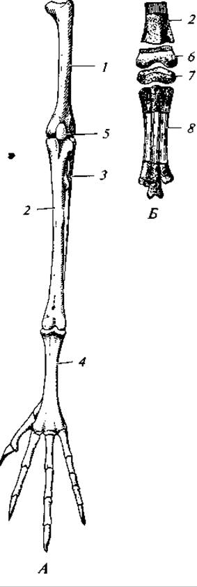
Fig. 133. Skeleton of a pigeon's hindlimb (A) and part of a nestling's hindlimb (B):
1 — femur; 2 — tibia; 3 — reduced Fibula; 4 — tarsometatarsus; 5 — Patella; 6 — fused proximal tarsal elements that later fuse with the tibia; 7 — fused distal tarsal elements that later fuse with the bases of the metatarsals; 8 — fusing Metatarsal Bones
Due to the presence of specialized muscles, tendons, and skin membranes, the bones of the wing are interconnected in such a way that the entire wing cannot be fully extended in a straight line, and its segments remain at an angle to one another. The limb segments can move in only one plane — the plane of the wing — allowing it to fold and unfold.
Hindlimbs and Pelvic Girdle. The hindlimbs and their girdle also possess a number of distinctive features associated with the fact that during locomotion, the entire weight of the body is supported by the hindlimbs. The strength of the pelvis is ensured by the fusion of the large iliac bones along their entire length with the complex sacrum. Very large ischial bones also fuse with the ilia. Conversely, the pubic bones are small, appearing as thin rods attached to the outer margin of the ischium. All three pelvic bones participate in The formation of the acetabulum. On the ventral side, the left and right halves of the pelvis are widely spaced, which is related to the laying of large, hard-shelled eggs.
The femur is of typical structure. The lower leg consists of two typical bones, but only the tibia develops fully, while the fibula is vestigial and fuses with the former. The proximal row of Tarsal Bones fuses with its distal part. This fusion is so complete that in adult birds, the sutures are entirely obliterated. The limb segment following the lower leg is called the tarsometatarsus. In the adult bird, it consists of a single long bone. However, embryonically, this bone arises from the fusion of the metatarsal bones and the lower (distal) row of tarsal bones. Consequently, the ankle joint in birds (as in reptiles) is located between the two rows of tarsal bones rather than between the tarsus and the lower leg, as in amphibians. This joint is more accurately termed the intertarsal joint (Fig. 133).
Birds most commonly have four toes, less frequently three, and in only one case (the African ostrich) — two.
Digestive System. All modern birds lack teeth; their function as an instrument for capturing and holding food is performed by the horny sheaths covering the upper and lower jaws. The shape of the beak varies greatly and is in direct correlation with The Nature of the food and the Methods of obtaining it. For example, in birds of prey, most of the beak is covered with a thick horny sheath and is hooked at the tip. Granivorous birds have a conical beak adapted for cracking the hard seed coats. Anseriformes have a flattened beak equipped with lamellae that function as a straining apparatus. In pelicans, a large skin pouch is suspended between the rami of the lower Mandible, serving to scoop up fish.
A Tongue is attached to the floor of the Oral Cavity, and its shape is equally diverse. In birds of prey, it is short and stiff; in waterfowl, fleshy and flattened; in woodpeckers and wrynecks, very long and slender. In nectar-feeding birds (such as hummingbirds and honeycreepers), the tongue is highly mobile and rolls into a tube through which they suck nectar.
Salivary Glands vary in development among birds and are almost absent in some species (e.g., nightjars). Saliva moistens food, facilitating swallowing. Salivary gland secretion is especially abundant in swiftlets (genus *Collocalia*), which build their nests — popularly known as "birds' nests" — from saliva that hardens in the air. In some birds, the enzyme amylase is present in the saliva, so carbohydrate Digestion begins already in the mouth.
The long Esophagus in certain birds (such as raptors, gallinaceous birds, and pigeons) forms an expansion known as the crop. It serves for the temporary storage of swallowed food. In pigeons, the walls of the crop during the nestling-rearing period secrete a fatty, curd-like substance — the so-called "crop milk" (containing over 10% protein and 12–15% fat) — with which the birds feed their young. The secretions of the esophageal walls are also used to feed nestlings in tubenoses, flamingos, and certain other birds.
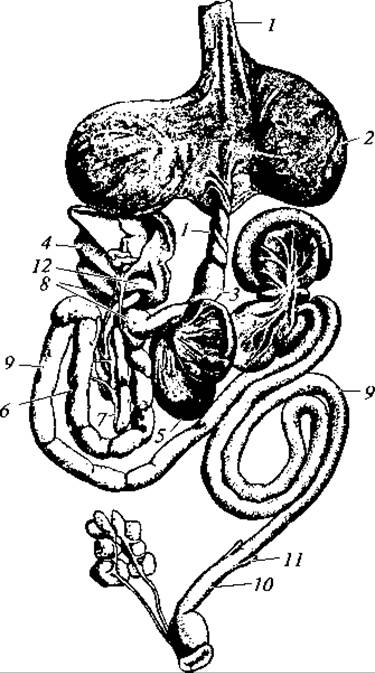
Fig. 134. Digestive system of a pigeon:
1 — esophagus; 2 — crop; 3 — proventriculus (glandular Stomach); 4 — Liver; 5 — gizzard (muscular stomach); 6 — duodenum; 7 — Pancreas; 8 — Bile ducts; 9 — Small Intestine; 10 — Large Intestine; 11 — caeca; 12 — Spleen
The esophagus leads into the thin-walled glandular stomach, where food is acted upon by digestive secretions. The glandular stomach is followed by the thick-walled muscular stomach (gizzard), whose inner surface is lined with a tough, horn-like cuticle. Here, food is ground down by the contractions of the powerful muscular walls (up to 30 contractions per second) and stones present in The Stomach cavity, which act as millstones. The degree of development of the gizzard correlates with the nature of the diet. It is least developed in insectivorous and carnivorous birds, well developed in granivorous species, and reaches its maximum development in gallinaceous birds, which feed on coarse vegetative plant parts (conifer needles, twig tips, buds). In granivorous birds, pressures of up to 20–30 kg/cm2 can be generated within the gizzard. Digestion is rapid; for instance, in the house sparrow, grain is digested in 3–4 hours, beetles in 1 hour, and caterpillars in 15 minutes.
The small intestine is relatively long. The pancreas lies within the duodenal loop. The well-demarcated posterior section of the intestine is relatively short and is not differentiated into a colon and rectum. At the junction of the small and large intestines, most birds have two small caeca. The short large intestine opens into the cloaca, on the dorsal side of which many birds have a blind pouch — the bursa of Fabricius — which apparently acts as an endocrine gland and participates in the body's immunological defense. It decreases in size with age.
The relative length of the intestine varies and depends on the dietary habits. Thus, in the African ostrich, the intestine is 20 times the body length; in the cormorant and kite, 11–12 times; in diurnal raptors, 7–8 times; and in most insectivores, 4–7 times.
The liver is large and bilobed. A Gallbladder is present in most species (though absent in the pigeon). The bile duct opens into the duodenum (Fig. 134).
Thus, the General characteristics of the Avian Digestive System are as follows: 1) absence of teeth, functionally replaced partly by the horny beak (for capturing and holding food) and partly by the gizzard (for mechanical grinding of food); 2) relatively short intestinal length (whereas in herbivorous mammals the intestine is 20 times the body length, in herbivorous birds such as gallinaceous birds it is only about 8 times longer than the body); 3) division of the stomach into two compartments, necessitated by the inability to chew food in the Oral Cavity and The Need for intensive chemical Processing due to the shortened intestine; 4) lack of Differentiation of the hindgut into a colon and rectum, which should apparently be regarded as an adaptation for weight reduction, since The primary function of the rectum is limited to the temporary storage of fecal matter and Water reabsorption; 5) presence of the bursa of Fabricius, a specialized endocrine gland.
Respiratory system. The respiratory Organs are highly specialized and adapted to an aerial lifestyle to a greater extent than any other internal organ system.
The glottis leads into the Trachea, the upper part of which forms the Larynx, supported by an unpaired cricoid cartilage and paired arytenoid cartilages. This laryngeal structure in birds is known as the cranial (or upper) larynx and does not function as a vocal organ. That function is performed by the syrinx (lower larynx), which is unique to birds. It is located at the bifurcation of the trachea into the two Bronchi and represents an expansion supported by cartilaginous rings (Fig. 135). Projecting into the lumen of the syrinx from its lateral walls are the external tympanic membranes, while the internal tympanic membranes project from below at the tracheal branching point. By virtue of specialized syringeal muscles, these membranes can alter their position and tension, which accounts for the variety of sounds produced.
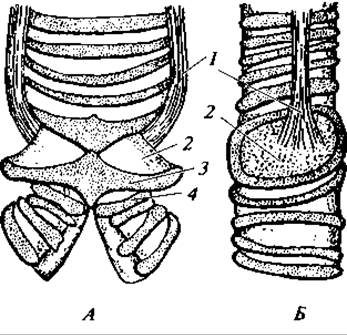
Fig. 135. Syrinx (lower larynx) of a pigeon:
anterior view (A) and lateral view (B); 1 — tracheobronchial muscles; 2 — external tympanic membrane; 3 — last tracheal ring; 4 — first bronchial ring
The Upper Respiratory Tract plays a crucial role in thermoregulation. It has been established that when ambient temperatures rise, avian respiration becomes markedly faster and shallower. Simultaneously, there is a pronounced vasodilation of the Blood Vessels in the oral and pharyngeal cavities, resulting in enhanced heat dissipation from the bird's body.
The Lungs of birds are not hollow sacs, as in amphibians and partly in reptiles, but dense spongy structures attached to the dorsal wall of the thoracic cavity. Upon entering the lungs, the bronchi branch dichotomously multiple times, and their main branches pierce right through the lungs to open into the air sacs (see below). The bronchial branches are interconnected by fine channels known as tertiary bronchi or parabronchi. The walls of the parabronchi feature small depressions interwoven with blood capillaries, which is precisely where the blood is oxygenated.
As mentioned, some of the bronchial branches extend beyond the lungs proper and expand into massive, thin-walled air sacs whose volume is roughly 10 times that of the lungs.
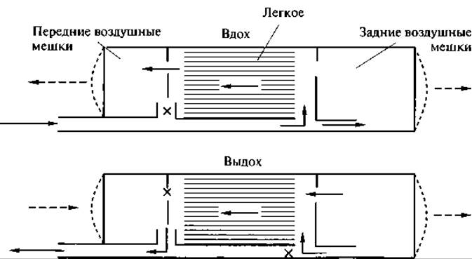
Fig. 136. Diagram of airflow in the avian respiratory system (after K. Schmidt-Nielsen, 1976).
Solid arrows indicate the direction of airflow, dashed arrows show the expansion or contraction of the air sacs, and the 'x' mark denotes points where airflow is blocked during a given phase of respiration.
The air sacs are located between various Internal Organs, with their extensions running between muscles beneath the skin and penetrating pneumatic bones. Birds possess several air sacs: two cervical, one interclavicular, two to three pairs of thoracic, and one pair of very large abdominal sacs.
The Significance of the air sacs is profound and multifaceted. Their primary role is to drive the respiratory mechanism. As they expand, air is forcefully drawn through the central bronchi into the posterior air sacs. During the first inhalation, oxygen-rich air enters the posterior air sacs. During the first exhalation, air with a high oxygen content moves from the posterior air sacs into the lungs. It is during this phase of respiration that the primary oxygenation of the blood occurs.
During the second inhalation, air moves from the lungs into the anterior air sacs. During the second exhalation, air is expelled from the anterior air sacs to the outside. Thus, airflow in the avian respiratory system always moves in a single direction (Fig. 136): from the posterior air sacs through the lungs into the anterior air sacs and out of the body.
In addition to supporting respiration (moving air through the lungs), the air sacs serve other Functions. For instance, during intensive muscle exertion in flight, they protect the bird from overheating, as relatively cool air flows around virtually all internal organs and partially over the musculature. The air sacs also reduce friction between organs during flight. Finally, they decrease body density, increase intra-abdominal pressure, and facilitate defecation.
Respiration rates vary among species. In a resting pigeon, the average breathing rate is 26 breaths per minute, increasing to 77 while walking, and reaching 400 during flight. Pulmonary ventilation thus exceeds metabolic gas exchange requirements by 2.5 times, serving to dissipate excess heat via pulmonary evaporation. It should be noted that heat production during flight is 8 times higher than at rest. Small birds generally have a higher respiration rate than large ones: the average respiratory rate per minute is 30–43 for a duck, and 90–100 for small passerines.
Small birds have a higher metabolic rate and consume significantly more oxygen than large ones. For example, a hummingbird with a body mass of 3 to 7 g consumes 4 to 10 ml of oxygen per hour per gram of body weight; the Siberian jay (mass 71 g) consumes 1.75 ml, the pigeon (mass 150 g) consumes 0.98 ml, and the emu (mass 38 kg) consumes 0.023 ml. These Examples confirm the general rule of an inverse relationship between body size and metabolic intensity in homoeothermic animals. For comparison, in phylogenetically lower reptiles, this value is only 0.1–0.3 (L. Prosser, 1977).
Circulatory organs. The primary feature of the Circulatory system in birds is the complete Separation of arterial and venous blood, which is determined by The structure of their heart and the efferent system of arterial arches (Fig. 137).
The Heart is four-chambered, consisting of two atria and two ventricles. The relative size of the avian heart compared to other vertebrates is large, which is associated with a High Metabolic Rate, especially during flight. For instance, the cardiac index (heart mass/body mass, %) in a duck is 0.6, whereas in a rabbit of similar mass it is only 0.2. Small birds have a relatively larger heart mass than large ones due to their more intensive METABOLISM (which, in turn, stems from the less favorable body volume-to-surface area ratio in smaller animals regarding heat loss). For example, in a bullfinch with a body mass of 23 g, the relative heart mass is 1.3%, in a lesser redpoll (mass 13 g) it is 1.6%, and in a coal tit (mass 8 g) it is 1.8%.
There is also a correlation between relative heart mass and the vigor of movement. In a strong flyer like the Eurasian hobby, heart mass averages 1.7% of body mass; in a weaker flyer like the common kestrel, it is 1.2%; and in a poor flyer like the Eurasian magpie, it is only about 0.9%.
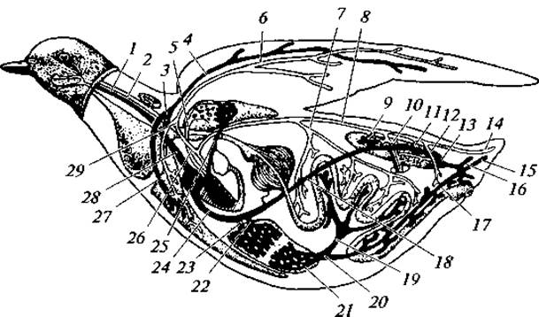
Fig. 137. Blood Circulation in the pigeon:
1 — jugular vein; 2 — carotid artery; 3 — subclavian Veins; 4 — brachial vein; 5 — Pulmonary veins; 6 — brachial artery; 7 — visceral artery; 8 — dorsal aorta; 9 — renal Arteries; 10 — sciatic artery; 11 — renal artery; 12 — iliac artery; 13 — internal iliac vein; 14 — middle caudal artery; 15 — caudal vein; 16 — tail vein; 17 — coccygeomesenteric vein; 18 — INFERIOR VENA CAVA; 19 — mesenteric vein; 20 — HEPATIC PORTAL VEIN; 21 — hepatic portal system; 22 — right hepatic vein; 23 — left hepatic vein; 24 — inferior vena cava; 25 — pulmonary artery; 26 — tracheobronchial artery; 27 — thoracic vein; 28 — SUPERIOR VENA CAVA; 29 — Subclavian Artery
The avian heart functions more vigorously than that of lower terrestrial vertebrates. For example, the marsh frog has a heart rate of 40–50 beats per minute, compared to an average of 730 beats per minute in the bullfinch. The dependence of heart rate on body size is clearly evident. In a pigeon weighing 250 g, the average heart rate is 248 beats per minute; in the European greenfinch (mass 22 g), it is 697; in the European goldfinch (mass 13 g), it is 754; and in the coal tit (mass 8 g), it is 1037. Heart rate also depends on the bird's state of activity. For instance, in a resting pigeon, the heart rate averages 165 beats per minute, rising to 550 during flight. When diving, birds experience bradycardia, i.e., a decrease in heart muscle contraction frequency. In the white-throated dipper, 5 seconds after immersion in water, the heart rate drops to 73% of the initial rate, down to 48% after 10 seconds, and to 42% after 15 seconds.
A progressive evolutionary trait in birds is the overall increase in blood volume. In teleost Fishes, blood mass accounts for approximately 3% of body mass; in tailless amphibians, it is 6%; and in birds, it reaches 9%. The oxygen capacity of avian blood is roughly doubled compared to that of reptiles. All these organizational features serve as a vital prerequisite for elevating the overall metabolic rate in birds.
A single right aortic arch departs from the left ventricle, soon branching into paired anonymous arteries, which in turn divide into paired carotid and subclavian arteries. This is how the head, Pectoral Girdle, and forelimbs are supplied with blood. The aortic arch curves around the right side of The Heart and extends beneath the spine toward the caudal region of the body as the dorsal aorta. Unpaired visceral and mesenteric arteries branch off the dorsal aorta to supply the stomach and intestines. Paired femoral and sciatic arteries supply blood to the abdominal wall muscles, pelvic organs, and the hindlimbs with their girdle.
The common pulmonary artery, carrying venous blood, arises from the right ventricle. It divides into two short branches: the left and right pulmonary arteries.
Venous blood from the head collects into paired jugular veins. Merging with the paired subclavian veins, they form the left and right anterior venae cavae, which empty blood into the right atrium. The venous System of the trunk region resembles that of reptiles. The main difference is that the abdominal vein, which in reptiles forms the hepatic portal system along with several smaller veins, is functionally replaced in birds by the coccygeomesenteric vein. Furthermore, the renal portal system is partially reduced. The posterior vena cava empties into the right atrium.
Due to the complete separation of the SYSTEMIC AND PULMONARY circuits, all organs are supplied with pure arterial blood. This circumstance, combined with rapid blood circulation and vigorous gas exchange, results in a high body temperature in birds, averaging 42°C. In large birds, it is typically 38–40°C, whereas in small species it can reach 45°C.
Further confirmation of the high metabolic level in birds is found in their blood pressure. For example, in the pigeon it is 135/105 mm Hg, compared to 80/60–14/10 mm Hg in squamate reptiles (L. Prosser, 1977).
An important and fundamentally new physiological trait of birds is The Diversity of their thermoregulation mechanisms, encompassing both chemical and physical thermoregulation. The former involves altering metabolic intensity—i.e., The rate of heat production—in response to ambient temperature and the quantity and quality of food consumed. For instance, a drop in external temperature from 33 to 10°C causes a threefold increase in oxygen consumption in the house sparrow. Physical thermoregulation involves modifying the rate of heat loss. Of considerable importance in birds is so-called thermal panting (or polypnea): accelerated breathing leads to increased heat dissipation via exhaled air and moisture evaporation from the respiratory organs and tracts. Through this mechanism, small birds can dissipate about half of the heat accumulated in their bodies. In large birds, heat dissipation in this manner can even exceed heat production. Consequently, animals such as ostriches and even pigeons can withstand ambient temperatures of 51°C with almost no overheating.
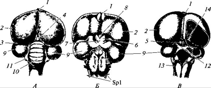
Fig. 138. Brain of a pigeon:
view from the dorsal (A) and ventral (B) sides, with opened ventricles and removed Cerebellum (C); 1 — olfactory lobes of the Forebrain; 2 — cerebral hemispheres; 3 — Diencephalon; 4 — Pineal Gland (epiphysis); 5 — optic lobes of the Midbrain; 6 — optic tracts; 7 — optic chiasm; 8 — infundibulum with Pituitary Gland; 9 — optic lobes of the midbrain; 10 — cerebellum; 11 — lateral cerebellar projections; 12 — optic commissure; 13 — Medulla Oblongata; 14 — corpora striata; Spl — first pair of Spinal Nerves
Nervous system. The Central Nervous System of birds is more complex than that of reptiles. This is due to a higher level of vital activity and more complex interactions between birds and their living conditions. Nervous-reflex activity and adaptive behavior in birds are quite diverse and sophisticated. Morphologically, this is due to the relatively large size of the brain. In reptiles, the brain mass is roughly equal to that of the Spinal Cord; in birds, the brain is always larger: 1.5 times larger in gallinaceous birds and 2.5 times larger in pigeons. Its enlargement is driven by The Development of the cerebral hemispheres (Fig. 138). For example, The ratio of the mass of the forebrain to the mass of all other brain regions is 1:1 in gallinaceous birds, 2:1 in birds of prey, and up to 3:1 in passerines and parrots. However, just like in reptiles, most of the forebrain is formed by the corpora striata and the expansion of the archipallium in the floor and walls of the Lateral ventricles. Conversely, the pallial roof is relatively weakly developed compared to mammals, for instance, and is represented by a thin layer of Nerve Cells. The olfactory lobes are small, which is associated with the underdevelopment of the olfactory organs. In the diencephalon, on its upper surface covered by the cerebral hemispheres, lies the pineal gland, while a large pituitary gland is located on the floor behind the optic chiasm. The cerebellum is very large, consisting predominantly of a median lobe—the vermis, marked by characteristic transverse furrows. The development of the cerebellum is associated with complex movements during flight that require coordination. Due to the development of the cerebellum, the optic lobes of the midbrain are strongly displaced laterally. There are eleven pairs of Cranial Nerves.
The spinal cord, similar to that of reptiles, features enlargements in the Regions of the brachial and lumbar nerve plexuses.
Sense Organs. The Organ of Hearing comprises three divisions; as in reptiles, there are inner and middle ears. The cochlear duct is better developed and separated from the saccule by a constriction. The Eustachian tubes open into the Pharynx. There is a single auditory ossicle in the Middle ear. Birds possess a distinct External ear. The tympanic membrane lies somewhat deeper than the skin surface, at the bottom of a small funnel-shaped depression that represents the external auditory meatus. In some nocturnal birds, such as owls, the cavity of this depression is large and bordered by a skin fold that enhances hearing sensitivity, while the surrounding feathers ensure better sound capture and directionality. Birds have acute hearing, within the range of 30 Hz – 30 kHz, and The Organ of hearing, along with the eyes, is one of their most vital sense organs. Often, birds detect danger primarily through their sense of hearing. During hunting, owls orient themselves mainly by sound, locating the sound source from a distance of 20 – 25 m with an accuracy of up to 1°, and catching a running mouse almost without a miss.
It has been established that some birds, such as the cave swiftlet (Collocalia fuciphaga), possess the ability for echolocation. They emit sound pulses with a frequency of 1.5 – 4.5 kHz repeating 9 – 10 times per second. This bird can fly in complete darkness within caves, successfully avoiding collisions with other birds and surrounding objects. It is believed that penguins use echolocation when hunting in water, and curlews in fog.
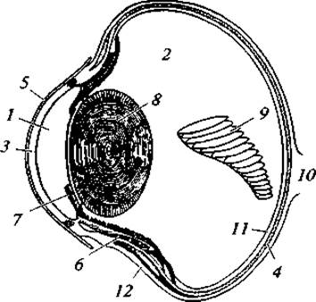
Fig. 139. Cross-section of the eye of a bird of prey:
1 — anterior chamber; 2 — posterior chamber filled with vitreous body; 3 — cornea; 4 — choroid; 5 — sclera (connective tissue tunic); 6 — ciliary muscle; 7 — iris; 8 — lens; 9 — pecten; 10 — Optic nerve; 11 — retina; 12 — sclera
The organs of Vision in birds are exceptionally well developed (Fig. 139). Unlike amphibians, reptiles, and mammals, there are no bird species with underdeveloped eyes. The eyeballs are relatively large, especially in species active at twilight and at night, or in those that spot prey from a distance. The ratio of Eyeball mass to total body weight in species with different biology is as follows: geese (feeding on grass) — 1/570; magpies (feeding on slowly moving prey, such as insects, which they search for nearby) — 1/70; falcons (feeding on birds, usually caught in flight, or insects spotted from afar) — 1/40; owls (feeding at night mainly on small rodents) — 1/30. In various bird species, there are from 50 to 300 thousand photoreceptors (rods and cones) per 1 mm2 of the retina, and up to 500 thousand to 1.5 million in the area of acute vision. Different combinations of rods and cones in various species allow them to distinguish either many details of an object or its contours under low illumination. The most sensitive spot on the human retina—the macula—contains 200 thousand photoreceptors. By this measure, diurnal birds of prey surpass humans in visual acuity by 8 times (Fig. 140). The peregrine falcon, for example, reacts to a moving bird at a distance of up to 1,100 m; the merlin—up to 800 m.
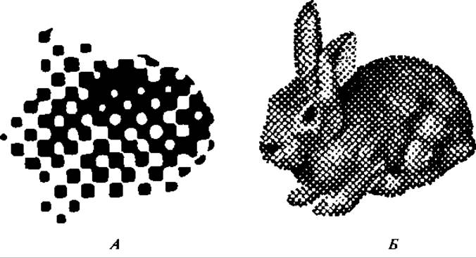
Fig. 140. Retinal perception of a rabbit image by a human
(A) and a hawk (B)
In most birds, vision is nearly monocular. The field of view of each eye is approximately 150°, while the binocular visual field is only 30 – 50°. In owls, vision is largely binocular, which ensures precise distance determination to the prey.
Accommodation in the avian eye is achieved in two ways: 1) by changing the shape of the lens through the action of the ciliary muscle, and 2) by altering the distance between the lens and the retina, caused by the action of annular Muscles surrounding the sclera that change the shape of the eyeball. In addition to the upper and lower eyelids, there is a third eyelid, or nictitating membrane, attached by its base to the inner (anterior) margin of THE EYE AND capable of drawing across the entire eye like a window blind.
Olfaction is poorly developed. Apparently, only some birds are capable of perceiving scents well, such as the New Zealand kiwis. Compared to other species, the SENSE OF SMELL is better developed in Procellariiformes, shorebirds, ducks, and vultures. Morphologically, the olfactory organs of birds differ from those of reptiles by a greater differentiation of the nasal conchae.
Excretory organs. The excretory organs are arranged quite similarly to those of reptiles. In avian embryos, trunk Kidneys (mesonephros) form, which are subsequently replaced, as in other amniotes, by pelvic kidneys (metanephros).
The relative size of The Kidneys in birds is larger than in reptiles and even mammals. For instance, in the starling it accounts for 1.2% of body weight, in terns — 1.6%, and in the merganser — 2.6%. The enlargement of the kidneys is directly linked to the very intense overall metabolism in birds. This is also evidenced by the large number of nephrons, numbering in the tens of thousands. In the structure of nephrons, as in reptiles, a reduction of the vascular glomerulus is characteristic; urine production occurs largely due to the secretory activity of the convoluted tubules. The main product of Protein metabolism, as in reptiles, is uric acid. This type of protein metabolism is undoubtedly caused, firstly, by the terrestrial development of the embryo within an egg, where it is virtually devoid of the ability to take up water from the external environment, and secondly, by the necessity of sequestering metabolic products within a closed egg system. Naturally, in such a situation, a urea-based type of metabolism could not emerge, as it requires a very high water expenditure during urination. Furthermore, uric acid has the lowest toxicity, allowing this metabolic product to be stored within the egg system throughout the entire developmental period. Thus, birds provide yet another example where a uricotelic type of metabolism arises in the "closed" egg system, rather than a ureotelic one as found in fishes, amphibians, and mammals.

Fig. 141. Reproductive organs of a male (A) and female (B) bird:
1, 3 — Testes; 2 — renal lobes; 4 — Ureters; 5 — vas deferens; 6 — cloaca; 7 — ovarian follicles (Graafian follicles); 8 — oviduct funnel; 9 — Uterus; 10 — albumin-secreting region of the oviduct; 11 — opening of the intestine
Urine passes through the urinary tracts very quickly, which is apparently related to the poor solubility of uric acid and the potential for urinary salts to clog the ducts. The absence of a Urinary Bladder in birds is also associated with this. The explanation sometimes given in textbooks linking the absence of the bladder to weight reduction is unfounded. Water loss during urination in birds is negligible because water is reabsorbed from the urine back into the body within the cloaca. This circumstance, along with the absence of any significant cutaneous evaporation, results in birds having an extremely low demand for water intake. Many species do not drink water at all, such as most birds of prey and certain passerines.
Reproductive organs. The testes are a pair of bean-shaped bodies located above the upper lobe of the kidneys and suspended by a mesorchium (Fig. 141). The size of the testes varies by season. In the chaffinch, for example, the volume of the testes increases 1,125-fold during the breeding season, and 1,500-fold in the starling. Weakly defined epididymes, homologous to the anterior parts of the embryonic trunk kidneys, adjoin the inner margins of the testes. Vas deferens tubes extend from the epididymes, running parallel to the ureters and opening into the cloaca. In some species, the vas deferens forms enlargements before entering the cloaca—Seminal Vesicles that serve as a sperm reservoir.
Copulatory organs are present in only a few species. Corresponding cloacal protrusions are found in Anseriformes, tinamous, and ratites. In herons, storks, and bustards, they are rudimentary. The majority of birds lack copulatory organs, and internal Fertilization is achieved by bringing the cloacal openings of the male and female into close proximity, whereby sperm is transferred into the female's reproductive tract.
The FEMALE Reproductive System, with very few exceptions, is strikingly asymmetric and consists of the left Ovary and left oviduct (see Fig. 141). The right ovary develops only occasionally in diurnal raptors, owls, loons, parrots, rails, and gallinaceous birds, and is particularly common in parrots. However, the right ovary very rarely functions. Curiously, in most cases, ova matured in the right ovary are shed into the left oviduct. The reduction of the right half of the reproductive system in female birds is apparently due to the laying of relatively large, hard-shelled eggs and their prolonged passage. The egg takes 1 to 2 days to move through the oviduct.
The avian ovary is an irregular, granular body lying anterior to the left Kidney. Its size varies considerably depending on the maturity and size of the ova developing within it. The oviduct is a long tube, one end of which opens via a funnel into the body cavity, while the other (lower) end opens into the cloaca. A mature ovum, rich in yolk, passes from the body cavity into the funnel of the oviduct, where it is fertilized and transported further down. The oviduct proper consists of several sections. The initial section, along almost its entire length (except for the uppermost part), is very rich in glands that secrete albumen, coating the passing egg in a thick layer. In chickens, the egg spends 3 to 6 hours in this section. In the next, narrower section, the egg is covered with two parchment-like shell membranes. This is followed by the so-called uterus—a section rich in glands that form the calcareous eggshell, its pigment, and a thin cuticle. The egg remains in the uterus for 12 to 20 hours. The final section of the oviduct (the Vagina) is short and muscular; from it, the egg passes into the cloaca and then to the outside.
The total passage time of an egg through the oviduct is about 24 hours in chickens and 41 hours in pigeons.
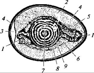
Fig. 142. Structure of a bird's egg:
1 — chalaza; 2 — shell; 3 — air Cell; 4 — outer shell membrane; 5 — fluid albumen; 6 — dense albumen; 7 — germinal disc; 8 — light yolk; 9 — dark yolk
The Egg. As is clear from the description above, the true ovum is only what is commonly called the yolk, which is covered by the primary egg membrane. Following fertilization, a zygote is formed, which begins to cleave as the egg moves through the oviduct, forming a multicellular germinal disc. When the egg is laid, Cell Division in the germinal disc pauses and resumes with the onset of incubation. The remaining structures that Supplement the egg (such as the albumen) are membranes produced no longer by the ovary, but by the oviduct (Fig. 142).
The yolk consists of tiny vesicles of dark yolk interspersed with egg plasma, or light yolk. The layers of light and dark yolk are arranged concentrically. The yolk stores nutrients and water consumed by the developing embryo during its formation. The COMPOSITION OF THE yolk varies among species, notable for its high fat and water content. Unlike the eggs of most reptiles, avian eggs do not absorb water from the external environment during embryonic development; the entire supply of water necessary for development is contained within the egg itself.
Oxidation of fat in the yolk yields water. Chicken yolk contains 50% water, 23% fats, 16% Proteins, and 11% lipoids; goose yolk contains 44% water, 36% fats, 17% proteins, and 3% other substances.
The albumen is a secondary egg membrane of semi-liquid consistency. Its primary function is to protect the egg proper (i.e., the yolk) from mechanical damage and sudden jolts, while also serving as an important source of water during embryonic development. The composition of chicken albumen is 87% water, 12% proteins, and 1% other substances. The albumen is enclosed externally by two thin membranes that diverge slightly at the blunt end of the egg to form an air cell, which accommodates changes in egg volume with temperature fluctuations. From the shell membrane at the poles of the egg, thickened layers of albumen extend through the albumen mass toward the yolk in the form of twisted cords known as chalazae. Their inner ends attach to the yolk membrane. As a result, the yolk does not float freely in the albumen, but is suspended by the chalazae, which protect it from jolts. Furthermore, due to the large mass of the nutritive yolk, its vegetative pole settles downward regardless of the egg's position, while the germinal disc remains on top, twisting the chalazae in the process. This adaptation is crucial for incubation, ensuring the germinal disc is always in the optimal position for warming.
The shell consists primarily of calcium bicarbonate (89 – 97%) along with trace amounts of other salts and organic matter. Its main function is to protect the egg from mechanical damage; it permits gas exchange while preventing bacterial penetration. As a rule, birds that nest in the open and lay large eggs have thicker shells. During embryonic development, the shell thins as it is partially utilized to build the skeleton. For instance, the mass of calcareous shell elements in a chicken egg decreases by nearly 8% during incubation, while the lime content in the developing embryo increases by 500% by the 20th day of incubation.
Embryonic gas exchange within the egg is facilitated by the porosity of the shell. A chicken egg has over 7,000 pores, which are concentrated mainly at the blunt end. As incubation progresses and gas exchange intensifies, the pore openings enlarge.
The outermost layer of the egg is the cuticular layer, formed by proteinaceous secretions deposited after the egg passes through the uterus. The function of the cuticle is to protect the egg and embryo from bacterial invasion.
Last update: 13/08/2026
Editorial and Educational Adaptation: This material has been compiled based on the primary/original source text. The project team performed an editorial review, corrected technical inaccuracies, structured sections, and adapted the content for an educational format.
What was processed:
- elimination of formatting defects (OCR errors, structural breaks, corrupted characters);
- editorial organization of content;
- standardization of terminology in accordance with academic sources;
- verification of factual statements against the original source text.
All mentions of the author, publication year, and origin of the primary text have been preserved in accordance with the source.