Fundamentals of Biochemistry - Filippovich, Y. B. 1999
Protein Metabolism
Protein Biosynthesis
Structure/149.html">The problem of METABOLISM/35.html">Protein Biosynthesis is one of the two most crucial and pressing challenges in modern natural science: while fundamentally new ways of harvesting energy in inanimate nature will be discovered thanks to advances in elementary particle physics, the ultimate solution to controlling life itself in living nature will be achieved through understanding the chemistry and biology of protein bodies.
The Study of protein Structure and Biosynthesis serves as a focal point where the solutions to vital biological questions converge: elucidating the laws of heredity and Variability, controlling the GROWTH AND DEVELOPMENT of organisms, identifying the causes of various diseases, and developing treatments for them. It is quite logical, therefore, that physicists refer to the 20th century as the age of the atom, whereas biochemists call it the age of protein.
Protein biosynthesis in the living Organism proceeds at a very high intensity. The average assembly rate of polypeptide chains in bacterial Cells is 16–17 amino acid residues per second, in Yeast 7–10, and in mammals 5–7. The synthesis time (in seconds) for a globin molecule in rabbit reticulocytes is 20, for Ovalbumin in the chicken oviduct 80, and for total rat Liver Proteins 80. In 1 minute, a rabbit reticulocyte synthesizes 5 ∙ 104 globin molecules, a chicken oviduct Cell synthesizes 6 ∙ 105 ovalbumin molecules, and a giant cell from the posterior region of the silk gland in the silkworm synthesizes 38 ∙ 1011 Silk Fibroin molecules.
The history of concepts regarding The Mechanism of protein biosynthesis. The path traversed in resolving one of the central PROBLEMS OF MODERN biochemistry—the mechanism of protein biosynthesis—is highly instructive and contradictory.
The earliest hypothesis was the reversal of proteolysis, which dates back to the late 19th century. In 1886, A. Ya. Danilevsky observed The formation of protein-like substances upon treating a concentrated solution of peptones (produced by the peptic Digestion of proteins) with gastric juice Enzymes. Later, the range of enzymes capable of reversing proteolysis was expanded (Trypsin, Pepsin, Papain, cathepsins), as were the proteins whose partial hydrolyzates could undergo such reversal (albumins, globulins, fibrin, casein, etc.). The products resulting from the reversed Protein Hydrolysis reaction were named plasteins.
Although it is now clear that plastein-formation reactions are unrelated to natural protein biosynthesis, they continue to attract researchers' attention to this day because they have found Practical Application in converting non-food proteins into edible ones and in synthesizing Peptides, notably aspartame (the methyl ester of L-α-aspartyl-L-phenylalanine), which is used as a sucrose substitute (being 100 times sweeter than sucrose) in the confectionery industry (under the commercial name slastilin):
Class="center">
A definite contribution to The Development of modern concepts regarding the mechanism of protein biosynthesis was made by studies on The biosynthesis of quasi-peptide bonds (i.e., bonds that are not true peptide bonds but closely resemble them), carried out in the 1950s in A number of laboratories, including our own laboratory headed by A. E. Braunshtein. Conducted on compounds such as glutamine, acetanilide, Glutathione, and hippuric acid, these studies proved the enzymatic nature of their formation reactions, the necessity of energy supply via oxidative processes, and, most importantly, the involvement of ATP in the biosynthesis of quasi-peptide bonds.
Equally significant were the results derived from developing the transpeptidation hypothesis as a potential variant of the peptide bond biosynthesis mechanism. In our country, research in this direction was intensively pursued in the 1950s by V. N. Orekhovich and co-workers. Studying transpeptidation reactions first demonstrated that The transfer of aminoacyl or peptidyl groups to the amino group of Amino Acids can occur not only from an amide or peptide bond, but also from an ester bond. It subsequently turned out that this very mechanism underlies the transpeptidation reaction within the ribosome.
The amino acid substitution hypothesis, which emerged during the same period alongside the Introduction of radioactive tracer Methods into biochemistry, did not exert any substantial influence on the Development of concepts concerning the mechanism of protein biosynthesis.
A turning point in the development of approaches to elucidating the mechanism of protein bodies biosynthesis was the establishment of its coupling with RNA biosynthesis (J. Brachet, 1941; T. Caspersson, 1941). This led to the formulation of the template model of protein biosynthesis, which forms the foundation of modern views in this field. The template mechanism of polymer biosynthesis, ensuring the error-free Replication of their Primary Structure, represents one of the most distinctive features of living matter. It serves as an exemplary illustration of those fundamentally new regularities that accompany the ORIGIN AND EVOLUTION of the biological form of the motion of matter: the inefficient mechanism of ordinary chemical synthesis, based on random molecular collisions, is replaced here by directed, template-specific synthesis; its rate exceeds that in disordered systems by billions of times.
Of fundamental importance in resolving the question of the protein biosynthesis mechanism was the discovery of its localization within the cellular ribosomal apparatus and the creation of cell-free systems where Ribosomes were the sole structure supporting protein biosynthesis (see p. 280). Elucidating their Structure and function has yielded invaluable information regarding the stages of template Protein Biosynthesis and the subtle molecular mechanisms that accompany it.

Fig. 93. General scheme of template biosynthesis of protein bodies (explained in text)
As will be shown below, alongside the template mechanism, nature also utilizes a multienzyme pathway of protein and peptide biosynthesis, where the specific ordering of amino acids is ensured by the spatial arrangement of catalytically active proteins within a multienzyme complex.
Template mechanism of protein biosynthesis. The general scheme of template biosynthesis of protein bodies is shown in Fig. 93. It comprises three preparatory processes—The transport of matter, energy, and information to the ribosome—and the core central process, which is the assembly of polypeptide chains within the ribosome. One of the elements in this scheme (the upper right portion of the figure)—the Transcription (rewriting) of information regarding The sequence of amino acid residues in the synthesized protein molecule—was discussed earlier. It is known that the information for this is encoded in the cellular genetic apparatus by the sequence of deoxyribonucleotide residues within the DNA molecule. Once transcribed into a sequence of ribonucleotide residues within the informative part of the mRNA molecule (synthesized on DNA AS A template), this information regarding the Primary structure of the protein is delivered to the ribosome. Here it is translated from a polynucleotide sequence into the Amino Acid Sequence of the newly forming protein within the ribosomal apparatus. The other two processes—the transport of matter (18 Proteinogenic Amino Acids and two amides) and the transport of energy required for peptide bond synthesis (the upper left portion of the figure), as well as the most complex process—the assembly of the polypeptide chain in the active, translating ribosome (the central portion of the figure)—require detailed characterization, which is provided below.
Amino Acid Activation and their transport to the ribosome. The synthesis of a peptide bond from free amino acids proceeds with the absorption of energy amounting to approximately 12 kJ/mol. Consequently, the idea was long ago put forward that protein biosynthesis is coupled with oxidative processes (A. V. Blagoveshchensky, M. P. Yurgenson, 1937) or with The breakdown of high-energy bond-containing compounds (F. Lipmann, 1941). The Development of the latter approach led to the Discovery of the enzymatic process of amino acid activation. In 1955, M. Hoagland first proposed the now universally accepted two-stage scheme for this reaction.
Its First stage consists of the interaction of an amino acid with ATP, resulting in the formation of aminoacyl adenylate and the release of pyrophosphate. The hydrolytic breakdown of the latter, facilitated by pyrophosphatase, ensures the irreversibility of the aminoacyl adenylate formation reaction. The chemistry of this activation stage was examined earlier (Chapter III) in the characterization of ligases that catalyze C—O bond synthesis (see p. 137). The Second Stage involves the transfer of the amino acid from the resulting aminoacyl adenylate to the terminal adenosine of the acceptor stem of tRNA:
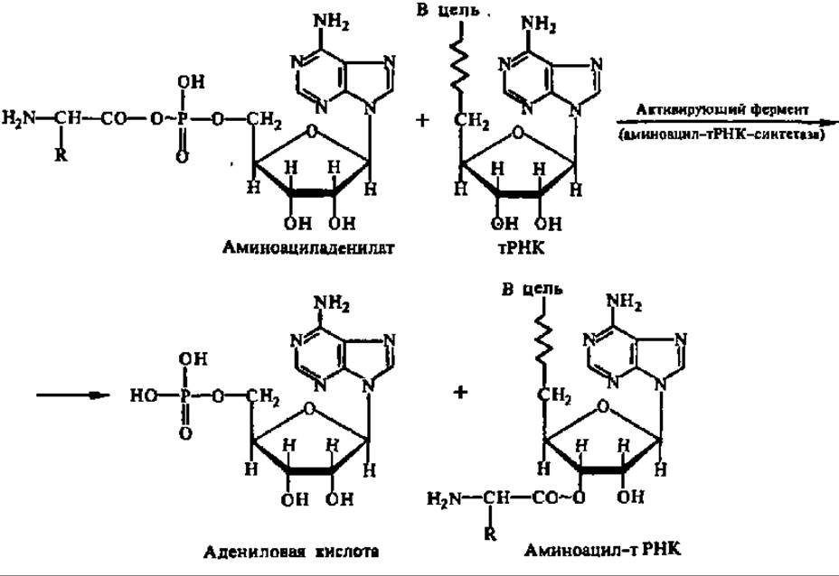
As can be seen from the structural formula of aminoacyl adenylate, it represents an anhydride of an Amino Acid and the phosphoric acid residue of adenosine-5'-phosphate, with the oxygen donor for the anhydride bond being the carboxyl OH-group of the amino acid. Being anhydrides, free aminoacyl adenylates readily react with amino acids even at very low concentrations (10-3 M) near a neutral medium reaction (pH 7.4). The reaction proceeds without enzyme participation and is accompanied by the formation of peptide bonds. They acylate free, accessible amino groups of the protein molecule with equal vigor. At the same time, when bound to the enzyme, aminoacyl adenylates are extremely inert, and there can be no question of them being utilized for Protein Synthesis directly from within the complex.
The Specificity of the amino acid activation reaction According to the aforementioned scheme is close to absolute, and is evaluated as superspecificity. Only amino acid isomers of the natural L-series are activated; D-amino acid isomers do not enter the reaction. However, certain homologs of L-Amino acids are activated alongside proteinogenic L-amino acids.
Catalytic acceleration of the activation of each proteinogenic amino acid is carried out by its own specific enzyme dedicated solely to that amino acid. These enzymes have been discovered in representatives of various animal classes, plants (including Algae), and microorganisms, meaning they are ubiquitous. This indicates their critical role in protein biosynthesis.
Activating enzymes are isolated at low Temperature from the so-called pH 5 fraction of the supernatant. The latter is obtained after ultracentrifugation of a diluted cell homogenate at 105,000 g for 1 hour, As a result of which all Structural elements of the cellular contents are pelleted. Upon acidifying the supernatant with CH3COOH (since HCl denatures enzymes) to pH 5.3, a precipitate forms containing a mixture of amino acid-activating enzymes. Therefore, they were initially termed pH 5 enzymes. In accordance with modern nomenclature, they are called Aminoacyl-tRNA synthetases (aaRSases).
Recent studies indicate that aminoacyl-tRNA synthetases exist within cells as high-molecular-weight complexes known as chodosomes. With a sedimentation coefficient of 26–29S and an M of ~1.4 megadaltons, these complexes comprise several aaRSases (specific, for example, for Ala, Gly, Glu, Leu, and Ser, or for Lys, Met, Arg, Leu, and Trp) as well as enzymes that modify aaRSases and regulate their activity—such as protein Kinases, methyltransferases, ADP-ribosyltransferases, phosphoprotein Phosphatases, and others.
The enzymes that activate amino acids are exceptionally labile and easily denatured with a loss of activity. Even ultrasonic Cell Disruption during the Isolation of the enzymatic preparation and brief storage lead to their inactivation. For the normal functioning of amino acid-activating enzymes, the presence of Mg2+ in the reaction medium is required, because ATP binding occurs via the imidazole radical of the Histidine residue in the active center of the enzyme through the magnesium ion.
The molecular weights of most aminoacyl-tRNA synthetases (arsynthetases, more than 50 of which have been isolated in a highly purified state) are close to 100,000, although in some cases they reach 200,000 or more (such as glycyl- and phenylalanyl-tRNA synthetases). The molecules of most aminoacyl-tRNA synthetases are dimers or tetramers (occasionally trimers), and only in isolated cases (for example, alanyl-, arginyl-, aspartyl-, valyl-, and isoleucyl-tRNA synthetases) are they represented by a single polypeptide chain comprising approximately 1,000 amino acid residues. Characteristically, in aminoacyl-tRNA synthetases with a dimeric structure, the subunits function cooperatively: the release of the synthesized aminoacyl-tRNA from one subunit occurs at the moment when a tRNA molecule binds to the other subunit following ATP and the amino acid. Consequently, each subunit in the aminoacyl-tRNA synthetase molecule possesses three structurally distinct binding sites for the listed substrates, with the binding of each preceding substrate facilitating the acceptance of the subsequent one. Therefore, during tRNA aminoacylation, ternary enzyme-substrate complexes are readily formed. ATP is bound least firmly by aminoacyl-tRNA synthetases (Ks = 10-4 M), amino acids are bound an order of magnitude better (Ks = 10-5 M), and tRNA is bound most strongly (Ks = 10-8 M). Aminoacyl-tRNA synthetases are the "slowest" enzymes; their molecular activity ranges from several dozen to several hundred catalytic acts per minute.
Based on studies of the active centers of aminoacyl-tRNA synthetases using various methods and investigations into The kinetics of the amino acid activation reactions they accelerate, the mechanism of their action has begun to be elucidated. ATP is the first to bind to the aminoacyl-tRNA synthetase, adenylylating a histidine residue in the active center of the enzyme with the release of pyrophosphate:

The amino acid activated through the mediation of the adenylylated aminoacyl-tRNA synthetase interacts with it, resulting in the formation of an aminoacyl adenylate. The latter, possessing an exceptional ability to aminoacylate any radicals containing a labile hydrogen atom, transfers the aminoacyl group to the histidine radical of the enzyme's active center, thereby forming the aminoacylated enzyme:
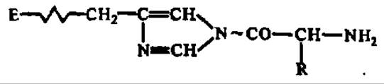
Following the binding of the corresponding tRNA by the enzyme, the aminoacyl group is transferred to the OH group of the adenosine residue at its acceptor end (for the reaction equation, see p. 281). In some aminoacyl-tRNA synthetases, The Role of the histidine residue in carrying out the aforementioned stages of amino acid activation can be played by a glutamic acid radical.
During the Formation of the enzyme-substrate complex from the aminoacyl-tRNA synthetase and tRNA, both undergo conformational rearrangements that facilitate the aminoacylation reaction. However, The most significant and intriguing aspect of the interaction between the aminoacyl-tRNA synthetase and tRNA is their ability to recognize each other flawlessly in accordance with absolute specificity for a particular amino acid. This is apparently based on multi-point interactions between the enzyme and tRNA, with a crucial role in recognition belonging to the tRNA anticodon.
For a long time, it was believed that the aminoacyl group is always attached via the OH group of the 3'-carbon atom of the ribose of the terminal adenosine in the tRNA molecule. It turned out that sometimes the hydroxyl of the 2'-carbon atom of the ribose residue can perform this same function. This is observed during the activation of phenylalanine, leucine, and isoleucine with the participation of their respective aminoacyl-tRNA synthetases. In contrast, seryl- and threonyl-tRNA synthetases ensure the attachment of aminoacyl groups exclusively to the 3'-carbon atom of ribose, whereas tyrosyl- and cysteinyl-tRNA ligases attach them to both the 2'- and 3'-carbon atoms. It has also been established that the aminoacyl group in aminoacyl-tRNA is capable of migrating from the 2'- to the 3'-carbon atom of ribose and vice versa, until equilibrium is established:

It is believed that aminoacylation of tRNA at either the 2'- or 3'-position of the terminal adenosine ribose residue prevents errors during the activation of stereochemically similar amino acids (e.g., valine and Threonine). However, even if an error occurs—that is, if a different amino acid attaches to the tRNA instead of the specific one—the aminoacyl-tRNA synthetase can correct the mistake itself, as it possesses The ability to hydrolyze non-specific aminoacyl adenylates foreign to it.
Thus, throughout the activation process, the strictly selective attachment of each amino acid to its specific tRNA is consistently ensured, along with the generation of a pool of aminoacyl-tRNAs that directly supply energy-rich amino acid residues to the ribosome.
That this is indeed the case—that aminoacyl-tRNAs are the sole source of amino acid residues in protein biosynthesis—has been proven in experiments using aminoacyl-tRNAs containing 14C-labeled amino acids: as incubation in the protein-synthesizing system proceeds, the content of the labeled amino acid in the aminoacyl-tRNA preparation decreases, while mirroring this, it increases proportionally in the synthesized protein (Fig. 94).
The activation of amino acids is accompanied by their coding (encryption): upon attaching to the corresponding tRNA, the amino acid receives a code or cipher in the form of a three-nucleotide residue sequence in the anticodon loop of the tRNA that is strictly specific only to that amino acid (see p. 216). This base triplet is called the anticodon. It corresponds to a complementary codon within the mRNA. The interaction of mRNA codons with aminoacyl-tRNA anticodons determines the order of amino acid residues in the protein synthesized via the template mechanism. This fundamentally important proposition has been experimentally proven and is known as the "Chapeville experiment" (named after one of the scientists who participated in it). In this experiment, cysteinyl-tRNA was converted into alanyl-tRNA by reduction at the aminoacyl residue:


Fig. 94. Demonstration of the incorporation of 14C-leucine into protein (1) from 14C-leucyl-tRNA (2); see text for explanation
The alanyl-tRNACys synthesized in this manner contains an Alanine residue attached to a tRNA bearing a Cysteine anticodon. When it was introduced into the protein-synthesizing system, cysteine positions in the polypeptide chain of the resulting protein were occupied by alanine residues. This unequivocally indicates that it is the tRNA that encodes the positioning of its attached aminoacyl residue into a predetermined Location within the newly forming protein molecule. Therefore, aminoacyl-tRNA synthetases were at one time called codases or cipherases; however, the Nomenclature Committee of the International Union of Biochemistry recommended against using these terms, and they are no longer in current use.
Template mechanism of polypeptide chain assembly. Protein biosynthesis in all structural elements of The Cell (Nucleus, Mitochondria, Chloroplasts, Endoplasmic reticulum, etc.) takes place on ribosomes. Therefore, it is specifically through the Study of the Structure and properties of ribosomes, as well as the mechanism of their interaction with the compounds serving as precursors for protein biosynthesis, that the most impressive results have been achieved in uncovering the regularities of protein body formation.
Structure and properties of ribosomes. Ribosomes are the smallest ribonucleoprotein bodies. They are contained mainly in the Cytoplasm of plant, animal, and bacterial cells in numbers amounting to tens of thousands in each cell.
Ribosomes are isolated by differential centrifugation of the cell content obtained after cell homogenization. It is known that this approach allows the cell content to be separated into a series of fractions (see Chapter I). Ribosomes are primarily associated with the membranes of The endoplasmic reticulum. Therefore, during the Fractionation of Cell contents, they are sedimented together with lipoprotein membrane fragments—microsomes.
To isolate ribosomes, microsomes are treated with deoxycholate, a reagent that dissolves Lipids and consequently disrupts microsomes; ribosomes are then sedimented in isotonic Buffer solutions by centrifugation at 100,000 g for 1 hour. Ribosomes can also be isolated upon the disruption of other subcellular particles (nuclei, Plastids, mitochondria). Currently, ribosome isolation has been accomplished from numerous objects representing the animal and plant kingdoms, as well as microorganisms. The shape and structure of ribosomes isolated from different sources are similar, but according to their sedimentation coefficient values, they are divided into two classes: 70S and 80S. The former are found in all prokaryotes and are also present in the nuclei, mitochondria, and chloroplasts of eukaryotes; the latter are localized exclusively in the cytoplasm of eukaryotes.
70S and 80S ribosomes are stable at Mg2+ concentrations close to 0.001 M. When the Mg2+ content is increased to 0.01 M, ribosomes form dimers or aggregate into larger structures, whereas when lowered to 0.0001 M, they dissociate into subparticles: 70 (80)S ⇄ 30 (40)S + 50 (60)S. This dissociation-association process of ribosomes and their subparticles is also controlled by Polyamines (see p. 211) and Changes in the concentration of other divalent cations.
Being exclusively ribonucleoproteins (also called RNP particles), ribosomes are composed of nearly equal amounts of RNA and Protein, which vary depending on the ribosome class (Table 22) with a clearly expressed tendency toward a slight predominance of protein within them.
Table 22 Composition of ribosomes
Ribosome class |
70S |
80S |
||
Ribosomal subparticles |
30S |
50S |
40S |
60S |
RNA |
16S |
23S and 5S |
18S |
28S, 5S |
and 5.8S |
||||
Number of proteins |
21 |
34 |
31 |
41 |
Molecular weights of proteins, thousand daltons |
12—65 |
9—31 |
10—44 |
10—54 |
RNA/protein ratio, % |
37/63 |
36/64 |
54/46 |
41/59 |
Besides proteins and Nucleic Acids, ribosomes contain trace amounts of other substances: Mg2+ and Co2+, di- and polyamines, a number of other cations (Fe3+, Zn2+, Al3+, Ba2+, Sr2+, Ni2+, Cr2+, NH4), as well as latent Ribonuclease.
In their Native State—that is, within living cells—ribosomes are heavily hydrated: for instance, 1 g of dry matter from reticulocyte ribosomes binds 2.7 g of Hydration Water. This indicates the exceptionally high porosity of native ribosomes.
Protein and RNA molecules within ribosomal particles are held together by very weak bonds: incubation with 0.5–1 M salt solutions at low temperature is sufficient to dissociate the protein from the nucleic acid. A similar dissociation occurs, for example, at pH 12 during Electrophoresis. Protein–nucleic acid interactions in ribosomes are predominantly electrostatic in nature. The high content of positively charged basic amino acids in total ribosomal proteins (12% Lysine, 11% Arginine, and 3% histidine), combined with the large number of negatively charged internucleotide phosphates in Ribosomal RNAs, provides ideal conditions for extensive ionic bonding between them.
The structure and function of high-molecular-weight (16–18S and 23–28S) and low-molecular-weight (5S and 5.8S) rRNAs were discussed earlier (see Chapter VI). Regarding the structure and function of ribosomal proteins, substantial new information has emerged in recent years. As shown in Table 22, both small (30–40S) and large (50–60S) ribosomal subunits contain a strictly defined number of proteins that vary widely in molecular weight. Proteins localized in the small (30–40S) ribosomal subunits are designated by the letter S (from "small"), while those in the large subunits (50–60S) are designated by L (from "large").
Thanks to modern Protein Chemistry techniques, all proteins from E. coli ribosomes have been isolated in a highly purified, homogeneous state. Their primary structures have been fully determined, and for many of them, secondary and tertiary structures are also known. Typically, ribosomal protein molecules are asymmetric, globular, and compact, featuring a significant proportion of a-helices (Fig. 95). Some of these proteins, enriched in hydrophobic amino acids in specific Regions of the polypeptide chain, readily form dimers and even oligomers, as well as interacting with one another to form complexes within the ribosomal subunits.
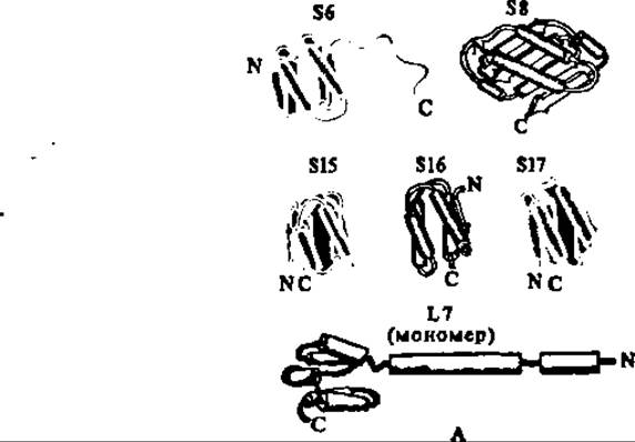

Fig. 95. Tertiary structures of proteins S6, S8, S15, S16, S17, and L7 (A) and the atomic model of protein L7 (B):
Helical regions are represented by cylinders, and ß-structures by arrows. In protein S6, the N-terminal fragment 105–135 lacks a tertiary structure. In the atomic model of ribosomal protein L7, numbers indicate the positions of N- and C-terminal amino acid residues in the a-helical segments of the polypeptide chain, while the oval between a-helices 51–59 and 93–101 denotes the cavity that accommodates a-helix 4–11 upon dimer formation.

Fig. 96. Spatial Structure of the E. coli 70S ribosome (model):
The 30S subunit is shown from the front; its dimensions are ~23 nm in length and 12 nm in width. The 50S subunit is shown from the rear; its dimensions are approximately 20–23 nm in all directions. The lateral projections (the "finger" on the right and the lateral lobe on the left) contain proteins L7/L12 and L1, respectively. The subunits are associated "HEAD-to-head" and lateral projection to lateral lobe ("platform to L-ridge"). The 30S subunit covers only part of the 50S subunit, leaving free the region adjacent to the L7/L12 stalk, which likely houses the functionally important centers of the ribosome. Apparently, the association of the 30S and 50S subunits creates a gap or cleft in the region between the head and body of the 30S subunit and the Base of the central protuberance of the 50S subunit, which is believed to accommodate the mRNA molecule.
Over the past decade, major breakthroughs have been achieved in elucidating the three-dimensional Organization of ribosomes as a whole and mapping the localization of individual structural elements within their subunits (see Table 22). Special recognition is due to the Soviet school of biochemistry—specifically, researchers at the Institute of Protein Research in the Pushchino Biological Center of the Russian Academy of Sciences (Moscow Region)—where fundamental studies of the cellular ribosomal apparatus were conducted under the leadership of Academician A. S. Spirin. The topology of ribosomes is illustrated in Fig. 96. It is now evident that the spatial architecture of ribosomes is dictated by the Tertiary Structure of the 16–18S and 23–28S rRNAs, which determines the overall shape and design of the 30–40S and 50–60S ribosomal subunits, respectively. This is particularly striking when analyzing data on the architecture of the E. coli 30S ribosomal subunit obtained by A. S. Spirin and co-workers. Their findings demonstrate that the morphological model of the 30S subunit is predetermined by the 16S rRNA in a compact conformation, onto which the S1–S21 proteins identified in this subunit interact and anchor at specific positions (Fig. 97). The E. coli 50S subunit is constructed in a similar fashion.
Data on the localization of specific functional centers within the ribosome are of paramount importance for understanding the mechanism of protein synthesis. These centers orchestrate the key stages of polypeptide chain biosynthesis: binding aminoacyl-tRNA, binding protein factors required for ribosomal function, decoding The nucleotide sequence specifying the amino acid order in the nascent protein, peptide bond formation, releasing deacylated tRNA after peptide bond formation, translocating peptidyl-tRNA from the aminoacyl to the peptidyl site while advancing mRNA by one nucleotide triplet, and hydrolyzing the ester bond between the polypeptide and tRNA upon Translation termination. Therefore, it is fitting to conclude this Overview of MODERN CONCEPTS OF ribosome structure with a Discussion of the functional ribosome model (G. Wittmann et al., 1974), which highlights its active centers (Fig. 98).
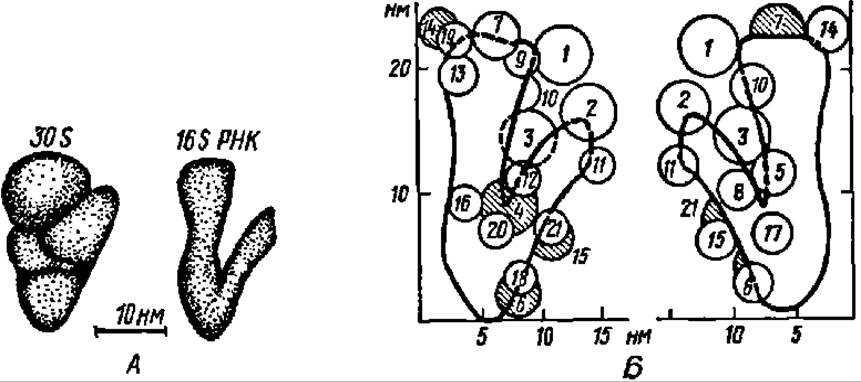
Fig. 97. Spatial STRUCTURE OF THE E. coli 30S ribosomal subunit:
A — morphological model; B — arrangement of proteins and RNA according to the model (proteins in the Background are shaded).
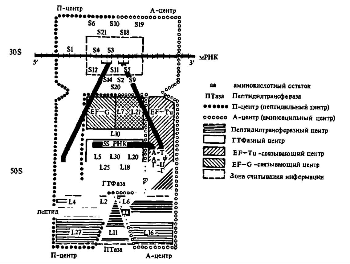
Fig. 98. Functional model of the E. coli 70S ribosome.
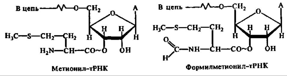
Bacterial protein biosynthesis is initiated with the participation of three protein initiation factors: IF-1, IF-2, and IF-3. IF-3 is a protein with a Molecular Weight of 21,000–23,500. It induces Conformational Changes in the 30S ribosomal subunit that facilitate the binding of IF-1, IF-2, GTP, mRNA, and formylmethionyl-tRNA. The recruitment of the latter ensures the delivery of the first (N-terminal) amino acid—formylmethionine—which starts the polypeptide chain of any bacterial protein. Subsequently, this N-terminal formylmethionine residue may be cleaved off during post-translational protein Processing. The tRNA species that delivers formylmethionine during bacterial protein synthesis differs from the tRNA that transfers Methionine during Polypeptide chain elongation; thus, distinct tRNAfMet and tRNAMet species exist:
IF-3 is the first factor to bind to the 30S ribosomal subunit, triggering the initiation phase of protein synthesis by sequestering the 30S subunit from the pool of free 30S and 50S subunits generated during 70S ribosome dissociation. It also promotes the formation of the mRNA-binding site on the 30S subunit.
IF-1 and IF-2 are proteins with molecular weights of 8,900–9,400 and 90,000–118,000, respectively. IF-1 stimulates the binding of IF-2 to the 30S ribosomal subunit and facilitates mRNA attachment. IF-2 plays a central role in anchoring formylmethionyl-tRNA to the 30S subunit and in GTP hydrolysis. Although GTP is essential for Translation initiation, its exact function in this process is not yet fully understood.
IF-1, IF-2, formylmethionyl-tRNA, and GTP bind to the 30S subunit as a complex (see Fig. 99). Once this complex is formed, mRNA joins the subunit. The interaction between the anticodon of formylmethionyl-tRNA and the start codon of mRNA (the AUG triplet) correctly positions the mRNA on the ribosome to ensure accurate reading of the nucleotide sequence encoding the primary structure of the synthesized protein. Thus, formylmethionyl-tRNA helps mRNA find the precise register on the 30S subunit required for translating the amino acid sequence, which highlights its critical role in bacterial translation initiation. In eukaryotes, this function is often fulfilled by a specialized tRNAMet species whose structure differs from the elongator tRNAMet that inserts methionine residues during peptide chain elongation.
At the same time, recent studies have demonstrated that an even greater role in stabilizing the proper positioning of mRNA on the 30S subunit is played by... The three MAIN STAGES OF template-directed protein biosynthesis—initiation, elongation, and termination—are enclosed in boxes; protein initiation and elongation factors are indicated by conventional symbols directly on the diagram; some symbols are explained separately in the upper right corner of the figure.
the Shine-Dalgarno sequence (short: AGGAG, or long: UAAGGAGGU) interacts with the complementary region at the 3'-end of 16S rRNA and is located 5 to 13 nt upstream of the start AUG codon.

Fig. 99. Scheme of protein biosynthesis in Escherichia coli (explained in the text):
The binding of mRNA to the complex consisting of the 30S subunit, initiation factors, and GTP is accompanied by the release of initiation factor IF-3, which has completed its function (see Fig. 99). The complex, now including the mRNA as well, strongly attracts the 50S subunit and associates with it to form the 70S ribosome. At this moment, IF-1 leaves the ribosome, having also fulfilled its role in stabilizing the mRNA within the ribosome. IF-2, still retained in the newly formed 70S ribosome and bound to GTP, accelerates the hydrolysis of GTP into GDP and inorganic phosphate, and is released from the ribosome along with the GTP hydrolysis products. The energy released during GTP hydrolysis is apparently necessary to drive conformational changes in the 70S ribosome, as a result of which formylmethionyl-tRNA is stabilized in the peptidyl center of the ribosome (see elongation phase 1 in Fig. 99). Such a ribosome is now capable of assembling a polypeptide chain of a specific structure, which is why it is called a translating (active) ribosome. Consequently, the elongation phase of protein synthesis takes place within the translating ribosome.
In Eukaryotic cells, the initiation of ribosomal protein synthesis also proceeds with the participation of eukaryotic initiation factors (eIFs). There are nine such factors: eIF-1, eIF-2, eIF-3, eIF-4A, eIF-4B, eIF-4C, eIF-4D, eIF-5, and eIF-6. All of them, except for eIF-2 and eIF-3, are single Polypeptides with molecular weights ranging from 15,000 (eIF-1) to 160,000 (eIF-5). Factor eIF-2 appears to be a dimer (MW = 86,000; $\alpha$-subunit = 38,000; $\beta$-subunit = 48,000); eIF-3 consists of multiple subunits with a total molecular weight exceeding 500,000.
Of the nine listed protein factors, eIF-2, eIF-3, and eIF-5 are absolutely essential for the initiation of protein biosynthesis in eukaryotes, while the remaining factors enhance the Functions of these three. Factor eIF-2 interacts via its $\beta$-subunit with methionyl-tRNA and via its $\alpha$-subunit with GTP, forming a complex with the 40S subunit. Its activity is regulated by phosphorylation of the $\alpha$-subunit mediated by a specific protein kinase. Factor eIF-3 also binds to the 40S subunit, which leads to the formation of the mRNA-binding site on it and stabilizes its association with the eIF-2 $\cdot$ methionyl-tRNA $\cdot$ GTP complex. Following the attachment of mRNA to the 40S subunit at the mRNA-binding site, and with the participation of factor eIF-5, the complex vigorously recruits the 60S subunit. This is followed by the release of all previously bound protein factors (including eIF-5), the hydrolysis of GTP, and the anchoring of methionyl-tRNA in the peptidyl center of the newly formed active 80S ribosome capable of translation. Factor eIF-6 ensures the dissociation of the 80S ribosome into 40S and 60S subunits, freeing the 40S subunit for a new initiation cycle.
Elongation of protein biosynthesis in bacterial cells is mediated by three protein elongation factors: EF-Tu, EF-Ts, and EF-G (representing Three types of Translation elongation factors). Mammals have two elongation factors: TF-1 and TF2 (translation factors 1 and 2). Bacterial EF-Tu (MW = 47,000) and EF-Ts (MW = 35,000) correspond to mammalian TF-1 (MW = 186,000), whereas bacterial EF-G corresponds to mammalian TF-2 (MW = 70,000). The EF-G of Escherichia coli has a molecular weight of 77,321.45 and is a polypeptide consisting of 701 amino acid residues whose primary structure has been elucidated.
The elongation process begins with the binding of aminoacyl-tRNA carrying the amino acid residue that must occupy the second position from the N-terminus of the protein molecule being synthesized on the ribosome. In Bacteria, this aminoacyl-tRNA forms a complex with EF-Tu and GTP, through which it attaches to the aminoacyl center of the translating ribosome in accordance with The Genetic Code (see below)—that is, via the interaction between complementary triplets of the tRNA anticodon and the mRNA codon positioned opposite the aminoacyl center of the ribosome (see Figs. 98 and 99). In mammals, this process occurs with the participation of TF-1.
According to modern data, codon-anticodon interaction is not the sole determinant in the Selection of appropriate aminoacyl-tRNAs for polypeptide chain assembly. The entire tRNA molecule is involved, as post-translational modification significantly alters its ability to be accepted by the ribosome. Ribosomal proteins S4, S9, S13, L2, and L7 are also recruited into the decoding process; the latter two, being localized in the peptidyl center of the ribosome, form a tetrameric complex with two tRNA molecules.
Both aminoacyl-tRNA and GTP bind to EF-Tu via free SH-groups of cysteine residues in its molecule, and the primary structure of the GTP (and correspondingly GDP) binding site spanning 42 amino acid residues has been deciphered. The activity of EF-Tu is regulated by phosphorylation and interaction with ppGpp.
Due to the Cleavage of GTP into GDP and inorganic phosphate, the aminoacyl-tRNA is brought into proximity with the formylmethionyl-tRNA located in the peptidyl center of the ribosome, while the EF-Tu $\cdot$ GDP complex and inorganic phosphate are released from the ribosome. Upon interacting with EF-Ts and GTP, the EF-Tu $\cdot$ GDP complex is converted back into the EF-Tu $\cdot$ GTP complex, which is then capable of binding the next aminoacyl-tRNA molecule (see Fig. 99).
Within the peptidyl center, a reaction takes place between formylmethionyl-tRNA and aminoacyl-tRNA, whereby the formylmethionine residue is transferred to the free NH2 group of the amino acid residue that is part of the aminoacyl-tRNA. As a result, dipeptidyl-tRNA is formed—meaning the first peptide bond in the future protein molecule is established—along with uncharged tRNAfMet (see Fig. 99). This process is termed the transpeptidation reaction. It is catalyzed by a corresponding enzyme, with transpeptidase activity inherent to ribosomal proteins L16, L11, and L6, and likely to a segment of 23S rRNA (see Fig. 98).
During the next elongation phase, the peptidyl-tRNA is transferred to the tRNAfMet site within the ribosomal peptidyl center, while the latter is released and moves to the ribosomal E-site, from where it is subsequently displaced by the incoming aminoacyl-tRNA and expelled from the ribosome.
This elongation step is known as translocation and is mediated by EF-G in bacteria and TF-2 in eukaryotes, accompanied by the mandatory hydrolysis of another GTP molecule (see Fig. 99). As a result of translocation, the dipeptidyl-tRNA occupies a position in the ribosomal peptidyl center, whereas the aminoacyl center is completely vacated and becomes ready to accept a new aminoacyl-tRNA complexed with EF-Tu or TF-1 and GTP. Crucially, during translocation, the peptidyl-tRNA moves into the ribosomal peptidyl center along with the mRNA molecule to which it is tethered via codon-anticodon interactions (see Fig. 98). This movement spans precisely one nucleotide triplet; consequently, the next consecutive codon of the mRNA molecule is positioned opposite the aminoacyl center, determining which aminoacyl-tRNA will enter the aminoaryl center to supply the next amino acid residue to the nascent protein chain.
Termination of Protein synthesis in the ribosome likewise involves three protein factors: RF-1, RF-2, and RF-3 in bacteria, and a single termination factor R (derived from "recognize") in higher organisms. An E. coli cell contains approximately 500 molecules of each of these proteins. The protein factors RF-1 and RF-2 have a molecular mass of 45,000 each and are capable of recognizing the stop codons that terminate polypeptide chain assembly in the mRNA molecule: factor RF-1 recognizes UAG and UAA, whereas factor RF-2 recognizes UGA and UAA. Factor RF-3 (also referred to as S-protein) stimulates the activity of factors RF-1 and RF-2.
As soon as a stop codon of the mRNA molecule reaches the appropriate position in the ribosomal aminoacyl center following a translocation event, one of the termination factors, RF-1 or RF-2, binds to it.
1 pppGpp — 3'-pyrophosphoguanosine-5'-pyrophosphate.
This blocks the binding of an aminoacyl-tRNA molecule, especially since no tRNAs possess anticodons complementary to stop codons. The binding of factor RF-1 or RF-2 to mRNA triggers the peptidyl esterase activity of ribosomal proteins—specifically proteins L11 and L16 located in the 50S subunit (see Fig. 98)—thereby hydrolyzing the ester bond linking the newly synthesized polypeptide to the tRNA. Consequently, the protein synthesized within the ribosome is released. Simultaneously, the tRNA and mRNA are liberated, and the 70S ribosome dissociates into 30S and 50S subunits that re-enter the general pool of ribosomes and subunits, ready to be recruited for a new cycle of protein biosynthesis (see Fig. 99). Template-driven protein biosynthesis termination also requires a GTP molecule. In bacteria, GTP acts as an allosteric regulator of termination factor activity, whereas in animals, it is hydrolyzed into GDP and inorganic phosphate.
It was long believed that eukaryotes possessed a single release factor (eRF) responsible for recognizing stop codons. This factor was characterized (50 kDa) and sequenced. However, a family of related proteins designated as eRF1 has recently been described. These proteins have been isolated from humans, frogs, and yeast, sequenced (comprising 428, 437, and 437 amino acid residues, respectively), shown to interact with all three stop codons, and found to be GTP-independent, thereby enhancing The fidelity of translation termination:
The multienzyme mechanism of peptide biosynthesis. The multienzyme pathway of peptide biosynthesis was pioneered by F. Lipmann and co-workers (1968). It has now been established that at least five peptide Antibiotics—gramicidin, tyrocidine, bacitracin, cyclosporin, and myobacillin—are synthesized entirely independently of nucleic acids, including tRNA, while still ensuring the error-free assembly of polypeptide chains with defined primary structures.
The biosynthesis of gramicidin S is mediated by a multienzyme system consisting of two protein fractions with molecular masses of 280,000 and 100,000. These fractions were isolated from a protein extract of Bacillus brevis and separated using Sephadex G-200 Gel filtration into the two aforementioned complementary protein fractions. When combined in the presence of amino acids, ATP, and Mg2+, they catalyze the in vitro Synthesis of the cyclic decapeptide gramicidin S (Fig. 100).
The light protein fraction ($M_r = 100,000$) racemizes and activates D-phenylalanine, whereas the heavy fraction ($M_r = 280,000$) activates the remaining four L-amino acids. Amino acid activation by these enzymes proceeds in two stages: first, aminoacyl adenylates of the respective amino acids are formed via a reaction between the free Amino Acids and ATP accompanied by the release of pyrophosphate; second, the aminoacyl groups are transferred from the aminoacyl adenylates to the HS-groups of cysteine residues within the enzyme polypeptide chain, yielding amino acid thioesters. In this state, the light fraction—carrying the D-phenylalanine activated at its COOH group—associates with the heavy fraction, which carries L-Proline, L-valine, L-Ornithine, and L-leucine activated at their respective COOH groups. This complex initiates the sequential biosynthesis of peptide bonds between these amino acids in the exact order they appear in the gramicidin S molecule. The direct transfer of the D-phenylalanine residue from its thioester linkage (on the light protein fraction) to the $ ext{NH}_2$ group of the L-proline residue—linked via a thioester bond to a subunit of the heavy protein fraction—is mediated by the aminoacyl carrier protein (ACP), which is a component of the multienzyme complex. This is a relatively low-molecular-weight protein (approximately 20,000) containing pantetheine as a prosthetic group:

The D-phenylalanine residue is first transferred to the HS-group of pantetheine and subsequently to the $ ext{NH}_2$ group of L-proline. This establishes the first peptide bond in the future gramicidin molecule. Next, the D-Phe-L-Pro dipeptide residue is transferred from the thioester linkage (on the heavy protein fraction subunit) to the HS-group of the ACP pantetheine, and from there to the $ ext{NH}_2$ group of the valine residue. This peptidyl transferase reaction repeats until the pentapeptide D-Phe→Pro→Val→Orn→Leu is fully synthesized (see Fig. 100). Because an identical, adjacent multienzyme complex simultaneously synthesizes another pentapeptide of the same sequence, the two pentapeptides join in a head-to-tail fashion to form the cyclic decapeptide molecule of gramicidin S (see Fig. 100).
The biosynthesis of another peptide antibiotic, tyrocidine, follows a mechanism analogous to the one discussed above:

Here, the multi-enzyme system carries out the complete assembly of a cyclic decapeptide; that is, 10 peptide bonds are synthesized sequentially in accordance with the primary structure of the peptide. This multi-enzyme complex has a more complex structure, as it consists of three protein fractions: 1) M = 100,000 — racemizes (and activates) D-phenylalanine; 2) M = 230,000 — activates L-proline; 3) M = 460,000 — activates D-phenylalanine, asparagine, glutamine, Tyrosine, valine, ornithine, and leucine, and also racemizes phenylalanine.

Fig. 100. Multi-enzyme mechanism of biosynthesis of half of the gramicidin molecule (explanation in the text)
It has recently been proven that the biosynthesis of mycosubtilin (a cyclic 13-membered peptide), bacitracin A, and cyclosporin A (both 11-membered cyclic peptides) is mediated by a multi-enzyme complex.
F. Lipmann attempted to integrate the template and non-template models of protein biosynthesis into a unified concept. Although not all details of the latter have been elucidated, it is certain that, from an evolutionary perspective, the multi-enzyme pathway of assembling polypeptides of a given structure preceded the ribosomal pathway of their biosynthesis, and both pathways currently coexist in microbes. The striking analogy between the activity of multi-enzyme peptide biosynthesis systems and the functioning of higher fatty acid synthetase indicates that THE PRINCIPLE OF multi-enzyme biosynthesis for fairly complex compounds is widely utilized in nature.
Coding of protein biosynthesis. Elucidating the question of which specific nucleotide triplet within mRNA encodes the incorporation of a particular amino acid into a protein represents one of the most fascinating chapters in modern biochemistry and molecular biology.
How can a four-letter code (based on the number of bases in mRNA, i.e., A — adenine, G — guanine, C — cytosine, and U — uracil) be translated into a twenty-letter code (based on the number of amino acids making up a protein molecule)?
If each combination of NUCLEOTIDES in mRNA is assigned the ability to encode THE POSITION OF a single amino acid in a protein, a doublet code is hardly feasible (the number of nucleotide pairs is less than the number of amino acids constantly found in proteins), a quadruplet code is unrealistic (the number of combinations far exceeds the number of amino acids), whereas a triplet code best satisfies the numerical ratio of possible codons to protein amino acids. These calculations are based on the fact that combining 4 nucleotides in pairs yields 16 combinations, in groups of three — 64, in groups of four — 256 combinations, and so on:
Doublet code |
Triplet code |
Quadruplet code |
|||
(42 = 16) |
(43 = 64) |
(44 = 256) |
|||
AA UU GG CC |
AAA |
UUU |
GGG |
CCC |
AAAA UUUU GGGG CCCC |
AU UA GA CA |
AAU |
UUA |
GGA |
CCA |
AAAU UUUA GGGA CCCA |
AG UG GU CU |
UAA |
AUU |
AGG |
ACC |
etc. |
AC UC GC CG |
AUA |
UAU |
GAG |
CAC |
|
AAG |
UUG |
GGU |
CCU |
||
GAA |
GUU |
UGG |
UCC |
||
AGA |
UGU |
GUG |
CUC |
||
AAC |
UUC |
GGC |
CCG |
||
CAA |
CUU |
CGU |
GCC |
||
ACA |
UCU |
GCG |
CGC |
||
AUG |
UAG |
GAU |
CAU |
||
AGU |
UGA |
GUA |
CUA |
||
AGC |
UAC |
GAC |
CAG |
||
ACG |
UCA |
GCA |
CGA |
||
AUC |
UGC |
GUC |
CUG |
||
ACU |
UCG |
GCU |
CGU |
||
Through a series of ingenious experiments, the triplet Nature of the protein synthesis code was proven experimentally. First, the qualitative composition of nucleotide residues comprising one or more codons responsible for the incorporation of a specific amino acid into a protein was established. A decisive role here was played by the observation of M. Nirenberg (1961), who, using polyuridylic acid as mRNA, demonstrated for the first time that the incorporation of phenylalanine into a polypeptide chain is coded by the UUU triplet. Polyuridylic acid, poly(U), was introduced into a cell-free protein-synthesizing system composed of washed (mRNA-depleted) ribosomes, a complete set of tRNAs, aminoacyl-tRNA synthetases, and amino acids (some of which were labeled with 14C or 15N), ATP and its generating compounds (phosphoenolpyruvate, acetyl phosphate, and similar substances), GTP, Mg2+, and Mn2+. In this system, using poly(U) as a template, the synthesis of peptides consisting solely of phenylalanine residues took place. Thanks to the application of an almost complete set of synthetic homo- and heteropolyribonucleotide templates—obtained using polynucleotide phosphorylase—in S. Ochoa's laboratory, the work on identifying the qualitative composition of codons for all amino acids was completed within a year.
The greatest difficulty lay in determining the sequence of nucleotides in the codons that dictate the position of an amino acid within a protein molecule: given a known qualitative composition of a codon, it remained unclear which specific alternation of nucleotide residues within it (e.g., ACU, CAU, UAC, AUC, CUA, or UCA) actually encodes the given amino acid during protein biosynthesis on the ribosome. However, this difficulty was also overcome after it was discovered that synthetic trinucleotides, like mRNA, can specifically bind aminoacyl-tRNAs to ribosomes. Furthermore, H. Khorana's laboratory synthesized polyribonucleotides of defined structure (with a known sequence of nucleotide residues) and used them to experimentally determine the primary structure of codons for each amino acid in cell-free protein-synthesizing systems.
As a result, by the end of 1965, complete data on the genetic code of protein synthesis in the cellular ribosomal apparatus had been obtained (Table 23). As can be seen from the table, out of 64 triplets, 61 encode the sequence of amino acid incorporation into the polypeptide chain during its biosynthesis on the ribosome. Three triplets (UAA, UAG, and UGA) do not participate in coding. Nevertheless, they play an essential role in protein biosynthesis. It is precisely these triplets that are recognized by protein termination factors the moment they reach the ribosomal aminoacyl center (as the mRNA moves through the ribosome). As is well known, this leads to the completion of protein molecule synthesis.
Table 23 Protein synthesis code
First letter |
Second codon letter |
Third letter |
First letter |
Second codon letter |
Third letter |
||||||
of codon |
U |
C |
A |
G |
of codon |
of codon |
U |
C |
A |
G |
of codon |
Phe |
Ser |
Tyr |
Cys |
U |
Ile |
Thr |
Asn |
Ser |
U |
||
Phe |
Ser |
Tyr |
Cys |
C |
Ile |
Thr |
Asn |
Ser |
C |
||
U |
Leu |
Ser |
A |
A |
Ile |
Thr |
Lys |
Arg |
A |
||
Leu |
Ser |
Trp |
G |
Met |
Thr |
Lys |
Arg |
G |
|||
Leu |
Pro |
His |
Arg |
U |
Val |
Ala |
Asp |
Gly |
U |
||
Leu |
Pro |
His |
Arg |
C |
Val |
Ala |
Asp |
Gly |
C |
||
C |
Leu |
Pro |
Gln |
Arg |
A |
G |
Val |
Ala |
Glu |
Gly |
A |
Leu |
Pro |
Gln |
Arg |
G |
Val |
Ala |
Glu |
Gly |
G |
||
A detailed study of The properties of the protein synthesis code has shown that it is triplet, continuous, non-overlapping, degenerate, and universal.
The triplet nature of the code has been proven through a series of targeted experiments. These include: comparing the number of Mutations in the T4 phage genome with the appearance of mutants and their reversion to the wild type upon Treatment of the bacteriophage with a chemical mutagen—acridine orange (F. Crick); determining the minimum length of an oligouridylic acid fragment capable of binding phenylalanyl-tRNA (M. Nirenberg); synthesizing oligoribonucleotides of defined structure and studying their coding properties when used as mRNA in a cell-free protein-synthesizing system (H. Khorana); and analyzing the distribution of Amino Acid Substitutions in homologous proteins (G. Wittmann).
It would seem that no doubts remained that the interaction of nucleotide triplets (codons) in mRNA with nucleotide triplets (anticodons) in tRNA uniquely determines the binding of the corresponding aminoacyl-tRNA to the ribosome. However, facts have recently accumulated demonstrating that only two of the three nucleotide residues in a codon are of essential importance for coding (U. Lagerqvist, 1978). Thus, in Table 23, which summarizes the data on the protein synthesis code, it is easy to notice the existence of codon families that differ only in their third letter—which in a number of cases is recognized ambiguously. Initially, to explain this phenomenon, THE CONCEPT OF imperfect adherence to the complementarity rule between nitrogenous bases in the codon and anticodon was employed (The Wobble Hypothesis). Currently, the two-out-of-three hypothesis is increasingly favored. Thus, the protein synthesis code is essentially quasi-doublet.
The continuity of the protein synthesis code implies that all the constituent codons are arranged in mRNA—which encodes the biosynthesis of a given protein—in a strict sequence adjacent to one another, without being separated by any other mono- or oligonucleotide insertions.
The non-overlapping nature of the protein synthesis code means that none of the nucleotides of one codon form a part of another (neighboring) codon.
The degeneracy of the code amounts to its redundancy for Certain amino acids. It can be seen from Table 23 that the incorporation of both leucine and argininine into the polypeptide chain is provided by six different codons; the vast majority of Other Amino Acids — by four or two codons, and in isolated cases (methionine and Tryptophan) — by a single codon. Naturally, as a consequence of code degeneracy, there are corresponding sets of tRNAs that interact with the set of codons for a given amino acid.
Finally, the universality of the protein synthesis code is determined by the fact that it is identical for all living things on Earth: from the simplest phage or bacterium to the pinnacle of creation — man, and has remained unchanged for over 3 billion years. However, the protein synthesis code in mitochondria differs significantly from that of PROKARYOTES AND EUKARYOTES, and its origin remains a mystery. Furthermore, in Ciliates and Mycoplasmas, it also departs from the canonical code, with termination codons performing a coding function.
To these five Properties of the code, one should add noise Immunity, the presence of punctuation marks (initiation and termination triplets), series-linkage (see codon series in Table 23), Symmetry (a 180° rotation does not disrupt the arrangement of strong and weak bases), the correlation between the composition and arrangement of bases in a codon and the polarity and size of amino acids, and, finally, unambiguity (each codon corresponds to a single amino acid).
Is coding always carried out with absolute precision, or is miscoding possible, resulting in the incorporation of an amino acid into a polypeptide chain contrary to the codon structure? It is believed that a certain level of miscoding is evolutionarily programmed; in cases where a mutation alters one of the codons in mRNA, it can be translated as unchanged ("saving-grace miscoding"). This helps cells, including bacterial ones, survive unfavorable mutations. The existence of miscoding has also been demonstrated under non-physiological conditions in Peptide Synthesis experiments using cell-free protein-synthesizing systems. For instance, alongside massive incorporation of phenylalanine into the polypeptide chain on poly(U), the presence of leucine and isoleucine, as well as decreasing amounts of Serine, tyrosine, and valine, was noted in the resulting peptides. Clearly, these amino acids enter the peptide fraction as a result of miscoding caused by the quasi-doublet nature (two out of three) of the genetic code and the possibility of imperfect codon-anticodon matching during translation (the wobble hypothesis!). Miscoding is enhanced by increasing concentrations of Mg2+, putrescine, spermidine, and ethanol in the medium, as well as by lowering the pH and incubation temperature. Under physiological conditions, its level is 10-4–10-3 errors per codon, which to a certain extent increases cellular flexibility and potential (synthesis of protein variants, etc.). The issues of miscoding during protein synthesis are successfully studied at the Institute of Protein Research in Pushchino under the guidance of Academician A. S. Spirin.
In recent years, the problem of coding protein biosynthesis has been discussed from yet another fundamental perspective. Based on studies demonstrating the ability of certain proteins to ensure their own multiplication, accumulation, and realization of their inherent pathological (infectious) process without the participation of nucleic acids, V. A. Kordium put forward the hypothesis that a new form of biological information exists—namely, the self-replicating spatial structure of a protein.
Last update: 06/08/2026
Editorial and Educational Adaptation: This material has been compiled based on the primary/original source text. The project team performed an editorial review, corrected technical inaccuracies, structured sections, and adapted the content for an educational format.
What was processed:
- elimination of formatting defects (OCR errors, structural breaks, corrupted characters);
- editorial organization of content;
- standardization of terminology in accordance with academic sources;
- verification of factual statements against the original source text.
All mentions of the author, publication year, and origin of the primary text have been preserved in accordance with the source.