BIOCHEMISTRY: A TEXTBOOK FOR MEDICAL UNIVERSITIES - E.S. Severin - 2004
SECTION 13. HEME METABOLISM AND IRON METABOLISM
Heme serves as the prosthetic group for numerous Proteins, including Hemoglobin, Myoglobin, mitochondrial ETC Cytochromes, and cytochrome P450, which is involved in microsomal oxidation. The Enzymes catalase, peroxidase, and cytochrome c oxidase also contain heme as a coenzyme.
Because all body Cells contain heme-containing proteins, heme synthesis occurs in virtually all cells—with the exception of mature erythrocytes, which, as is well known, lack a protein-synthesizing system.
During heme degradation in reticuloendothelial system (RES) cells, the Bile pigment bilirubin is formed. Subsequent Catabolism of bilirubin in the Liver, intestines, and Kidneys yields the final heme degradation products stercobilin and urobilin, found in feces and urine, respectively. The iron released during heme breakdown is reused for the synthesis of iron-containing proteins.
I. Structure and Biosynthesis of Heme
A. Structure of Heme
Heme consists of a ferrous iron ion and a porphyrin ring (Fig. 13-1). The core structure of Porphyrins is porphin. Porphin is composed of four pyrrole rings linked together by methene bridges (Fig. 13-1). Depending on The structure of the substituents on the pyrrole rings, several types of porphyrins are distinguished: protoporphyrins, etioporphyrins, mesoporphyrins, and coproporphyrins. Protoporphyrins are precursors to all Other types of porphyrins.
Class="center">Fig. 13-1. Structure of porphin (A), protoporphyrin IX (B), and hemoglobin heme (C). Porphin is a cyclic structure consisting of four pyrrole rings linked by methene bridges. Protoporphyrin IX contains four methyl groups, two vinyl groups, and two propionic acid residues. In hemoglobin heme, Fe2+ forms two covalent and two coordinate bonds with the nitrogen atoms of the pyrrole rings of protoporphyrin IX.

The heme moieties of different proteins may contain various types of porphyrins (see Section 6). The heme of hemoglobin contains protoporphyrin IX, which features 4 methyl groups, 2 vinyl groups, and 2 propionic acid residues. Iron in heme is in the reduced state (Fe+2) and is bound by two covalent and two coordinate bonds to the nitrogen atoms of the pyrrole rings. Upon iron oxidation, heme is converted into hematin (Fe3+). The highest concentrations of heme are found in erythrocytes (packed with hemoglobin), Muscle cells (containing myoglobin), and liver cells (due to their high content of cytochrome P450).
B. Biosynthesis of Heme
Heme is synthesized in all Tissues, but most rapidly in the Bone Marrow and liver (Fig. 13-2). In the bone marrow, heme is required for hemoglobin synthesis in reticulocytes, whereas in hepatocytes, it is used for The production of cytochrome P450.
Fig. 13-2. Biosynthesis of heme. The numbers on the diagram indicate the enzymes: 1 — 5-aminolevulinate synthase; 2 — 5-aminolevulinate dehydratase; 3 — porphobilinogen deaminase; 4 — uroporphyrinogen III cosynthase; 5 — uroporphyrinogen decarboxylase; 6 — coproporphyrinogen III oxidase; 7 — protoporphyrinogen oxidase; 8 — ferrochelatase. Letters denote substituents on the pyrrole rings: M — methyl, V — vinyl, P — propionic acid residues, A — acetyl, PLP — Pyridoxal phosphate. The iron-storage protein ferritin serves as the cellular iron donor.

The initial reaction of heme biosynthesis—The formation of 5-aminolevulinic acid from Glycine and succinyl-CoA (Fig. 13-3)—takes place in the mitochondrial matrix, where succinyl-CoA (one of the substrates for this reaction) is produced via The Citric Acid Cycle. This reaction is catalyzed by the pyridoxal-dependent enzyme 5-aminolevulinate synthase.
Fig. 13-3. Reaction forming 5-aminolevulinic acid.

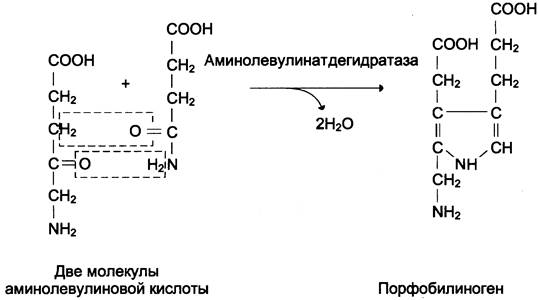
From the Cell/35.html">Mitochondria, 5-aminolevulinic acid is transported into the Cytoplasm. The intermediate stages of heme synthesis occur in the cytoplasm: the Condensation of 2 molecules of 5-aminolevulinic acid into a molecule of porphobilinogen (Fig. 13-4), the deamination of porphobilinogen to yield hydroxymethylbilane, the enzymatic conversion of hydroxymethylbilane into uroporphyrinogen III, and the decarboxylation of the latter to form coproporphyrinogen III. Hydroxymethylbilane can also be converted non-enzymatically into uroporphyrinogen I, which is subsequently decarboxylated to coproporphyrinogen I. From the cytoplasm, coproporphyrinogen III re-enters the mitochondria, where the final reactions of heme synthesis take place. Through two consecutive oxidative steps, coproporphyrinogen III is converted into protoporphyrinogen IX, and protoporphyrinogen IX into protoporphyrin IX. The enzyme ferrochelatase inserts ferrous iron into protoporphyrin IX, converting it into heme (Fig. 13-2). The iron-storage protein ferritin serves as the iron source for heme synthesis. Once synthesized, heme combines with the α- and β-polypeptide chains of globin to form hemoglobin. Heme also regulates globin synthesis: when The rate of heme synthesis drops, globin synthesis in reticulocytes is inhibited.
Fig. 13-4. Reaction forming porphobilinogen.
C. Regulation of Heme Biosynthesis
The regulatory step of heme synthesis is catalyzed by the pyridoxal-dependent enzyme 5-aminolevulinate synthase. The reaction rate is regulated allosterically and at the level of enzyme Translation.
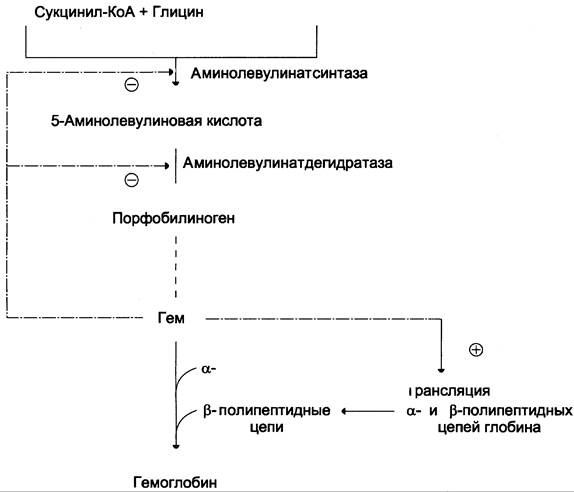
Heme acts as an allosteric inhibitor and corepressor of 5-aminolevulinate synthase synthesis (Fig. 13-5).
Fig. 13-5. Regulation of heme and hemoglobin synthesis. Through negative feedback, heme inhibits 5-aminolevulinate synthase and 5-aminolevulinate dehydratase while acting as a translational inducer for the α- and β-chains of hemoglobin.
In reticulocytes, the synthesis of this enzyme at the translational stage is regulated by iron. The initiation region of the mRNA encoding the enzyme contains a nucleotide sequence that forms a hairpin loop known as the iron-responsive element (IRE) (Fig. 13-6).
Fig. 13-6. Regulation of aminolevulinate synthase synthesis. A — at high iron concentrations in reticulocytes, iron binds to the iron-binding protein, decreasing the affinity of this protein for the iron-responsive element (IRE) of the mRNA encoding aminolevulinate synthase. Translation initiation protein factors bind to the mRNA and initiate the translation of aminolevulinate synthase. B — at low iron concentrations in reticulocytes, the iron-binding protein exhibits a high affinity for the IRE and interacts with it. Translation initiation protein factors are unable to bind to the mRNA, and translation is halted.
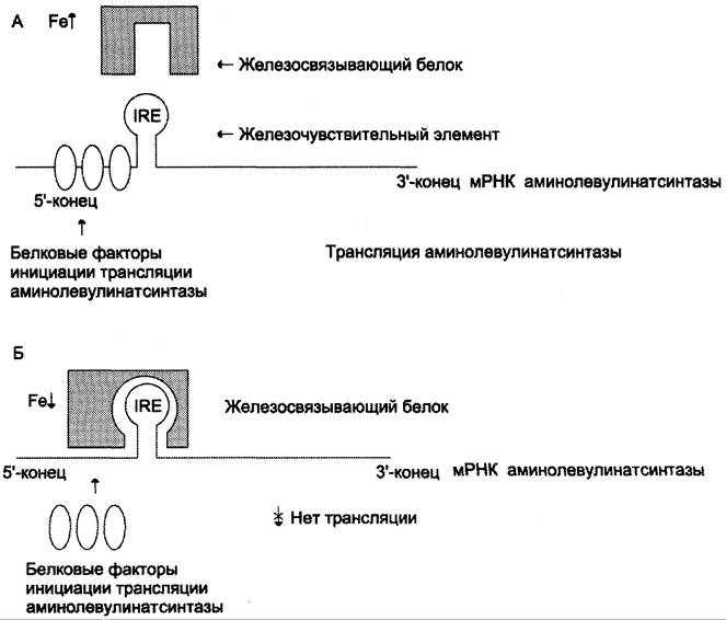
At high intracellular iron concentrations, iron forms a complex with Cysteine residues of the regulatory iron-binding protein. The interaction of iron with the regulatory iron-binding protein reduces the affinity of this protein for the IRE in the mRNA encoding aminolevulinate synthase, thereby allowing translation to proceed (Fig. 13-6, A). At low iron concentrations, the iron-binding protein attaches to the iron-responsive element located in the 5'-untranslated region of the mRNA, suppressing the translation of aminolevulinate synthase (Fig. 13-6, B).
Aminolevulinate dehydratase is also allosterically inhibited by heme; however, since The activity of this enzyme is nearly 80-fold greater than that of aminolevulinate synthase, this inhibition is of little physiological significance.
Pyridoxal phosphate deficiency and drugs that act as its structural analogs decrease the activity of aminolevulinate synthase.
G. Disorders of Heme Biosynthesis: Porphyrias
Inherited and acquired disorders of heme synthesis characterized by elevated levels of porphyrinogens and their oxidation products in tissues and Blood, along with their excretion in urine, are referred to as porphyrias (derived from the Greek word for purple).
Inherited porphyrias are caused by Genetic Defects in enzymes involved in heme synthesis, with the exception of aminolevulinate synthase. These conditions are characterized by impaired heme production. Since heme Functions as an allosteric inhibitor of aminolevulinate synthase, the activity of this enzyme increases, leading to the accumulation of heme synthesis intermediates, namely aminolevulinic acid and porphyrinogens.
Depending on the primary site of the pathological process, inherited porphyrias are classified as hepatic or erythropoietic. Erythropoietic porphyrias involve the accumulation of porphyrins in normoblasts and erythrocytes, whereas hepatic porphyrias involve their accumulation in hepatocytes.
Severe forms of porphyria present with neuropsychiatric disorders, impaired reticuloendothelial system function, and Skin lesions. Porphyrinogens are colorless and non-fluorescent, but upon exposure to light, they are readily converted into porphyrins, which exhibit intense red fluorescence under ultraviolet light. In sun-exposed skin, interaction between porphyrins and oxygen generates singlet oxygen. Singlet oxygen accelerates Lipid Peroxidation in cell membranes and causes cell destruction, which is why porphyrias are frequently accompanied by photosensitivity and ulceration of exposed skin areas. The neuropsychiatric symptoms associated with porphyrias are attributed to the neurotoxic properties of aminolevulinate and porphyrinogens.
Mild forms of inherited porphyrias may sometimes remain asymptomatic; however, the administration of drugs that act as Inducers of aminolevulinate synthase can precipitate an acute attack. Well-known inducers of aminolevulinate synthase include sulfonamides, barbiturates, diclofenac, voltaren, Steroids, and gestagens. In some cases, symptoms do not manifest until Puberty, when elevated PRODUCTION OF β-steroids induces the synthesis of aminolevulinate synthase. Porphyrias are also observed in lead poisoning because lead inhibits aminolevulinate dehydratase and ferrochelatase. Certain halogenated herbicides and insecticides act as inducers of aminolevulinate synthase, and exposure to these agents leads to symptoms of porphyria.
Last update: 06/08/2026
Editorial and Educational Adaptation: This material has been compiled based on the primary/original source text. The project team performed an editorial review, corrected technical inaccuracies, structured sections, and adapted the content for an educational format.
What was processed:
- elimination of formatting defects (OCR errors, structural breaks, corrupted characters);
- editorial organization of content;
- standardization of terminology in accordance with academic sources;
- verification of factual statements against the original source text.
All mentions of the author, publication year, and origin of the primary text have been preserved in accordance with the source.