LEHNINGER PRINCIPLES OF BIOCHEMISTRY - VOL. 2. BIOENERGETICS AND METABOLISM - 2014
PART II. BIOENERGETICS AND METABOLISM
Class="center">If you get an idea, you shouldn't reject it just because it doesn't fit into the framework of the prevailing theory.
Claude Bernard, An Introduction to the Study of Experimental Medicine, 1813
The most important and remarkable feature of the acceptance of the chemiosmotic hypothesis, in my view, is the altruism and magnanimity with which former opponents of this hypothesis not only agreed with it, but unanimously elevated it to the rank of a theory.
Peter Mitchell, Nobel Lecture, 1978
19. OXIDATIVE PHOSPHORYLATION AND PHOTOPHOSPHORYLATION
Oxidative Phosphorylation is the culmination of energy-yielding METABOLISM in aerobic organisms. All oxidative processes in the degradation of CARBOHYDRATES, Fatty acids, and Amino Acids converge at this final stage of cellular respiration, where The energy released during oxidation is channeled into ATP synthesis. Photophosphorylation is the process by which photosynthetic organisms capture solar energy—the primary energy source for the Earth's biosphere—and direct it toward ATP synthesis. The vast majority of organisms synthesize the ATP they require primarily through OXIDATIVE PHOSPHORYLATION AND photophosphorylation.
In Eukaryotic Cells, Oxidative phosphorylation takes place in Cell/35.html">Mitochondria, whereas photophosphorylation occurs in Chloroplasts. During oxidative phosphorylation, both in the light and in the dark, O2 is reduced to H2O via Electron transfer from the Coenzymes NADH and FADH2. In photophosphorylation, The oxidation of H2O to O2 is driven exclusively by light energy, utilizing NADP+ as the ultimate electron acceptor. Despite these differences, these two highly efficient energy-conversion processes share remarkably similar molecular mechanisms.
Modern concepts regarding the Mechanism of ATP synthesis in Mitochondria and chloroplasts stem from the hypotheses put forward by Peter Mitchell in 1961. Mitchell proposed that the proton concentration gradient across a membrane, generated by oxidation reactions, serves as an energy reservoir. This chemiosmotic theory became a foundational cornerstone of 20th-century biology. The chemiosmotic theory successfully accounts for oxidative phosphorylation and photophosphorylation, as well as diverse energy-transducing pathways such as active membrane transport and bacterial flagellar motility.
Oxidative phosphorylation and photophosphorylation operate via closely related mechanisms in three fundamental aspects. First, both processes are driven by the flow of electrons through a chain of membrane-bound carriers. Second, the exergonic release of Free energy during "downhill" electron transfer is coupled to the "uphill" transport of protons across a proton-impermeable membrane, resulting in the storage of electron free energy as a transmembrane Electrochemical Potential (p. 551, vol. 1). Third, the spontaneous flow of protons back down their concentration gradient from the intermembrane space through specialized protein channels provides the free energy required for ATP synthesis. This reaction is catalyzed by ATP synthase, a membrane protein complex that couples proton flux to the phosphorylation of ADP.
We begin this chapter by exploring oxidative phosphorylation. First, we examine the electron transfer chain, the Organization of its carriers into large functional complexes embedded within The inner mitochondrial membrane, and the pathways of electron flow alongside the coupled proton translocation. Next, we look closely at the remarkable enzyme complex that operates via a "rotational catalysis" mechanism, converting proton-motive energy into ATP. Finally, we discuss The regulatory mechanisms that coordinate oxidative phosphorylation with the various Catabolic pathways of fuel oxidation.
Following our Structure/133.html">Discussion of mitochondrial oxidative phosphorylation, we turn to the key aspects of photophosphorylation, focusing first on Light absorption by Photosynthetic Pigments, the light-driven electron transfer mechanisms from H2O to NADP+, and the Molecular Basis of coupling electron and proton translocation. We will also highlight the evolutionary conservation and underlying similarities between the mechanisms of ATP Synthesis in chloroplasts and mitochondria.
OXIDATIVE PHOSPHORYLATION
19.1. Electron Transfer Reactions in Mitochondria
In 1948, Eugene Kennedy and Albert Lehninger first demonstrated that oxidative phosphorylation in eukaryotic cells takes place within mitochondria. This landmark discovery ushered in the modern era of Bioenergetics. Mitochondria, much like Gram-negative Bacteria, are bounded by a double membrane (Fig. 19-1). The outer mitochondrial membrane is relatively permeable to small molecules (Mr < 5000) and ions, which diffuse freely through aqueous channels formed by integral Proteins called porins. The inner membrane, by contrast, is impermeable to most small molecules and ions, including H+ protons; only specific substances can cross it via dedicated transport proteins. The inner membrane harbors the Components of the Respiratory Chain and ATP synthase.
Albert L. Lehninger, 1917–1986

Fig. 19-1. Structure of a mitochondrion. The inner membrane is folded into cristae, which dramatically increase its surface area. For instance, the inner mitochondrial membrane of a rat Liver cell contains about 10,000 sets of electron-transfer systems and ATP synthase molecules, whereas that of a cardiac Muscle cell contains roughly three times as many due to a much higher density of cristae. The intra-mitochondrial pool of coenzymes and metabolic intermediates is physically and functionally segregated from the cytosolic pool. Although mitochondria in invertebrates, plants, and Protozoa share an identical basic architecture, they vary widely in shape, size, and the degree of inner membrane folding.
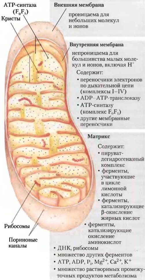
The mitochondrial matrix, enclosed by the inner membrane, contains the Pyruvate dehydrogenase complex and the Enzymes responsible for The Citric Acid Cycle, fatty acid β-oxidation, and amino acid oxidation—essentially all the pathways of fuel oxidation, with the exception of Glycolysis, which takes place in the Cytosol. Due to its selective permeability, the inner membrane shields the metabolic intermediates and Enzymes of the cytosol from those operating within the matrix. It also contains specialized transport systems that shuttle pyruvate, fatty acids, amino acids, or their α-keto derivatives into the matrix, where they feed into The Citric Acid cycle. Similarly, specific transporters export newly synthesized ATP from the matrix while importing fresh supplies of ADP and inorganic phosphate.
Electrons Are Carried by Universal Electron Acceptors
Oxidative phosphorylation begins with The entry of electrons into the respiratory chain. The vast majority of these electrons are generated by the action of various dehydrogenases, which harvest electrons from distinct catabolic pathways and deliver them to universal electron acceptors—specifically, nicotinamide NUCLEOTIDES (NAD+ or NADP+) or flavin nucleotides (FMN or FAD).
Nicotinamide nucleotide-linked dehydrogenases catalyze reversible reactions that can be generally represented as follows:
Reduced substrate + NAD+ ⇄ Oxidized substrate + NADH + H+
Reduced substrate + NADP+ ⇄ Oxidized substrate + NADPH + H+
Most dehydrogenases involved in catabolic processes utilize the coenzyme NAD+ as an electron acceptor. Some of these enzymes are localized in the cytosol, others in the mitochondrial matrix, while some exist in both cytosolic and mitochondrial isoenzyme forms (Table 19-1).
Table 19-1. Some Important Reactions Catalyzed by NAD(P)H-Dependent Dehydrogenases
Reaction* |
Localization** |
NAD-dependent dehydrogenases |
|
α-Ketoglutarate + CoA + NAD+ ⇄ succinyl-CoA + СО2 + NADH + Н+ |
M |
L-Malate + NAD+ ⇄ oxaloacetate + NADH + Н+ |
M and C |
Pyruvate + CoA + NAD+ ⇄ acetyl-CoA + СО2 + NADH + Н+ |
M |
Glyceraldehyde-3-phosphate + Pi + NAD+ ⇄ 1,3-bisphosphoglycerate + NADH + Н+ |
C |
Lactate + NAD+ ⇄ pyruvate + NADH + Н+ |
C |
β-Hydroxyacyl-CoA + NAD+ ⇄ β-ketoacyl-CoA + NADH + Н+ |
M |
NADP-dependent dehydrogenases |
|
Glucose-6-phosphate + NADP+ ⇄ 6-phosphogluconate + NADPH + Н+ |
C |
NAD- or NADP-dependent dehydrogenases |
|
L-Glutamate + Н2O + NAD(P)+⇄ α-ketoglutarate + NH+4 + NAD(P)H |
M |
Isocitrate + NAD(P)+ ⇄ α-ketoglutarate + СO2 + NAD(P)H + Н+ |
M and C |
* These reactions and their corresponding enzymes are discussed in detail in Chapters 14–18.
** M — mitochondria, C — cytosol.
NAD-dependent dehydrogenases remove two hydrogen atoms from the substrates being oxidized. One hydrogen atom is transferred to NAD+ as a hydride ion (:H), while the other is released into the medium as an H+ ion (see Fig. 13-24). Both NADH and NADPH are Water-soluble electron carriers that can reversibly dissociate from their respective dehydrogenase complexes during a reaction. NADH delivers electrons derived from catabolic reactions to the beginning of the respiratory chain—the NADH dehydrogenase complex described below. NADPH, on the other hand, primarily supplies electrons for anabolic processes. Within cells, the NADH and NADPH pools are strictly segregated, maintaining distinct redox potentials. This compartmentalization is achieved by keeping The ratio of the reduced form to the oxidized form relatively high for NADPH and relatively low for NADH. Neither NADH nor NADPH can cross the inner mitochondrial membrane; however, the electrons they carry are able to cross the membrane freely in both directions.
Flavoproteins contain flavin nucleotides (FMN or FAD) tightly and sometimes covalently bound to a protein moiety (see Fig. 13-27). The oxidized forms of flavin nucleotides can accept either a single electron from a substrate to form a semiquinone intermediate, or two electrons to yield FADH2 or FMNH2. Electron transfer is driven by the fact that the reduction potential of the flavoprotein is more positive than that of the compound being oxidized. Unlike the NAD and NADP coenzymes, the standard reduction potential of flavin nucleotides depends on the specific protein to which they are bound. Interactions with the Functional groups of the protein distort the electron orbitals of the flavin ring, thereby altering the relative stability of its oxidized and reduced forms. Consequently, the standard reduction potential corresponds to the flavoprotein functioning in that particular reaction, rather than to isolated FAD or FMN. Thus, a flavin nucleotide is more accurately viewed as an integral part of the flavoprotein's Active Site than as a freely diffusing substrate or product of the electron-transfer reaction.
Because flavoproteins participate in one- or two-electron transfers during redox reactions, they appear to serve as versatile transducers between two-electron pathways (such as dehydrogenations) and one-electron pathways (such as quinone-to-hydroquinone reductions), as discussed in detail below.
Electrons flow through a chain of membrane-embedded carriers
The mitochondrial respiratory chain consists of a series of sequentially acting electron carriers, many of which are integral Membrane Proteins containing prosthetic groups capable of accepting and donating one or two electrons. During oxidative phosphorylation, electron transfer can occur via three distinct mechanisms: first, by direct electron transfer, such as the REDUCTION OF Fe3+ to Fe2+; second, by The transfer of a hydrogen atom (H+ + e-); and third, by the transfer of a hydride ion (:H-) carrying a pair of electrons. In redox reactions, a charge equivalent to a single electron is termed a reducing equivalent.
In addition to NAD and flavoproteins, the respiratory chain utilizes a hydrophobic quinone (ubiquinone) and two classes of iron-containing proteins: Cytochromes and iron-sulfur proteins. Ubiquinone (coenzyme Q, or simply Q) is a lipid-soluble benzoquinone characterized by a long isoprenoid side chain (Fig. 19-2). Functional analogs of mitochondrial coenzyme Q include plastoquinone in plant chloroplasts and menaquinone in bacteria. Ubiquinone can accept a single electron to form a semiquinone radical (•QH), or two electrons to become fully reduced to ubiquinol (QH2, see Fig. 19-2). Like flavoproteins, ubiquinone acts as a bridge between two-electron Donors and one-electron acceptors. Due to its small size and hydrophobic nature, ubiquinone diffuses rapidly within The Lipid Bilayer of the inner mitochondrial membrane, shuttling reducing equivalents between less mobile, membrane-embedded Protein Complexes. Because it transports both electrons and protons, ubiquinone plays a central role in coupling electron flow in the respiratory chain to the pumping of protons from the mitochondrial matrix into the intermembrane space.
Figure 19-2. Ubiquinone (Q, or coenzyme Q). Complete reduction of ubiquinone requires two electrons and two protons, proceeding in two sequential steps via a semiquinone radical intermediate.

Cytochromes are proteins characterized by distinctive absorption bands in the visible region of the spectrum, which are conferred by their iron-containing heme prosthetic groups (Fig. 19-3). Mitochondria contain three Major Classes of cytochromes—designated a, b, and c—which can be distinguished by their unique absorption spectra. The reduced forms of all cytochromes contain Fe2+ and exhibit three characteristic absorption bands in the visible spectrum (Fig. 19-4). The main visible absorption band of cytochrome a is shifted toward longer wavelengths (around 600 nm), that of cytochrome b is near 560 nm, and that of cytochrome c is near 550 nm. Within a given class of cytochromes, subscripts are often used to indicate the exact wavelength of the absorption maximum, such as b562.
Figure 19-3. Prosthetic groups of cytochromes. The prosthetic group of all cytochromes consists of a porphyrin macrocycle formed by four five-membered nitrogen-containing rings. Four nitrogen atoms are coordinated with a central iron ion in either the reduced (Fe2+) or oxidized (Fe3+) state. Iron protoporphyrin IX is found in cytochrome b, Hemoglobin, and Myoglobin (see Fig. 4-16, Vol. 1). Heme c in cytochrome c is covalently attached to the protein via thioether linkages to two Cysteine residues. Heme a in cytochrome a features a long isoprenoid side chain attached to one of the pyrrole rings. The strong visible light absorption characteristic of Hemes is due to their extensive system of conjugated double bonds (highlighted in pink).

In cytochromes a and b, the heme group is held tightly within the Protein Structure by non-covalent interactions. In contrast, the heme of cytochrome c is anchored to the polypeptide chain via covalent thioether bonds formed with two conserved cysteine residues (Fig. 19-3). Much like flavoproteins, the standard reduction potentials of the heme iron in cytochromes are modulated by the microenvironment created by surrounding amino acid side chains, resulting in unique redox potentials for each cytochrome. Cytochromes a, b, and several c-type cytochromes are integral Proteins of the inner mitochondrial membrane. A notable exception is mitochondrial cytochrome c, which is a water-soluble peripheral protein bound electrostatically to the outer face of the inner mitochondrial membrane.
Figure 19-4. Absorption spectra of the oxidized (red curve) and reduced (blue curve) forms of cytochrome c. Greek letters α, β, and γ designate the absorption bands of the reduced form.

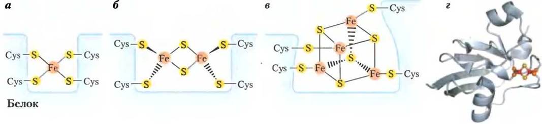
In iron-sulfur proteins, the iron atoms are not incorporated into a heme ring; instead, they are coordinated either to inorganic sulfide ions, to the sulfur atoms of cysteine residues in the polypeptide chain, or to both. The simplest iron-sulfur centers consist of a single iron atom coordinated to four cysteine SH groups, whereas more complex centers contain two or four iron atoms (Fig. 19-5). In the Rieske iron-sulfur proteins (discovered by John S. Rieske), a single iron atom is coordinated by two Histidine residues rather than cysteines. All iron-sulfur proteins participate in one-Electron transfer reactions, during which a single iron atom within the cluster undergoes reversible oxidation or reduction. Mitochondria contain at least eight distinct iron-sulfur proteins. The reduction potentials of these centers span a wide range, from approximately -0.65 V to +0.45 V, depending heavily on their local protein environment.
Figure 19-5. Structures of iron-sulfur centers. (a) A simple iron-sulfur center in which a single iron ion is coordinated by sulfur atoms from four cysteine residues of the protein. (b and c) More complex iron-sulfur centers, where each iron atom is bonded to both inorganic sulfide and cysteine sulfur atoms. (d) The spatial arrangement of iron atoms (red spheres), inorganic sulfur atoms (yellow spheres), and cysteine sulfur atoms (orange spheres) in the Fe2S2 center of ferredoxin from the cyanobacterium *Anabaena* 7120 (PDB ID 1FRD). Note that standard nomenclature for iron-sulfur centers specifies only the number of iron and inorganic sulfur atoms (e.g., Fe2S2 indicates two iron and two inorganic sulfur atoms). In reality, each iron atom in this cluster is coordinated by four sulfur atoms—two inorganic sulfides and two cysteine thiols. The standard reduction potential of iron in these centers is determined by cluster geometry and specific interactions with the surrounding protein matrix.
Electron flow through the mitochondrial respiratory chain can be summarized as follows: electrons pass from NADH, succinate, or other primary electron donors to flavoproteins and ubiquinone, and subsequently through a sequence of iron-sulfur proteins and cytochromes to molecular oxygen. We will now examine the experimental Methods used to study electron transfer in the respiratory chains of mitochondria and chloroplasts.
One fundamental approach relies on the experimental measurement of standard reduction potentials for the various electron carriers, which constitute redox couples (see Table 19-2). Because electrons flow spontaneously from systems with lower (more negative) $E'^{\circ}$ values to those with higher (more positive) $E'^{\circ}$ values, electron transfer from oxidizable substrates to oxygen proceeds via a series of intermediate carriers in order of increasing reduction potential. The overall pathway of electron flow in the mitochondrial chain was established by determining the standard reduction potentials of its components in the sequence: NADH → Q → cytochrome b → cytochrome c1 → cytochrome c → cytochrome a → cytochrome a3 → O2. It is important to note that the actual reduction potentials of biological redox systems inside The Cell depend on the local concentrations of their oxidized and reduced forms and are not necessarily identical to values measured under standard biochemical conditions (see Equation 13-5 on p. 47).
Table 19-2. Standard reduction potentials of some conjugate oxidation-reduction pairs playing a crucial role in oxidative metabolism
Oxidation-reduction reactions (half-reaction) |
E'°, V |
2Н+ + 2е- —> Н2 |
-0,414 |
NАD+ + Н+ +2е- —> NАDН |
-0,320 |
NАDР+ + Н+ + 2е- —> NАDРН |
-0,324 |
NADH dehydrogenase (FМN) + 2Н+ + 2е- —> NADH dehydrogenase (FМNH2) |
0,30 |
Ubiquinone + 2Н+ + 2е- —> ubiquinol |
0,045 |
Cytochrome b (Fе3+) + е- —> cytochrome b (Fе2+) |
0.077 |
Cytochrome c1 (Fе3+) + е- —> cytochrome c1 (Fе2+) |
0,22 |
Cytochrome c (Fе3+) + e- —> cytochrome c (Fе2+) |
0,254 |
Cytochrome a (Fе3+) + е- —> cytochrome a (Fе2+) |
0,29 |
Cytochrome a3 (Fе3+) + е- —> cytochrome a3 (Fе2+) |
0,55 |
1/2 O2 + 2Н+ + 2е- —> Н2O |
0,8166 |
The second experimental METHOD FOR DETERMINING the Sequence of electron carriers in the mitochondrial respiratory chain is based on truncating this chain. To achieve this, an electron donor is introduced into the mitochondrion in the absence of oxygen (the electron acceptor). Oxygen (O2) is then introduced into the system, and The rate of carrier oxidation is determined spectrophotometrically. The carrier closest to the end of the respiratory chain—that is, to O2—is oxidized first, followed by the second carrier from the end of the chain, and so on. The sequence of electron carriers in the mitochondrial respiratory chain established in this manner is in good agreement with the results obtained from standard reduction potential measurements.
The third method relies on studying the action of specific inhibitors that block electron transfer at specific sites along the respiratory chain. When an inhibitor is introduced into the Electron Transport Chain, it creates a region where the oxidation-reduction state of the carriers changes. In the presence of O2 and an electron donor, the electron carriers located before the blocked site become reduced, while those immediately after the blocked site become oxidized, which can be detected spectrophotometrically (Fig. 19-6). The electron carrier sequence determined by this method is consistent with the results of the previous two methods.
Fig. 19-6. Determination of the sequence of electron carriers in the mitochondrial respiratory chain. This method is based on studying The Effect of ELECTRON TRANSPORT INHIBITORS on the oxidation state of each carrier. In the presence of an electron donor and oxygen (O2) as the electron acceptor, the inhibitor suppresses electron transfer at specific segments of the respiratory chain, thereby altering the redox state of the carriers. Carriers located upstream of the blocked site are reduced (shown in blue), whereas those downstream of the blocked site are oxidized (shown in pink).

Electron carriers in the mitochondrial respiratory chain form multienzyme complexes
Electron carriers are organized into multienzyme complexes—structurally distinct supramolecular assemblies of protein molecules embedded within the inner membrane. Treatment of the inner mitochondrial membrane with a mild detergent has made it possible to isolate four complexes of functionally linked electron carriers. Each of these complexes catalyzes electron transfer at a specific segment of the respiratory chain (Table 19-3; Fig. 19-7). Complexes I and II catalyze electron transfer to ubiquinone from NADH and succinate, respectively. Complex III transfers electrons from the reduced form of ubiquinone to cytochrome c, whereas Complex IV completes the transfer of electrons along the respiratory chain by passing them from cytochrome c directly to oxygen.
The structural features and Functions of the protein complexes involved in electron transport within the mitochondrial respiratory chain are discussed below.
Table 19-3. Proteins involved in electron transport in the mitochondrial respiratory chain
Enzyme-protein complex |
Mass, kDa |
Number of subunits* |
Prosthetic groups |
NADH dehydrogenase (Complex I) |
850 |
43 (14) |
FMN, FeS |
Succinate dehydrogenase (Complex II) |
140 |
4 |
FAD, FeS |
Ubiquinone-cytochrome c oxidoreductase (Complex III) |
250 |
11 |
hemes, FeS |
Cytochrome c** |
13 |
1 |
heme |
Cytochrome oxidase (Complex IV) |
160 |
13 (3-4) |
hemes, CuA, CuB |
* Values in parentheses indicate the number of subunits in the homologous complex isolated from bacterial cells.
** Cytochrome c is not a component of the enzyme complex; it is a water-soluble peripheral membrane protein that moves freely between complexes III and IV.
Fig. 19-7. Isolation of enzyme complexes involved in electron transfer in the respiratory chain from mitochondria. The outer mitochondrial membrane is removed by treating mitochondria with the detergent digitonin. The inner membrane is separated from the mitochondrial matrix by osmotic Shock. The inner membrane fragments are gently solubilized in a second detergent, and protein complexes I–IV, which participate in mitochondrial electron transfer, as well as ATP synthase (sometimes referred to as complex V; see Table 19-3), are isolated from this solution by Ion-exchange Chromatography. In their isolated state, complexes I–IV catalyze the transfer of electrons from NADH or succinate to mobile carriers (Q and cytochrome c) and ultimately to molecular oxygen. Isolated ATP synthase exhibits only ATP-hydrolyzing (ATPase) activity in vitro and cannot catalyze ATP synthesis.

Complex I catalyzes electron transfer from NADH to ubiquinone. The interaction of complex I with complex II and ubiquinone during electron transfer is illustrated in Fig. 19-8. Complex I (NADH-ubiquinone reductase or NADH dehydrogenase) consists of 42 different polypeptide chains and contains FMN as a prosthetic group along with at least six iron-sulfur centers. High-resolution electron spectroscopy has revealed that complex I has an L-like shape, with one end embedded in the matrix and the other attached to the membrane (Fig. 19-9). Complex I catalyzes two simultaneous and coupled processes. First, the exergonic transfer of a hydride ion from NADH to ubiquinone, accompanied by the uptake of a proton from the matrix, which can be represented by the following equation:
NАDН + Н+ + Q —> NАD+ + QH2 (19-1)
Fig. 19-8. Electron transfer from NADH, succinate, fatty acid CoA derivatives, and glycerol 3-phosphate to ubiquinone. From NADH, electrons are transferred to a flavoprotein, then to a series of iron-sulfur proteins in complex I, and subsequently to coenzyme Q. From succinate, electrons are transferred via a flavoprotein to iron-sulfur centers in complex II and then to coenzyme Q. Glycerol 3-phosphate donates electrons to glycerol 3-phosphate dehydrogenase, a flavoprotein located on the outer surface of the inner mitochondrial membrane, from which electrons also pass to coenzyme Q. Acyl-CoA dehydrogenase (the first enzyme in the fatty acid β-oxidation cycle) transfers electrons to electron-transferring flavoprotein (ETF), which then passes them to coenzyme Q via ETF-ubiquinone oxidoreductase.

Fig. 19-9. Structure of NADH-ubiquinone oxidoreductase (complex I). Complex I mediates the transfer of a hydrogen ion from NADH to FMN, from which two electrons are passed through a series of FeS centers to the iron-sulfur protein N-2, located in the matrix-embedded domain of complex I. The matrix-extruding domain has been crystallized and studied by X-ray crystallography (PDB ID 2FUG); The structure of the membrane-embedded domain of complex I remains unknown. From the N-2 protein group, electrons are transferred to the membrane-anchored region of the complex, where they reduce ubiquinone to ubiquinol QH2, which then diffuses into the lipid bilayer of the membrane. The transfer of each pair of electrons is accompanied by the pumping of four protons out of the matrix. The mechanism coupling electron and proton transfer in complex I is not yet fully understood. It is hypothesized that, as in complex III, electrons and protons are transferred by coenzyme Q through a specialized cyclic pathway known as the Q-cycle (Fig. 19-12). A molecule of QH2 participates in the transfer of a pair of electrons twice. An electrochemical potential is established across the inner mitochondrial membrane As a result of proton pumping. The inner face of the inner mitochondrial membrane becomes negatively charged ("-"), while the outer face becomes positively charged ("+"). A portion of the free energy released during Electron Transport and utilized for ATP synthesis is stored in the form of this electrochemical potential.

Second, the endergonic process of pumping four protons out of the matrix and transferring them across the inner membrane into the intermembrane space, i.e., exclusively in one direction (vectorially). This process consumes a fraction of the energy carried by the electrons moving along the chain. Because each proton carries a positive charge, the proton-depleted matrix becomes negatively charged, whereas the proton-enriched intermembrane space becomes positively charged. To emphasize the vectorial transport of protons, the net equation is often written with appropriate subscripts indicating the Location OF THE proton: "+" for the positively charged side of the inner membrane facing the intermembrane space, and "-" for the negatively charged side adjacent to the matrix.
NADH + 5Н+«-» + Q —> NAD+ + QH2 + 4H+«+» (19-2)
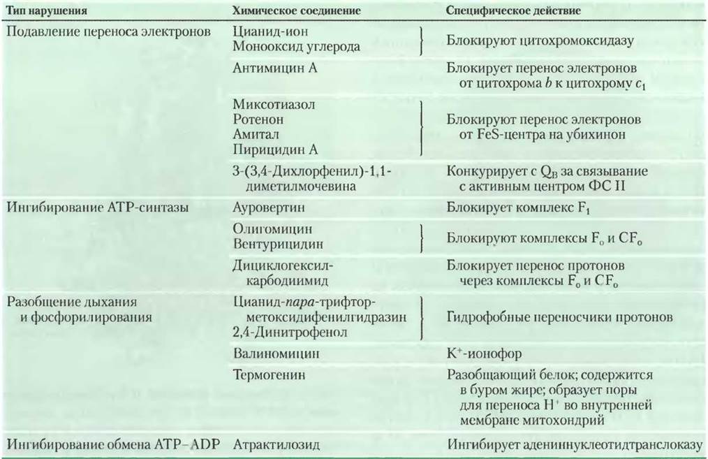
Certain chemical compounds block electron transfer from the iron-sulfur centers of complex I to ubiquinone, thereby halting the entire process of oxidative phosphorylation. Examples include amobarbital, a barbiturate drug; rotenone, a plant-derived compound used as an insecticide; and the antibiotic piericidin A (see Table 19-4).
Table 19-4. Chemical compounds that disrupt oxidative phosphorylation and photophosphorylation
In the inner mitochondrial membrane, the fully reduced form of ubiquinone, ubiquinol QH2 (Fig. 19-2), diffuses from complex I to complex III. In complex III, as a proton is transferred from the matrix to the intermembrane space, QH2 is oxidized to Q.
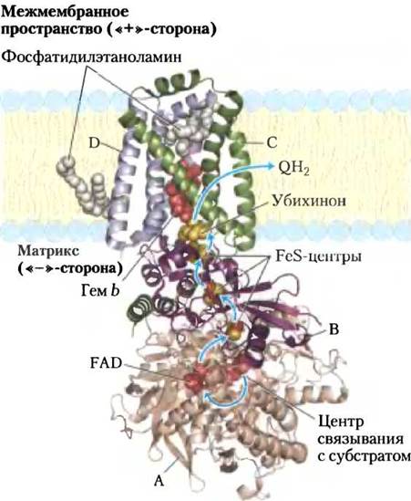
Complex II (succinate dehydrogenase), described in Chapter 16, transfers electrons from succinate to ubiquinone Q and is the only enzyme of the citric acid cycle that is tightly bound to the mitochondrial membrane (p. 198). Complex II is smaller than complex I and has a less complex structure. It comprises five prosthetic groups of two types and four subunits (Fig. 19-10). Subunits C and D of complex II are integral transmembrane proteins whose helices cross the membrane three times. These subunits incorporate type b heme groups and the binding site for ubiquinone, the final electron acceptor in the redox reactions catalyzed by complex II. Subunits A and B of complex II extend into the mitochondrial matrix (or into the cytosol in bacterial cells) and bear three Fe2S2-type iron-sulfur centers, as well as a covalently bound FAD coenzyme and the binding site for succinate, the substrate of succinate dehydrogenase. The total path length for electrons from the succinate binding site to FAD and then via the FeS centers to the ubiquinone binding site is no more than 40 Å. The distance between individual electron carriers in this chain does not exceed 11 Å, which is optimal for rapid electron transfer (Fig. 19-10).
Fig. 19-10. Structure of complex II (succinate dehydrogenase) (PDB ID 1NEK). This complex (showing the enzyme isolated from pig Heart) consists of two C subunits and one D subunit that span the membrane. Subunits B and A are located in the Cytoplasm. In polypeptide chain A, the succinate binding site is located downstream of the FAD prosthetic group. Subunit B includes three FeS centers. Ubiquinone is bound to subunit C. Heme b is located between subunits C and D. Two phosphatidylethanolamine molecules are bound so tightly to subunit D that they crystallize together with the protein. Electrons, whose path is indicated by blue arrows, move from succinate to FAD and subsequently through three iron-sulfur centers to ubiquinone. Heme b does not participate in electron transfer, but it prevents The formation of reactive oxygen species (ROS) in the event of an electron "leak."
Heme b in complex II apparently does not transfer electrons, but rather reduces the probability of Incomplete reduction of molecular oxygen to H2O, which occurs as a result of non-enzymatic transfer ("leakage") of electrons along the pathway from succinate to O2. Electron leakage leads to the formation of cell-toxic reactive oxygen species (ROS): hydrogen peroxide and the superoxide radical •O2-, as discussed in detail below. The generation of large amounts of ROS during succinate oxidation may be caused by point Mutations in The polypeptide chains of complex II near heme b or the quinone binding site. Individuals carrying such mutations develop an inherited disease known as paraganglioma—benign tumors of the HEAD and Neck, typically localized around the carotid artery, an organ highly sensitive to Blood oxygen levels.
■ In the respiratory chain, electrons from all substrates other than succinate—which are oxidized by mitochondrial membrane dehydrogenases—are fed into ubiquinone, bypassing complex II. During the first stage of fatty acid β-oxidation, catalyzed by the flavoprotein acyl-CoA dehydrogenase, electrons are transferred from the substrate to FAD, the prosthetic group of acyl-CoA dehydrogenase (see Fig. 17-8). The reduced form of acyl-CoA dehydrogenase then passes the electrons to a specific electron carrier, ETF, which in turn transfers them to ubiquinone via ETF-ubiquinone oxidoreductase (Fig. 19-8). Glycerol 3-phosphate, generated either during glycolysis via the reduction of dihydroxyacetone phosphate or from glycerol released during triacylglycerol breakdown, is oxidized by glycerol 3-phosphate dehydrogenase, a flavoprotein class enzyme (see Fig. 17-4). Glycerol 3-phosphate dehydrogenase is localized on the outer face of the inner mitochondrial membrane and, similarly to succinate dehydrogenase and acyl-CoA dehydrogenase, delivers electrons to ubiquinone (Fig. 19-8). The Role of glycerol 3-phosphate dehydrogenase in the so-called glycerol phosphate shuttle, which transfers reducing equivalents from cytosolic NADH into the mitochondrial matrix, is discussed in Section 19.2 (see Fig. 19-30). The action of all these enzymes is directed toward reducing ubiquinone to ubiquinol QH2, which is subsequently reoxidized by complex III.
Complex III (cytochrome bc1 complex, or ubiquinone-cytochrome c oxidoreductase) is the next link in the respiratory chain after complex II; it transfers electrons from ubiquinone to cytochrome c. Complex III also couples the transfer of electrons from ubiquinol QH2 to cytochrome c with The transport of protons from the matrix to the intermembrane space. The complete elucidation of the structures of the massive protein complex III (Fig. 19-11) and complex IV, whose properties are discussed below, was achieved through X-ray crystallography between 1985 and 1998. This marked a major milestone in understanding Electron transport along the mitochondrial respiratory chain and the biochemical functions of the respiratory complexes.
The functional unit of complex III is a dimer in which two cytochrome b monomer units surround a cavity in the middle of the membrane where ubiquinone can freely move from the matrix side (the Q«-» site in one monomer) to the intermembrane space (the Q«+» site in the other monomer) to transport electrons and protons across the inner mitochondrial membrane (Fig. 19-11b).
Fig. 19-11. Cytochrome bc1 (complex III). Complex III comprises two identical monomers combined into a functional assembly. Each monomer contains 11 distinct subunits: (a) The functional core of each monomer consists of three subunits: cytochrome b (green helix), encompassing hemes bH and bL; the Rieske iron-sulfur protein (purple spheres) with two Fe2S2 iron-sulfur centers; and cytochrome c1 (blue helix), containing a single heme (PDB ID 1BGY). (b) Cytochrome c1 and the Rieske iron-sulfur protein protrude above the positively charged surface of the inner membrane, enabling them to interact with cytochrome c, which is located in the intermembrane space and is not a component of complex III. The two ubiquinone binding sites, Q«-» and Q«+», can bind two different inhibitors that block oxidative phosphorylation. Antimycin A, which blocks electron transfer from heme bH to ubiquinone, binds to the Q«-» site located close to heme bH on the negatively charged (matrix) side of the inner membrane. Myxothiazol, which blocks electron transfer from ubiquinol QH2 to the Rieske iron-sulfur protein, binds to the Q«+» site located near the Fe2S2 center and heme bL on the positively charged side of the inner membrane. The dimeric structure of complex III is essential for its catalytic activity. At the interface between the two monomers lie depressions, each housing the Q«+» site of one monomer and the Q«-» site of the other. Intermediates of ubiquinone redox transformations move within these clefts.
Complex III can exist in two Conformations (not shown). In one of them, the FeS center of the Rieske protein is oriented toward the heme of cytochrome c1, which acts as the electron acceptor for this protein, and is moved away from cytochrome b and the ubiquinol QH2 binding site, from which electrons are transferred to the iron-sulfur center of the Rieske protein. In the other conformation, the FeS center of the Rieske protein is turned away from cytochrome c1 toward cytochrome b. Because the localization of the Rieske protein within complex III depends on the oxidation state of its iron-sulfur center, complex III transitions between these two conformations during Redox reactions in the mitochondrial respiratory chain.

Based on structural data for complex III and biochemical research findings, a mechanism for electron and proton transfer across complex III has been proposed (the Q cycle). The overall equation for the redox reactions in the Q cycle (Fig. 19-12) can be written as follows:
QH2 + 2 Cyt c1 (ox.) + 2H+«-» → Q + 2 Cyt c1 (red.) + 4H+«+» (19-3)
Fig. 19-12. The two Stages of the Q cycle. The direction of proton movement through complex III is indicated by blue arrows. In The First stage (left), Q on the negatively charged side is reduced to form a semiquinone radical, which is converted to QH2 In the second stage (right). Two QH2 molecules are oxidized to Q on the positively charged side of the membrane, releasing two protons into the intermembrane space from each QH2 molecule (four protons released in total). Each QH2 molecule transfers one electron to cytochrome c1 via the FeS center of the Rieske protein, and a second electron to a Q molecule located closer to the negatively charged side of the membrane via cytochrome b. Through these two stages, a Q molecule is reduced to QH2. The reduction of a single Q molecule consumes two protons translocated from the matrix.
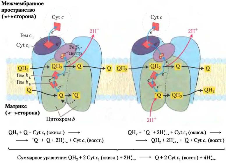
During these cyclic conversions, the two-electron carrier ubiquinone (QH2) passes electrons to single-electron carriers—cytochromes b562, b566, c1, and cytochrome c—while also pumping protons from the matrix into the intermembrane space.
As each pair of electrons traverses complex III, four Protons are pumped out of the matrix. The intricate pathway of electrons through complex III culminates in the oxidation of ubiquinol QH2 to Q and the reduction of two molecules of cytochrome c1.
Cytochrome c is a water-soluble intermembrane protein containing a single heme group. Following electron delivery from complex III to the heme of cytochrome c, cytochrome c migrates to complex IV and transfers its electrons to the binuclear copper active center of complex IV.
Complex IV (cytochrome c oxidase) transfers electrons from cytochrome c to O2, reducing it to H2O and thereby completing electron transport in the mitochondrial respiratory chain. Complex IV is a large protein molecule (13 subunits; Mr = 204,000) localized in the inner mitochondrial membrane. When isolated from bacterial cells, this complex consists of at least 3 to 4 subunits, as a smaller number disrupts its function (Fig. 19-13).
Fig. 19-13. Structure of cytochrome c oxidase (complex IV). This complex from bovine heart mitochondria consists of 13 subunits, though only the four core subunits are shown here (PDB ID 1OCC). (a) Complex IV contains four subunits, each being a dimer of two identical halves. Subunit I (yellow helix) contains hemes a and a3 and a CuB copper ion (green sphere). Heme a3 and CuB form a binuclear active Fe-Cu center. In subunit II (purple helix), two copper ions are linked to the protein moiety via -SH groups of cysteine residues, forming a binuclear CuA center analogous to the Fe2S2 centers in iron-sulfur proteins. The binuclear CuA center and the cytochrome c binding site are located in subunit II within a region protruding into the intermembrane space on the positively charged side of the inner membrane. Subunit III (lilac) is required for rapid proton translocation across subunit II. The function of subunit IV (green) remains unclear. (b) The binuclear CuA center. The copper ions are equivalent. Reduction yields a Cu1+Cu1+ center; oxidation yields a Cu1.5+Cu1.5+ center. The copper ions are coordinated by six amino acid residues: two His, two Cys, Glu, and Met.
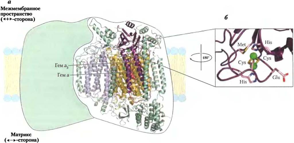
Mitochondrial subunit II of complex IV contains two Cu ions coordinated via -SH groups to two cysteine residues, forming a binuclear CuA center (see Fig. 19-13b) analogous to the Fe2S2 centers of iron-sulfur proteins. Subunit I contains two hemes (a and a3) and a CuB copper ion, which together form a second binuclear center that accepts electrons from heme a and subsequently passes them to molecular oxygen bound at the heme a3 site.
Electrons entering complex IV from cytochrome c travel from the CuA center to heme a, and then—via the a3-CuB center—to molecular oxygen (Fig. 19-14). Each time four electrons enter complex IV, it also takes up four H+ protons from the matrix across the negatively charged membrane surface; these protons are abstracted from substrates and are required to convert O2 into two molecules of H2O. The energy released in this redox process is utilized to eject protons into the intermembrane space (one proton per electron transferred through complex IV). This increases the electrochemical potential generated by the redox reactions and proton translocation in complexes I and III. The overall reactions taking place in complex IV can be written as follows:
4 Cyt c (red.) + 8H+«-» + O2 → 4 Cyt c (ox.) + 4H+«+» + 2H2O (19-4)
Fig. 19-14. Electron flow through complex IV. Subunits I, II, and III are Structural elements of complex IV that mediate electron transfer. In addition to these Polypeptides, complex IV contains 10 other subunits (shown in green). The pathway of electrons through complex IV begins with the transfer of electrons from each of two reduced molecules of cytochrome c (top) to the binuclear CuA center. From the CuA center, electrons are passed via heme a to the Fe-Cu center, which comprises the heme of cytochrome a3 and a CuB ion. Molecular Oxygen binds to heme a3 and is reduced by two electrons to yield the superoxide radical O2-2. Two more electrons from cytochrome c (four electrons are required in total to form H2O) convert O2-2 into two water molecules. The four required protons (H+) are abstracted from substrates in the matrix, from which they are pumped across by a mechanism that remains unknown.
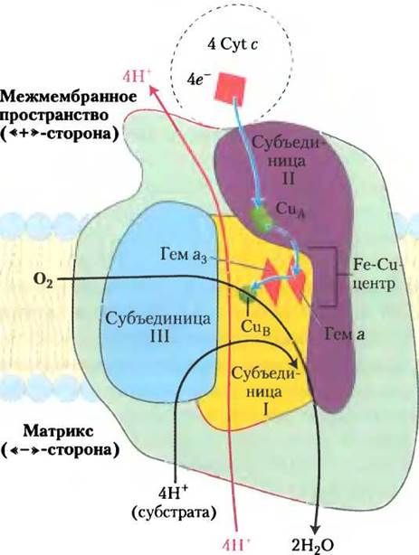
The complete reduction of molecular oxygen O2 takes place at the redox centers in several stages via sequential one-electron transfers, without the release of partially reduced intermediates such as hydrogen peroxide or the hydroxyl free radical—highly reactive species capable of damaging the cell. These aggressive intermediates remain weakly bound to the complex until the Formation of the water molecule is fully complete.
Mitochondrial complexes can form assemblies known as «respirasomes»
A wealth of data indicates that in healthy, intact mitochondria, the respiratory chain complexes are tightly associated within the inner membrane to form «respirasomes» — functional combinations of two or more electron-transfer complexes. For instance, when mitochondrial membranes are extracted under mild conditions, complex III is isolated together with complex I and remains bound to it even during native Electrophoresis. Similarly, complexes III and IV have been successfully isolated together, and electron crystallographic studies show that the size and shape of these complexes match the electron density distribution (Fig. 19-15). The kinetics of electron transport through multiple complexes must differ significantly between tight contact and complete Separation (the two limiting mechanisms). In the former case, where the complexes are in contact, electron transfer occurs through a solid-state medium. In the latter, the complexes function independently, and electrons are shuttled by ubiquinone and cytochrome c. Kinetic data favor electron transfer through a solid-state medium, thereby supporting the respirasome model.
Fig. 19-15. Model of a respirasome consisting of complexes III and IV. a — purified Yeast supercomplexes containing complexes III and IV, visualized by Electron Microscopy following uranyl acetate staining. This electron micrograph was produced by averaging hundreds of individual electron images. b — the structure of a single complex III molecule (red; from yeast) overlaid with the electron density map of two complex IV molecules (green; from bovine heart) to illustrate a plausible mode of interaction between these complexes within the respirasome. The micrograph is oriented perpendicularly to the plane of the structural overlay.
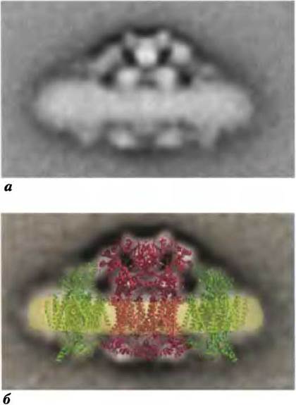
The lipid cardiolipin, which is abundant in the mitochondrial inner membrane (see Figs. 10-9 and 11-2), likely plays a crucial role in respirasome assembly. Its removal by detergents or its absence in certain yeast mutant cells leads to impaired Mitochondrial Electron Transport and a loss of affinity for respiratory chain enzymes.
Energy released during electron transfer in the respiratory chain is conserved as a proton gradient (electrochemical potential)
The transfer of two electrons from the reduced coenzyme NADH to molecular oxygen in the respiratory chain can be written as follows:
NADH + Н+ + 1/2 O2 —> NAD+ + Н2O (19-5)
This process is highly exergonic. The standard reduction potential for the NAD+/NADH redox pair is E'° = -0.320 V, and for the O2/Н2O pair, E’° = 0.816 V. The difference in standard potentials between these two systems is ∆E'° = + 0.816 - (-0.320) = 1.14 V.
The change in Standard Free Energy for the transfer of two electrons from the NAD+/NADH redox pair to the O2/Н2O pair can be calculated using Table 13-7 (p. 48):
∆G’° = -nF∆E'° (19-6)
= - 2 (96.5 kJ/V·mol) • (1.14 V)
= 220 kJ/mol (NADH)
This calculation assumes that the concentrations of NADH and NAD are each 1 M. However, in an actively respiring mitochondrion, the action of numerous dehydrogenases maintains the actual concentration ratio [NADH] / [NAD+] well above unity; consequently, the actual free energy change for reaction 19-5 must be considerably greater in absolute value than -220 kJ/mol. Similar calculations for the oxidation of succinate show that when electrons are transferred from the fumarate/succinate pair (E’° = 0.031 V) to oxygen, the standard free energy change is somewhat smaller than for NADH oxidation, yet it remains a large negative value of approximately -150 kJ/mol.
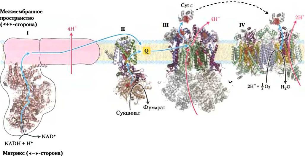
Most of the energy released during the transfer of electrons from NADH to oxygen is utilized to pump protons out of the matrix. For every pair of electrons delivered to oxygen, complex I pumps four protons out of the matrix, complex III pumps four H+, and complex IV pumps two H+ (Fig. 19-16). The coupling of vectorial electron and proton translocation can be described by the following equation:
NADH + 11 Н+«-» + 1/2 O2 —> NAD+ + 10Н+«+» + H2O (19-7)
The electrochemical potential, generated by both the concentration gradient of protons across the mitochondrial membrane and the separation of charges across the membrane, serves as a transient reservoir for the bulk of the energy released during respiratory chain electron transfer.
Fig. 19-16. Electron and proton fluxes through the four complexes of the mitochondrial respiratory chain. Electrons from NADH pass through complexes I and II to coenzyme Q. Reduced ubiquinol (QH2) acts as a mobile carrier of both electrons and protons, transferring electrons to complex III, which in turn passes them to another mobile component of the respiratory chain, cytochrome c. Complex IV subsequently transfers electrons from reduced cytochrome c to molecular oxygen (O2). Electron transfer through complexes I, III, and IV is coupled to the translocation of protons from the matrix into the intermembrane space. Coenzyme Q also funnels electrons into the respiratory chain from fatty acid β-oxidation (see Fig. 19-8). The structures shown originate from various sources: complex I from *Thermus thermophilus* (PDB ID 2FUG); complex II from pig heart (PDB ID 1ZOY); complex III from bovine heart (PDB ID 1BGY); cytochrome c from horse heart (PDB ID 1HRG); and complex IV from bovine heart (PDB ID 1OGC).
The energy (electrochemical potential) of the membrane gradient, referred to as the proton-motive force, consists of two components. The first is the chemical (potential) energy arising from the difference in H+ concentration between the mitochondrial matrix and the intermembrane space. The second is the electrical potential resulting from the charge separation across the mitochondrial inner membrane caused by the outward pumping of H+ ions. As a result, the matrix maintains a negative charge relative to the intermembrane space (Fig. 19-17).
Fig. 19-17. Generation of the proton-motive force. Energy from electron transfer establishes a difference in proton concentration [H+] between the two compartments of the mitochondrial space separated by the inner membrane, thereby creating a pH gradient (∆pH) and a Membrane Potential (∆Ψ). The net result—the proton-motive force (∆G)—can be calculated using the formula shown. For detailed explanations, see the text.
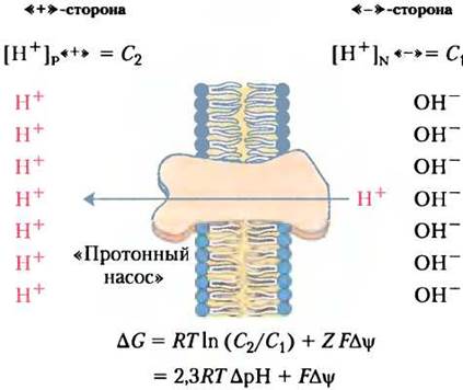
As discussed in Chapter 11, the change in free energy for electron transfer in the respiratory chain—when an electrochemical potential is generated by the translocation of H+ ions from the mitochondrial matrix to the intermembrane space—can be calculated using the following general equation:
∆G = RТIn(С2/С1) + ZF∆Ψ (19-8)
where C2 and C1 are the ion concentrations in the two compartments, with C2 > C1, Z is the charge of the ion (Z = 1 for a proton), and ∆Ψ is the membrane electrical potential in volts.
Assuming that proton translocation across the membrane occurs at 25 °C, substituting the appropriate variable values into Equation 19-8 yields the following expression:
In (С2/С1) = 2,3 (Ig [Н+]«+» - Ig[Н+]«-») = 2,3 (рН«-» - pH«+») = 2,3 ∆рН
∆G = 2,3 RT∆рН + F∆Ψ = (5,70 кДж/моль) ∆рН + (96,5 кДж/В • моль) ∆Ψ (19-9)
In actively respiring mitochondria, the experimentally determined membrane potential ranges from 0.15 to 0.20 V, and the matrix pH is approximately 0.75 units higher than that of the intermembrane space.
Box 19-1 Energetics of Electron Transport
Calculate The amount of energy converted into the proton gradient across the inner mitochondrial membrane during the transfer of a pair of electrons from NADH to oxygen in the respiratory chain. Assume that ∆Ψ = 0.15 V and ∆рН = 0.75.
Solution. Equation 19-9 can be used to calculate the free-energy change associated with the transport of 1 mol of protons across the inner mitochondrial membrane. Substituting ∆рН = 0.75 and ∆Ψ = 0.15 V into this equation yields ∆G = 19 kJ/mol (protons). According to Equation 19-7, the transfer of electrons from NADH to O2 is coupled to the translocation of 10 H+. Consequently, of the 220 kJ produced during the oxidation of 1 mol of NADH, nearly 200 kJ is "conserved" as an electrochemical proton gradient.
When protons flow spontaneously back into the matrix "down" their electrochemical gradient, free energy is released and can be used to perform work. In mitochondria, chloroplasts, and aerobic bacteria, the electrochemical energy of the proton gradient is harnessed to synthesize ATP from ADP and inorganic phosphate. The energetics and stoichiometry of this process are discussed in Section 19.2.
Oxidative Phosphorylation Generates Reactive Oxygen Species (ROS)
At certain stages of oxygen reduction in mitochondria, reactive oxygen free radicals can be formed, which have the potential to damage the cell. During electron transfer from QH2 to Complex III, as well as from Complex I to QH2, the radical ion •Q- is generated as an intermediate. It is quite likely that •Q- can transfer an electron to O2:
O2 + е- —> •O2-
The superoxide radical ion •O2- is a highly reactive species that can participate in further reactions to generate other free radicals, notably the even more reactive hydroxyl radical •OH (Figure 19-18).
Figure 19-18. Generation of ROS in mitochondria and mitochondrial defense mechanisms. When the rate of electron delivery to the respiratory chain is uncoupled from the rate of electron transfer along the chain, There is a marked increase in The production of superoxide radical ions •O2- at Complexes I and III, occurring when incompletely reduced ubisemiquinone radicals •Q- transfer an electron to O2 molecules. The superoxide radical acts on aconitase, which contains a [4Fe-4S] iron-sulfur cluster, leading to the release of Fe2+. In the presence of Fe2+, the Fenton reaction takes place, producing the highly reactive hydroxyl radical •OH. Reactions highlighted in blue protect cells against damage by superoxide radicals. Reduced Glutathione (GSH, see Figure 22-27) provides electrons for the reduction of H2O2 and oxidized Cys residues (-S-S-) in enzymes and other proteins; the reduced form of glutathione is regenerated from its oxidized form (GSSG) via NADPH-dependent reduction.

These reactive oxygen species can inflict significant damage on the cell by reacting with and impairing enzymes, Membrane Lipids, and Nucleic Acids. In actively respiring mitochondria, 0.1% to 4% of the oxygen forms •O2- radical ions, which is sufficient to cause cell death if these radicals are not rapidly detoxified. Factors that slow down the flow of electrons through the respiratory chain promote increased formation of superoxide radical ions, likely by extending the lifetime of •Q- in the Q-cycle.
Box 19-1. Foul-Smelling "Hot" Plants and Alternative Electron Transfer Pathways
Many flowering plants attract insect pollinators by releasing volatile compounds that mimic the natural food odors of the insects or their egg-laying sites. Plants pollinated by blowflies or beetles often use compounds mimicking the scent of dung and rotting flesh to attract these insects.
One family of foul-smelling plants is the Araceae (aroids), which includes philodendrons, jack-in-the-pulpits, and various species of skunk cabbage. These plants typically feature tiny flowers clustered on a vertical spadix surrounded by a modified leaf called a spathe. The spadix emits an odor reminiscent of rotting meat or manure. Furthermore, during pollination, the Temperature of the spadix rises—in some species, reaching 20–40 °C above ambient temperature. This heat generation (thermogenesis) facilitates the vaporization and dispersal of Aromatic Compounds and acts as an attractant in its own right (much as the active metabolic processes of putrefactive bacteria cause rotting meat or decomposing manure to warm up). Thanks to this thermogenic capacity, eastern skunk cabbage flowers emerge directly through the snow in early spring (Figure 1).
Figure 1. Eastern skunk cabbage.

How do plants generate this heat? Unlike animal mitochondria, which rely on conventional Electron Transport Chains, the mitochondria of plants, Fungi, and Unicellular Eukaryotes possess an alternative Respiratory system in which the energy of electron transfer is used to produce heat rather than to synthesize ATP. This alternative respiratory pathway has a specific architecture. First, it contains a cyanide-resistant QH2 oxidase that catalyzes electron transfer directly from ubiquinone to molecular oxygen, bypassing Complexes III and IV (Figure 2). Second, plant mitochondria contain a rotenone-insensitive NADH dehydrogenase (see Table 19-4) located on the outer surface of the inner mitochondrial membrane, which catalyzes electron transfer from matrix NADH directly to ubiquinone, bypassing Complex I and its proton pump. Third, plant mitochondria feature another NADH dehydrogenase on the inner face of the inner mitochondrial membrane, which transfers electrons from cytosolic NADPH or NADH to ubiquinone, also bypassing Complex I. The free energy released as electrons flow through this alternative respiratory chain is not channeled into ATP synthesis; instead, it is dissipated as heat, which plants utilize for various biological functions—such as melting snow around emerging shoots in skunk cabbage and dispersing foul odors to attract insect pollinators during blooming.
Figure 2. Electron transfer pathways in the inner mitochondrial membrane of plants. In plants, electrons may travel through Complexes I, II, and III, as in animal mitochondria, or via the alternative respiratory chain (indicated by blue arrows).

To prevent oxidative damage by •O2-, the cell employs several forms of the enzyme superoxide dismutase, which catalyzes the following reaction:
2 •O2- + 2 Н+ —> Н2O2 + O2
The resulting hydrogen peroxide (Н2O2) is detoxified through the action of glutathione peroxidase (Fig. 19-18). Glutathione reductase converts oxidized glutathione back to its reduced form by accepting electrons from NADPH, which is generated either by NADP-transhydrogenase (in mitochondria) or via the Pentose Phosphate Pathway (in the cytosol, see Fig. 14-20). Reduced glutathione also helps maintain protein sulfhydryl groups in their reduced state, thereby mitigating some of the damaging effects of oxidative stress (Fig. 19-18). NADP-transhydrogenase plays a vital role in this process by supplying the NADPH required for glutathione reductase activity.
In Plant Mitochondria, NADH Oxidation Occurs via Different Mechanisms
During periods of low light or in the dark, ATP synthesis in plant mitochondria proceeds through mechanisms entirely analogous to those in nonphotosynthetic organisms. In the light, however, mitochondrial NADH is generated primarily as a byproduct of converting Glycine—produced during Photorespiration—into Serine (see Fig. 20-21), a process that can be written as follows:
2 Glycine + NAD+ —> serine + СO2 + NH3 + NADH + Н+
As discussed in Chapter 20, plants accumulate NADH even when it is not required for ATP synthesis. Instead of being used for energy, this "surplus" NADH can be oxidized to NAD+ by transferring electrons directly from NADH to ubiquinone and subsequently to molecular oxygen, bypassing Complexes III and IV with their proton-pumping machinery. The Free energy of this electron transfer is dissipated as heat, which is of great physiological importance to certain plants (see Box 19-1). A distinctive feature of electron transfer in plant mitochondria is that plant QH2 oxidase, unlike cytochrome c oxidase (Complex IV), is not inhibited by cyanides.
Summary of Section 19.1 Electron-Transfer Reactions in Mitochondria
■ The chemiosmotic theory has been proposed to explain many energy-transducing processes in biological systems, including oxidative phosphorylation and photophosphorylation. The mechanism by which electron-transfer energy is coupled to ATP synthesis is fundamentally the same in both cases. The free energy released during Electron transport is conserved as an electrochemical gradient generated by the coupled "pumping" of protons (Н+) out of the matrix. The resulting proton-motive force then drives further proton translocation.
■ In mitochondria, hydride ions removed from substrates by NAD-linked dehydrogenases deliver their electrons to the respiratory chain, through which they are ultimately transferred to molecular oxygen (O2), reducing it to Н2O.
■ Shuttle systems transport reducing equivalents from cytosolic NADH into mitochondrial NADH. In all NAD-dependent dehydrogenation reactions, these reducing equivalents are channeled to mitochondrial NADH dehydrogenase (Complex I).
■ Reducing equivalents are transferred via a series of iron-sulfur centers to ubiquinone, which passes electrons to cytochrome b, the first electron carrier in Complex III. Within Complex III, electrons travel along two divergent pathways through two b-type cytochromes and cytochrome c1 to an iron-sulfur center. This iron-sulfur center then delivers electrons, one at a time via cytochrome c, to cytochrome c oxidase (Complex IV); this copper-containing enzyme also harbors cytochromes a and a3. The electrons accumulated by Complex IV are finally transferred to molecular oxygen (O2), reducing it to Н2O.
■ Electrons from various substrates enter the mitochondrial electron-transport chain through distinct entry points. For instance, during the oxidation of succinate by succinate dehydrogenase (Complex II), the flavoprotein of this complex transfers electrons to ubiquinone through a series of iron-sulfur centers. Similarly, electrons derived from Fatty acid oxidation are funneled to ubiquinone via the electron-transferring flavoprotein (ETF).
■ Potentially harmful reactive oxygen species (ROS) generated within mitochondria are neutralized by several protective enzymes, most notably superoxide dismutase and glutathione peroxidase.
■ In addition to the standard cyanide-sensitive pathway, plants, fungi, and unicellular eukaryotes possess an alternative, cyanide-resistant pathway for electron transfer involving NADPH.
Last update: 06/08/2026
Editorial and Educational Adaptation: This material has been compiled based on the primary/original source text. The project team performed an editorial review, corrected technical inaccuracies, structured sections, and adapted the content for an educational format.
What was processed:
- elimination of formatting defects (OCR errors, structural breaks, corrupted characters);
- editorial organization of content;
- standardization of terminology in accordance with academic sources;
- verification of factual statements against the original source text.
All mentions of the author, publication year, and origin of the primary text have been preserved in accordance with the source.