LEHNINGER PRINCIPLES OF BIOCHEMISTRY - VOL. 2. BIOENERGETICS AND METABOLISM - 2014
PART II. BIOENERGETICS AND METABOLISM
13. BIOENERGETICS AND BIOCHEMICAL REACTION TYPES
13.4. Oxidation-Reduction Reactions in Biological Systems
Much of METABOLISM centers on phosphoryl group transfers. However, another fundamental type of transfer is equally vital: the movement of electrons in oxidation-reduction reactions. In these reactions, one molecule loses electrons (becoming oxidized) while another gains electrons (becoming reduced). The work performed by living organisms is directly or indirectly powered by the flow of electrons through oxidation-reduction pathways. In nonphotosynthetic organisms, reduced compounds (nutrients) serve as the electron sources; in photosynthetic organisms, the initial electron donor is a molecule that enters an excited state upon absorbing light. The Metabolic pathways of electron transfer are complex. In enzyme-catalyzed reactions, electrons flow from various metabolic intermediates to specialized electron carriers. These carriers, in turn, pass the electrons to acceptors with a higher electron affinity, releasing energy in the process. Cells contain a variety of energy-transducing molecules that convert the energy of this electron flow into useful work.
We begin our Structure/133.html">Discussion by examining how electromotive force (emf) can perform work, then explore the theoretical foundations and experimental principles for measuring energy changes in oxidation reactions in terms of emf, and analyze the relationship between this force (expressed in volts) and The change in Free energy (in joules). Finally, we describe the chemical principles underlying oxidation-reduction processes and the structures of the most common specialized electron carriers that we will encounter repeatedly in the following chapters.
Electron Flow Can Accomplish Biological Work
Whenever we use an electric motor, turn on a light, run an electric heater, or rely on a spark in a car engine to ignite gasoline, we are harnessing the work done by a flow of electrons. The source of electrons in an electrical circuit can be a battery (electrochemical Cell) featuring two electrodes made of distinct chemical elements. Wires provide a pathway for electrons to flow from the chemical species at one pole of the battery, through the motor (the external circuit), to the other chemical species at the opposite pole. Because the Materials of the oppositely charged electrodes have different electron affinities, an electric current (a flow of electrons) spontaneously travels through the external circuit, driven by a force proportional to the difference in electron affinities between the electrode materials—that is, by the electromotive force (emf). An electromotive force (typically of several volts) is capable of performing work if an energy transducer, such as an electric motor, is included in the circuit. To perform useful work, the motor can be coupled to various mechanical devices.
Living cells also possess an internal biological "circuit" in which electrons are supplied by compounds such as glucose. During the enzymatic oxidation of glucose, the liberated electrons spontaneously move through a series of intermediate electron-carrying compounds (intermediates) to another chemical species, such as O2. This Electron transport is an exergonic process because O2 has a much higher electron affinity than the electron-carrying intermediates. The resulting emf powers numerous molecular energy transducers (Enzymes and other Proteins) that carry out biological work. For example, in Mitochondria, membrane-bound enzymes couple electron flow to the generation of a transmembrane pH gradient, which performs osmotic and electrical work. The resulting proton gradient possesses potential energy, sometimes referred to as the proton-motive force by analogy with electromotive force. Another enzyme, ATP synthase in The inner mitochondrial membrane, harnesses the proton-motive force to perform chemical work: the synthesis of ATP from ADP and Pi (driven by the spontaneous translocation of protons across the membrane). Similarly, in E. coli, membrane-localized enzymes convert emf into a proton-motive force, which is subsequently used to drive flagellar rotation. The electrochemical principles governing energy changes in a macroscopic circuit with a motor and a battery apply equally to the molecular processes involving electron flows in living cells.
Oxidation-Reduction Processes Can Be Represented as Half-Reactions
Although Oxidation and reduction never occur independently of one another, it is helpful when describing electron transfer to view a redox reaction as two separate half-reactions. For example, The oxidation of the ferrous ion Fe2+ by the cupric ion Cu2+
Fe2+ + Cu2+ ⇄ Fe3+ + Cu+
can be written as two half-reactions, where Fe2+ is the reducing agent and Cu2+ is the oxidizing agent:
Oxidation: Fe2+ ⇄ Fe3+ + e- (1)
Reduction: Cu2+ + е- ⇄ Cu+ (2)
In a redox reaction, the substance that gives up electrons (the electron donor) is called the reducing agent (reductant), and the substance that accepts electrons is called the oxidizing agent (oxidant). In this case, the divalent iron cation Fe2+ and the trivalent iron cation Fe3+ form a conjugate redox pair. Similarly, an acid and its corresponding base form a conjugate acid-base pair. Recall (see Chapter 2, Vol. 1) that acid-base reactions are generally expressed as:
proton donor ⇄ H+ + proton acceptor
A comparable general equation can be written for oxidation-reduction reactions:
electron donor ⇄ e- + electron acceptor
In half-reaction (1), Fe2+ is the electron donor and Fe3+ is the electron acceptor. The Fe2+ and Fe3+ ions constitute a conjugate redox pair.
Electron transfers in oxidation-reduction reactions between organic and Inorganic Compounds are fundamentally identical. In Chapter 7, we examined the oxidation of a reducing sugar (an aldehyde or ketone) by the cupric ion Cu2+ (see Fig. 7-10):
Class="center">
This reaction can be broken down into two half-reactions:

Because two electrons are removed from the carbon atom of the aldehyde group, the second half-reaction (the one-electron reduction of the divalent copper ion to the monovalent state) must be multiplied by a coefficient of 2 to balance the charges in the overall redox reaction.
Biological Oxidation processes frequently involve dehydrogenation as well
In living cells, carbon exists in several oxidation states (Fig. 13-22). When a bonding electron pair is shared between a carbon atom and another, non-carbon atom (typically H, C, S, N, or O), the electron pair is shifted toward the more electronegative atom. Electronegativity increases in the series H<C<S<N<O. For example, in methane, CH4, carbon is more electronegative than any of the four bonded hydrogen atoms; consequently, across all four C—H bonds, the C atom pulls all eight bonding electrons closer to itself (Fig. 13-22). In ethane, the electrons forming the C—C bond are equidistant from each carbon and shared equally between them, meaning that each carbon atom can be considered to have only seven, rather than eight, electrons associated with it. In ethanol, the C-1 atom is less electronegative than the bonded oxygen atom, so the O atom pulls both electrons of the C—O bond toward itself, leaving the C-1 atom with only five electrons. With each formal loss of electrons, a carbon atom undergoes oxidation; this occurs even in the absence of oxygen—for instance, when an alkane (—CH2—CH2—) is converted to an alkene (—CH=CH—), oxidation takes place, which represents a formal loss of electrons (i.e., negative charges), though it actually involves the loss of hydrogen atoms. In biological systems, oxidation frequently involves dehydrogenation, and many enzymes that catalyze oxidation reactions are called dehydrogenases. Note that the more reduced compounds in Fig. 13-22 (top) are richer in hydrogen than oxygen, whereas in the more oxidized compounds (bottom), the number of oxygen atoms increases while the number of hydrogen atoms decreases.
Fig. 13-22. Oxidation states of carbon in the biosphere. Pay attention to the carbon atoms marked in red and their bonding electrons. If a carbon atom is bonded to a less electronegative H atom, both bonding electrons (red) are shifted toward the carbon. If a carbon is bonded to another carbon, the electron pair lies exactly midway between them, although only one of the two electrons in that pair originated from the "red" carbon. If the "red" carbon is bonded to a more electronegative O atom, the bonding electrons are shifted toward the oxygen. The numbers in the right-hand Column indicate the number of electrons shifted toward the "red" carbon, which can be used to roughly estimate the oxidation state of carbon in a given compound (functional group): across this series of compounds, the oxidation state of carbon increases from top to bottom.

Not all biological oxidation-reduction reactions involve carbon. For example, The conversion of molecular nitrogen to ammonia entails the reduction of nitrogen atoms.
6H+ + 6e- + N2 —> 2NH3
The transfer of electrons from one molecule (an electron donor) to another (an electron acceptor) takes place via four different mechanisms.
1. Direct electron transfer. For example, the Fe2+/Fe3+ redox pair
can transfer electrons to the Cu+/Cu2+ pair:
Fe2+ + Cu2+ Fe3+ + Cu+
2. Transfer of hydrogen atoms (H+ + e-), since a hydrogen atom formally consists of a proton H+ and a single electron e-. In this case, the overall equation can be written as
AH2 ⇄ A + 2e- + 2H+
where AH2 is a hydrogen (or electron) donor. (Do not confuse this with acid dissociation reactions, which involve H+ ions but no electrons.) AH2 and A form a conjugate redox pair (A/AH2) that can react with another redox pair, B/BH2:
AH2 + B ⇄ A + BH2
3. Transfer of electrons from a donor to an acceptor in the form of a hydride ion (:H)- carrying two electrons, as seen in NAD-dependent dehydrogenases, which are discussed below.
4. Direct oxygen incorporation. Here, oxygen interacts with an organic reductant and is covalently incorporated into the reaction product. An example of such a reaction is the oxidation of a hydrocarbon to an alcohol:
R—CH3 + 1/2O2 —> R-CH2-OH
In this reaction, the hydrocarbon serves as the electron donor, and oxygen acts as the electron acceptor.
All four electron-transfer mechanisms are found in cells. In oxidation-reduction reactions, the term reduction equivalent is used to designate a charge transfer equivalent to 1 e-, regardless of whether the entity transferred is an electron itself, a hydrogen atom, a hydride ion, or an oxygen reaction resulting in an oxidized product. Because nutrient molecules are typically enzymatically dehydrogenated with the simultaneous loss of two reduction equivalents, and because each oxygen atom can accept two reduction equivalents, biochemists have agreed to define the unit of Biological oxidation as two reduction equivalents passing from a substrate to oxygen.
Reduction Potential as a Measure of Electron Affinity
If two conjugate redox pairs are present in a solution, the transfer of an electron from the electron donor of one pair to the electron acceptor of the other can occur spontaneously. The direction of such a reaction depends on the electron affinity of the electron acceptors in each redox pair. A measure of this affinity is the standard reduction potential E° (measured in volts), which can be determined experimentally (Fig. 13-23). Electrochemists have chosen the following half-reaction as the standard reference point for measurements:
H+ + e- —> 1/2 H2
Fig. 13-23. Measurement of the standard reduction potential E'° of a redox pair. The direction of electron flow is from the working electrode to the reference electrode. The most common reference electrode is the hydrogen electrode (pH 0). The electromotive force (emf) of the standard hydrogen electrode is defined as 0.00 V. At pH 7 for the working electrode, the E'° of the hydrogen electrode is -0.414 V. The direction of electron flow depends on the relative potentials of both electrodes (the so-called electron "pressure"). A salt bridge containing a saturated KCl solution provides a pathway for counterion movement between the working electrode and the reference electrode. From the observed emf and the emf of the reference cell, the investigator can determine the emf of the working half-cell containing the redox pair. By convention, the electrode that "acquires" electrons has a more positive reduction potential value.

This hydrogen electrode (referred to as a half-cell), at which this half-reaction takes place, is arbitrarily assigned a reduction potential of E'° = 0.00 V. When the hydrogen electrode (reference electrode) is connected via an external circuit to a working electrode (another half-cell) immersed in a solution containing standard concentrations of the oxidized and reduced forms (under standard conditions: 25 °C, 1 M solution concentrations, 101.3 kPa gas pressure), the flow of electrons in the external circuit is directed from the half-cell with the lower standard reduction potential to the half-cell with the higher potential. By convention, the electrode (half-cell) with the greatest "tendency" to gain electrons has a positive E° value (with a plus sign), whereas the one that donates electrons to the hydrogen half-cell has a negative E° value (with a minus sign). When two electrodes are connected through an external circuit, the electrode with the higher (more positive) E° value (higher reduction potential) will undergo reduction.
The reduction potential of a half-cell depends not only on The Nature of the chemical element, but also on its activity, which is related to its concentration in solution. More than 100 years ago, Walther Nernst derived an equation relating the standard reduction potential E° to the reduction potential E for an arbitrary concentration of oxidized and reduced elements in a cell.
The Nernst Equation
 (13-5)
where R is the universal gas constant, T is the absolute Temperature in Kelvin, n is the number of electrons transferred per molecule, and F is the Faraday constant (Table 13-1). At 298 K (25 °C) and after substituting the numerical values of the constants, we obtain
 (13-6)
Key Conventions.
For oxidation-reduction reactions, biochemists define standard conditions as for ∆G'°: pH 7 and 25 °C; the standard reduction potential is denoted as E'°. By convention, for any redox reaction, the potential difference ∆E'° is equal to the difference between the reduction potentials E'° of the electron acceptor and electron donor. ■
Table 13-7 lists the standard reduction potentials E'° that we will use—these apply only to systems at neutral pH (pH 7). Any E'° value in Table 13-7 is obtained as the potential difference between the standard hydrogen electrode (pH 0) and a conjugate redox pair at a concentration of 1 M and pH 7. Note that, According to the data in Table 13-7, in a circuit consisting of the conjugate pair 2Н+/Н2 at pH 7 and the standard hydrogen electrode (pH 0), electrons flow from the pH 7 half-cell to the standard half-cell (pH 0); thus, for the 2Н+/Н3 pair, the measured E'° value is -0.414 V.
Standard reduction potentials can be used to calculate free-energy changes
Why do biochemists use reduction potentials? They are convenient because if E values measured relative to the standard hydrogen electrode are known for any two half-cells, their reduction potentials relative to each other can be determined. This allows us to predict the direction of electron flow when two half-cells are connected by an external circuit or when the components of both half-cells are mixed in the same solution. Electrons tend to move toward the half-cell with the more positive E value, and the current is proportional to the difference in reduction potentials, ∆E. The energy of a spontaneous redox reaction is characterized by the change in Free Energy and is proportional to ∆E:
∆AG = -nF∆E or ∆G'° = -nFE'°
where n represents the number of electrons transferred in the reaction. Knowing E'° (obtained from the table of reduction potentials, Table 13-7) and the concentrations of the reactants, this equation can be used to calculate the free-energy change of a redox reaction.
Table 13-7. Standard reduction potentials of some biologically important half-reactions at pH 7.0 and 25 °C (298 K)
Half-reaction |
E'°, V |
1/2О2 + 2Н+ + 2е —> Н2O |
0,816 |
Fe3+ + e- —> Fe2+ |
0,771 |
NO-3+ 2Н+ + 2е- —> NO-2 + Н2O |
0,421 |
Cytochrome f (Fe3+) + е- —> cytochrome f (Fe2+) |
0,365 |
Fe(CN)63-(ferricyanide) + е- —> Fe(CN)64- |
0,36 |
Cytochrome a3(Fe3+) + е- —> cytochrome a3(Fe2+) |
0,35 |
O2 + 2Н+ + 2е- —> Н2O2 |
0,295 |
Cytochrome a (Fe3+) + е- —> cytochrome a (Fe2+) |
0.29 |
Cytochrome c (Fe3+) + е- —> cytochrome c (Fe2+) |
0,254 |
Cytochrome c1 (Fe3+) + е- —> cytochrome c1 (Fe2+) |
0,22 |
Cytochrome b (Fe3+) + е- —> cytochrome b (Fe2+) |
0,077 |
Ubiquinone + 2Н+ + 2е- —> ubiquinol + Н2 |
0,045 |
Fumarate2- + 2Н+ + 2е- —> succinate2- |
0,031 |
2Н+ +2е- —> Н2 (under standard conditions, pH 0) |
0,000 |
Crotonyl-CoA + 2Н+ + 2е- —> butyryl-CoA |
0,015 |
Oxaloacetate2- + 2H++ 2е- —> malate2- |
0,166 |
Pyruvate + 2Н+ + 2е- —> lactate- |
-0,185 |
Acetaldehyde + 2Н+ + 2е- —> ethanol |
0,197 |
FAD + 2Н+ + 2е- —> FADH2 |
-0,219а |
Glutathione + 2Н+ + 2е- —> 2 reduced glutathione |
-0.23 |
S + 2Н+ + 2е- = H2S |
-0,243 |
Lipoic Acid + 2Н+ + 2е- —> dihydrolipoic acid |
-0,29 |
NAD++ Н+ + 2е- —> NADH |
0,320 |
NADP++ Н+ + 2е- —> NADPH |
0,324 |
Acetoacetate + 2Н+ + 2е- —> β-hydroxybutyrate |
-0,346 |
α-Ketoglutarate + СO2 + 2Н + 2е- —> isocitrate |
-0,38 |
2Н+ + 2е- —> Н2 (at pH 7) |
0,414 |
Ferredoxin (Fe3+) + е- —> ferredoxin (Fe2+) |
-0,432 |
a For free FAD; FAD bound to a specific flavoprotein (e.g., succinate dehydrogenase) has a different E'° value, which depends on the protein environment.
Example 13-3. Calculation of ∆G'° and ∆G for a Redox Reaction
Calculate the standard free-energy change ∆G'° for the reaction in which acetaldehyde is reduced by the biological electron carrier NADH:
Acetaldehyde + NADH + Н+ —> ethanol + NAD+
Next, calculate the actual free-energy change ∆G if the concentrations of acetaldehyde and NADH are 1.00 M, and the concentrations of ethanol and NAD+ are 0.100 M.
(1) Acetaldehyde + 2Н+ + 2е —> ethanol
E'° = -0.197 V
(2) NAD+ + 2Н+ + 2е-~ —> NADH + Н+
E'° = -0.320 V
Recall that by convention, ∆E'° is the difference between the ∆E'° of the electron acceptor and that of the donor.
Solution. In this example, acetaldehyde is the electron acceptor (n = 2), accepting electrons from NADH.
∆Е’° = -0.197 V - (-0.320 V) = 0.123 V.
∆G'° = -nF∆Е'° = -2 • 96.5 kJ/(V • mol) • 0.123 V = -23.7 kJ/mol
This is the free-energy change for the redox reaction at pH 7 when acetaldehyde, ethanol, NAD+, and NADH are each present at 1.00 M concentrations.
To calculate ∆G when the concentrations of acetaldehyde and NADH are 1.00 M and those of ethanol and NAD+ are 0.100 M, we first determine E for both reductants (Equation 13-5):

From this, we can find ∆E and then use Equation 13-6 to calculate ∆G:
∆Е = -0.167 V - (-0.350) V = 0.183 V
∆G = -nF∆Е = -2 • 96.5 kJ/(V • mol) • 0.183 V = -35.3 kJ/mol
In this manner, the free-energy change of any redox reaction can be calculated at arbitrary concentrations of the redox pairs.
Specialized Electron Carriers Are Required for the Oxidation of Glucose to Carbon Dioxide in Cells
The principles of redox energetics discussed above apply to many metabolic reactions involving electron carriers. For example, in many organisms, the oxidation of glucose provides the energy for ATP synthesis. For the Complete oxidation of glucose,
С6Н12O6 + 6O2 —> 6СO2 + 6Н2O
∆С'° = -2840 kJ/mol. This energy is far greater than that required for ATP synthesis (about 50 to 60 kJ/mol; see Box 13-1). In cells, glucose is converted to CO2 not in a single energy-yielding step, but through a series of controlled reactions, several of which are oxidations. The free energy released in these oxidation steps is of the same order of magnitude as the energy required to synthesize ATP from ADP, allowing a portion of it to be conserved. Electrons liberated in these oxidation reactions are transferred to specialized electron-carrying Coenzymes such as NAD+ and FAD (discussed below).
Certain Coenzymes and Proteins Serve as Universal Electron Carriers
Numerous enzymes that catalyze cellular oxidations channel electrons from hundreds of different substrates onto just a few types of universal electron carriers. The reduction of these carriers during Catabolism results in the conservation of free energy released by substrate oxidation. NAD+, NADP+, FMN, and FAD are Water-soluble coenzymes that undergo reversible oxidation and reduction in many electron-transfer metabolic reactions. The NUCLEOTIDES NAD+ and NADP+ readily diffuse from one enzyme to another, whereas the flavin nucleotides FMN and FAD are usually very tightly bound to their target Flavoproteins, serving as prosthetic groups. Fat-soluble Quinones, such as ubiquinone and plastoquinone, act as electron carriers and proton Donors in the anhydrous environment of membranes. Iron-sulfur proteins and Cytochromes, with tightly bound prosthetic groups subject to reversible oxidation and reduction, also serve as electron carriers in numerous redox reactions. Some of these proteins are water-soluble, while others are peripheral or integral Membrane Proteins (see Fig. 11-6).
To conclude this chapter, we will examine The chemical properties of nucleotide coenzymes as well as the enzymes (dehydrogenases and flavoproteins) that utilize them. The Redox Properties of quinones, iron-sulfur proteins, and cytochromes are discussed in Chapter 19.
NADH and NADPH Act in Conjunction with Dehydrogenases as Soluble Electron Carriers
Nicotinamide adenine dinucleotide (NAD+ in its oxidized form) and its phosphorylated analog, nicotinamide adenine dinucleotide phosphate (NADP+), consist of two nucleotides joined through a phosphoanhydride bond between their phosphate groups (Fig. 13-24a). Because the nicotinamide ring resembles pyridine, these compounds are sometimes called pyridine nucleotides. The vitamin niacin serves as the source of the nicotinamide functional group in nicotinamide nucleotides.
Both coenzymes undergo reversible reduction of the nicotinamide ring (Fig. 13-24). As a substrate molecule is oxidized (dehydrogenated) with the loss of two hydrogen atoms, the oxidized form of the nucleotide (NAD+ or NADP+) accepts a hydride ion (:H-, equivalent to a proton and two electrons) and is reduced (to NADH or NADPH). The second proton released goes into the aqueous medium. A half-reaction can be written for each nucleotide cofactor:
NАD+ + 2е- + 2Н+ —> NADH + Н+
NАDР+ + 2е- + 2Н+ —> NАDРН + Н+
Figure 13-24. NAD and NADP. (a) Nicotinamide adenine dinucleotide (NAD+) and its phosphorylated analog NADP are reduced to NADH and NADPH, respectively, by accepting a hydride ion (i.e., two electrons and one proton) from the substrate being oxidized. The hydride ion can add to either the front (A face) or the back (B face) of the planar nicotinamide ring (see Table 13-8). (b) UV absorption spectra of NAD+ and NADH. Reduction of the nicotinamide ring gives rise to a new broad absorption band with a maximum at 340 nm. The formation of NADH in an enzymatic reaction can be readily followed by monitoring the appearance of absorption at 340 nm (molar extinction coefficient ε340 = 6,200 M-1cm-1).
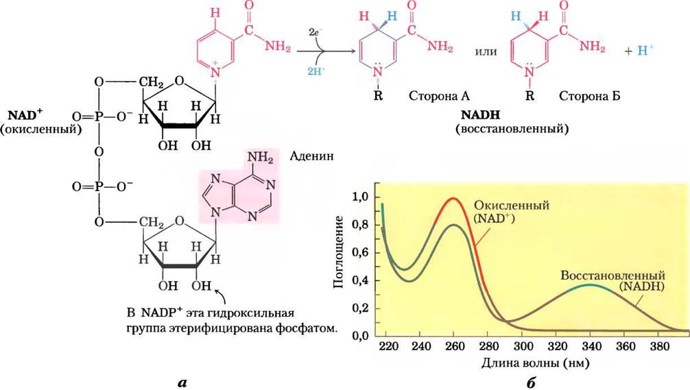
The reduction of NAD+ or NADP+ converts the benzenoid ring of the nicotinamide functional group (which bears a positive charge on the ring nitrogen) into a quinoid form (with no charge on the nitrogen). Note that the reduced nucleotides absorb light at 340 nm, whereas the oxidized forms do not (Fig. 13-24b). The plus sign in the Abbreviations NAD+ and NADP+ does not denote the net charge on these molecules (both are negatively charged); rather, it indicates that the nicotinamide ring is in its oxidized state, bearing a positive charge on the nitrogen atom. In the abbreviations NADH and NADPH, the symbol H denotes the added hydride ion. The designations NAD and NADP are used when referring to these nucleotides without specifying their oxidation state.
In most Tissues, the total concentration of [NAD+ + NADH] is approximately 10-5 M, and that of [NADP+ + NADPH] is about 10-6 M. In many Cells and Tissues, the NAD+(ox)/NADH(red) ratio is high, which favors the transfer of a hydride ion from substrate to NAD+ to form NADH. Conversely, NADPH(red) is normally present at much higher concentrations than its oxidized counterpart NADP+, favoring hydrogen transfer from NADPH to substrate. This reflects the specialized metabolic roles of the two coenzymes: NAD+ generally Functions in oxidations, typically in Catabolic pathways, whereas NADPH acts more frequently in reductions, almost invariably in anabolic pathways. Although some enzymes can utilize either coenzyme, most exhibit a marked preference for one over the other. Furthermore, the processes in which these two Cofactors participate are compartmentalized in different Organelles of Eukaryotic cells: the oxidation of energy-rich fuel molecules such as pyruvate, Fatty acids, and α-keto acids derived from Amino Acids occurs within the mitochondrial matrix, whereas reductive biosynthetic processes, such as fatty acid synthesis, take place in the Cytosol. This functional and spatial specialization enables The Cell to maintain two distinct pools of electron carriers with dedicated functions.
More than 200 enzymes are known to catalyze reactions in which NAD+ (or NADP+) accepts a hydride ion from a reduced substrate, or NADPH (or NADH) donates a hydride ion to an oxidized substrate. These reactions can be written in general terms as follows:
AH2 + NAD+ —> A + NADH + H+
A + NADPH + H+ —> AH2 + NADP+
where AH2 is the reduced substrate and A is the oxidized one. The general name for this class of enzymes is oxidoreductases, although they are commonly referred to as dehydrogenases. For example, Alcohol dehydrogenase catalyzes the initial catabolism of ethanol—its oxidation to acetaldehyde:

Note that one of the carbon atoms in ethanol has lost hydrogen; the alcohol is thereby oxidized to an aldehyde (to review the oxidation states of carbon, refer to Fig. 13-22).
In reduced NAD+ or NADP+, the hydride ion can theoretically be transferred to either face of the nicotinamide ring: the front (A) or the back (B), as shown in Fig. 13-24a. Studies with isotopically labeled substrates have shown that a given enzyme catalyzes either type A or type B transfer, but never both simultaneously. For example, Yeast alcohol dehydrogenase and vertebrate Heart Lactate dehydrogenase transfer a hydride ion to (or remove it from) the A face of the nicotinamide ring; they are therefore classified as type A dehydrogenases to distinguish them from another group of enzymes that transfer a hydride ion to (or remove it from) the B face (Table 13-8). Enzyme stereospecificity for one face or the other can be remarkably high; for instance, lactate dehydrogenase favors the A face over the B face by a factor of more than 5 • 107! This preference stems from the specific arrangement of functional groups in the enzyme molecule—involved in hydrogen bonding—relative to the —CONH2 group of the nicotinamide ring.
Table 13-8. Stereospecificity of Dehydrogenases Using NAD+ or NADP+ as Coenzymes
Enzyme |
Coenzyme |
Nicotinamide Ring Stereochemical Specificity (A or B) |
Book Page(s) |
Isocitrate dehydrogenase |
NAD+ |
A |
193 |
α-Ketoglutarate dehydrogenase |
NAD+ |
B |
194 |
Glucose-6-phosphate dehydrogenase |
NADP+ |
B |
109 |
Malate dehydrogenase |
NAD+ |
A |
199 |
NAD+ or NADP+ |
B |
269 |
|
Glyceraldehyde-3-phosphate dehydrogenase |
NAD+ |
B |
76 |
Lactate dehydrogenase |
NAD+ |
A |
91 |
Alcohol dehydrogenase |
NAD+ |
A |
93 |
Most dehydrogenases that utilize NAD or NADP bind the cofactor within a conserved protein domain known as the Rossman fold (Michael Rossmann first solved The structure of lactate dehydrogenase and described this structural motif). The Rossman fold typically consists of six parallel β strands linked to four α helices (Fig. 13-25).
Fig. 13-25. The Rossman fold. This structural motif is found in the NAD-binding region of many dehydrogenases. (a) Two structurally similar motifs, each containing three parallel β strands and two α helices (β-α-β-α-β). (b) The nucleotide-binding domain of lactate dehydrogenase (PDB ID 3LDH) with NAD in an extended conformation (ball-and-stick model) and the β-α-β-α-β Rossman fold motif (in varying shades of green); NAD is bound via Hydrogen Bonds and salt bridges.

The association between a dehydrogenase and NAD or NADP is relatively weak; the coenzyme readily dissociates and diffuses between enzymes, acting as a water-soluble electron carrier from one metabolite to another. For example, during alcohol production via glucose Fermentation in yeast cells, a hydride ion is removed from glyceraldehyde-3-phosphate by one enzyme (type B glyceraldehyde-3-phosphate dehydrogenase) and transferred to NAD+. The resulting NADH then leaves the enzyme surface and diffuses to another enzyme (type A alcohol dehydrogenase), which transfers the hydride ion to acetaldehyde, yielding ethanol:
(1) Glyceraldehyde-3-phosphate + NAD+ —> 3-phosphoglycerate + NADH + H+
(2) Acetaldehyde + NADH + H+ —> ethanol + NAD+
Sum: Glyceraldehyde-3-phosphate + acetaldehyde —> 3-phosphoglycerate + ethanol
Note that the overall reaction involves no net synthesis or consumption of NAD+ or NADH; the coenzymes function as true catalysts, being repeatedly released so that the total concentration of NAD+ + NADH remains unchanged.
Dietary Deficiency of Niacin, the Vitamin Precursor of NAD and NADP, Causes Pellagra
As noted in Chapter 6 and discussed further in subsequent chapters, most coenzymes are derivatives of Vitamins. The pyridine-like rings of NAD and NADP are derived from the vitamin niacin (nicotinic acid; Fig. 13-26), which in turn can be synthesized from Tryptophan. Humans cannot synthesize niacin in adequate amounts, particularly when consuming diets low in tryptophan (such as corn-based diets, which are very deficient in tryptophan). Niacin deficiency affects all NAD(P)-dependent dehydrogenases, leading to the severe disease pellagra (from the Italian pelle agra, meaning "sour Skin"; in dogs, the condition is known as "black Tongue"). The disease is characterized by the classic threeDs: dermatitis, diarrhea, and dementia, and is frequently fatal if untreated. A century ago, pellagra was widespread; in the southern United States, where corn served as a dietary staple, approximately 100,000 cases and nearly 10,000 deaths were recorded annually between 1912 and 1916. Joseph Goldberger demonstrated in 1920 that pellagra is caused by nutritional deficiencies, and in 1937 Frank Strong, D. Wayne Wooly, and Conrad Elvehjem discovered that niacin is the effective cure for "black tongue." The addition of this inexpensive compound to foods has virtually eradicated pellagra in developed nations. However, this victory is not absolute: pellagra still occurs among chronic alcoholists, whose intestinal niacin absorption is severely impaired and whose caloric intake is largely derived from alcoholic beverages virtually devoid of vitamins, including niacin. In certain regions, such as the Deccan Plateau in India, pellagra remains a public health concern, particularly among impoverished populations. ■
Fig. 13-26. Structures of niacin (nicotinic acid) and its derivative nicotinamide. In natural biological systems, these compounds are synthesized from tryptophan. Nicotinic acid was first isolated in the laboratory by the oxidation of natural nicotine—hence its name. Both nicotinic acid and nicotinamide cure pellagra, whereas nicotine (from cigarettes) has no therapeutic effect.


Flavin Nucleotides Are Tightly Bound to Flavoproteins
Flavoproteins (Table 13-9) are enzymes that catalyze oxidation-reduction reactions using flavin mononucleotide (FMN) or flavin adenine dinucleotide (FAD) as coenzymes (Fig. 13-27). These flavin nucleotide coenzymes are derived from the vitamin riboflavin. The condensed polynuclear structure of flavin nucleotides (the isoalloxazine ring) undergoes reversible reduction, accepting either one or two electrons (as one or two hydrogen atoms, each comprising one electron and one proton) from a reduced substrate. The fully reduced forms are designated FADH2 and FMNH2. When a fully oxidized flavin nucleotide accepts a single electron (one hydrogen atom), a semiquinone intermediate of the isoalloxazine ring is formed, designated FADH• and FMNH•. Because the functions of flavin nucleotides differ slightly from those of nicotinamide coenzymes (participating in both one- and two-electron transfers), flavoproteins are involved in a broader range of reactions than NAD(P)-linked dehydrogenases.
Table 13-9. Selected Enzymes (Flavoproteins) That Utilize Flavin Nucleotide Coenzymes
Enzyme |
Flavin nucleotide |
Page(s) in book |
Acyl-CoA dehydrogenase |
FAD |
Fig. 17-8 |
Dihydrolipoyl dehydrogenase |
FAD |
186 |
Succinate dehydrogenase |
FAD |
198 |
Glycerol-3-phosphate dehydrogenase |
FAD |
Fig. 19-30 and text |
Thioredoxin reductase |
FAD |
Fig. 22-39 and text |
NADH dehydrogenase (Complex I) |
FAD |
Fig. 19-1 and text |
Glycolate oxidase |
FAD |
Fig. 20-21 |
Fig. 13-27. Oxidized and reduced forms of FAD and FMN. FMN comprises the part of the FAD structure (in its oxidized form) shown above the dashed line. Flavin nucleotides accept two hydrogen atoms (two electrons and two protons) that are added to the flavin ring system. When FAD or FMN accepts only a single hydrogen atom, a semiquinone is formed, which is a stable radical.

Like the nicotinamide coenzymes (Fig. 13-24), flavin nucleotides undergo a shift in their principal absorption band upon reduction. Fully reduced flavoproteins (having accepted two electrons) typically exhibit an absorption maximum at 360 nm. Partially reduced flavoproteins (having accepted a single electron) display an additional absorption maximum at 450 nm, whereas the fully oxidized flavin system shows maxima at 370 and 440 nm. The absorption maxima of the intermediate one-electron-reduced radical form lie at 380, 480, 580, and 625 nm. These spectral characteristics can be utilized to study reactions involving flavoproteins.
In most flavoproteins, the flavin nucleotide is bound quite tightly to the protein, and in some enzymes—such as succinate dehydrogenase—it is attached covalently. Such tightly bound coenzymes are referred to as prosthetic groups. They do not function by diffusing as electron carriers from one enzyme to another; instead, they provide a mechanism by which the flavoprotein can temporarily hold electrons while catalyzing Electron transfer from a reduced substrate to an electron acceptor. A distinctive feature of flavoproteins is that the standard reduction potential (E′°) of the bound flavin nucleotide can vary, which is of profound importance. Due to strong interactions between the enzyme and the prosthetic group, the flavin ring acquires a reduction potential characteristic of that specific flavoprotein, which sometimes differs significantly from the reduction potential of the free flavin nucleotide. For example, for FAD bound to succinate dehydrogenase, E'° ≈ 0.0 V, whereas for free FAD, E'° = -0.219 V; for other flavoproteins, E'° ranges from -0.40 V to +0.06 V. Flavoproteins are highly complex structures; In addition to the flavin nucleotide, some of them also carry tightly bound inorganic ions (such as iron or molybdenum ions) capable of participating in electron transfer.
Some flavoproteins perform entirely unexpected functions by acting as light receptors. Cryptochromes are a family of flavoproteins widely distributed in eukaryotic organisms that mediate the effects of blue light on plant development and light-induced regulation of mammalian circadian rhythms (PHYSIOLOGICAL AND BIOCHEMICAL fluctuations with a 24-hour period). Cryptochromes are homologs of another flavoprotein family—photolyases. Photolyases, found in both PROKARYOTES AND EUKARYOTES, utilize the energy of absorbed light to repair chemical defects in DNA.
The Role of flavoproteins as electron carriers is discussed in Chapter 19 in the context of Oxidative Phosphorylation (in mitochondria) and Photophosphorylation (in Chloroplasts), while Chapter 15 [sic] will cover photolyase reactions.
Summary 13.4 Oxidation-Reduction Reactions in Biological Systems
■ In many organisms, the primary energy-yielding process involves the multistep oxidation of glucose to CO2, during which a portion of the oxidation energy is conserved as ATP as electrons are transferred to O2.
■ Biological oxidation-reduction reactions can be represented as two half-reactions, each characterized by a standard reduction potential E'°.
■ When two electrochemical half-cells, each containing the components of a half-reaction, are connected, electrons tend to flow toward the half-cell with the higher reduction potential—a process driven by the difference between the two reduction potentials (ΔE) and dependent on the concentrations of the oxidized and reduced forms.
■ The change in Standard Free Energy for a redox reaction is directly proportional to the difference in standard reduction potentials of the two half-cells: ΔG'° = -nFE'°.
■ Many BIOLOGICAL OXIDATION REACTIONS are dehydrogenations in which one or two hydrogen atoms (H+ + e-) are transferred from a substrate to a hydrogen acceptor. Oxidation-reduction reactions in living cells proceed with the participation of specialized electron carriers.
■ NAD and NADP are freely diffusible coenzymes for numerous dehydrogenases. Both NAD+ and NADP+ accept two electrons and one proton.
■ The flavin nucleotides FAD and FMN act as tightly bound prosthetic groups in flavoproteins. They are capable of accepting either one or two electrons and one or two protons. Flavoproteins also function as light receptors in cryptochromes and photolyases.
Last update: 06/08/2026
Editorial and Educational Adaptation: This material has been compiled based on the primary/original source text. The project team performed an editorial review, corrected technical inaccuracies, structured sections, and adapted the content for an educational format.
What was processed:
- elimination of formatting defects (OCR errors, structural breaks, corrupted characters);
- editorial organization of content;
- standardization of terminology in accordance with academic sources;
- verification of factual statements against the original source text.
All mentions of the author, publication year, and origin of the primary text have been preserved in accordance with the source.