Peptide Self-Regulation of Living Systems (Facts and Hypotheses) - Shataeva L. K. 2003
Peptides in Aqueous Solutions
Physicochemical Characteristics of Peptides
Redox Properties of Simple Peptides
It is well known that Peptides whose side groups are capable of participating in oxidation-reduction reactions—that is, in processes involving the acceptance and transfer of electrons—exhibit high biological activity. Respiratory chains and phosphorylation reactions are largely determined by the course of redox reactions.
Oxidizing and reducing agents always function as conjugate pairs, similarly to acid-base equilibria (Lehninger, 1974):
Class="center">[electron donor] = e- + [electron acceptor].
The equilibrium of this reaction is determined by the value of the redox potential. Currently, the standard potential is taken as that of the reaction
H2 = 2H+ + 2e-,
which at pH 7.0 in an aqueous solution is -0.42 V.
Measuring the redox potential makes it possible to estimate The change in Free energy during electron transfer. For example, The transfer of an electron-equivalent pair from an NAD+ molecule through the entire Respiratory Chain to an oxygen molecule (potential +0.82 V) releases 22.1 kJ/mol of chemical energy, which is sufficient to drive the synthesis of several moles of ATP from ADP.
The side groups of Cysteine and Histidine exhibit weak acidic properties: the pKa values of these groups are 8.3 and 6.0, respectively. However, the sulfur atom of cysteine and the nitrogen atom in the imidazole ring of histidine can accept and donate electrons depending on the state of the surrounding environment. The redox state of the system is determined by The ratio of the concentrations of the oxidized and reduced Components of the redox pair, such as Fe3+/Fe2+ or NAD+/NADH. The scheme illustrates the reduction of the nicotinamide ring of nicotinamide adenine dinucleotide (NAD+) by the sulfhydryl group of cysteine:
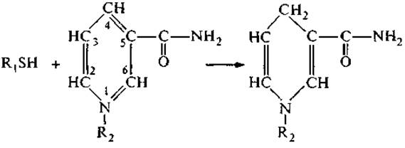
The transfer of a hydride ion (:H-) from the sulfur atom results in the reduction of the ring at positions 1 and 4.
Peptides containing histidine or cysteine residues act as Coenzymes or active sites of enzyme systems in catalytic reactions and contribute to many of the body's antioxidant systems. As integral components of membrane peptides, they are responsible for generating the cellular electrical potential and transmitting electrochemical signals to the external environment.
Many peptide regulators contain cysteine residues. Upon oxidation, their sulfhydryl groups form intramolecular S—S bonds, which form loops in the main peptide chain (tertiary Structure) and fix a specific conformation of the entire molecule. Mercaptoethanol and thioglycolic acid (reducing agents) break these bonds. The absence of antioxidants during the isolation of cysteine-containing peptides from natural sources invariably leads to the recovery of molecules with a more stable tertiary structure.
It is known that free cysteine in a neutral or mildly alkaline range is readily oxidized in the presence of oxygen or other oxidizing agents and dimerizes to form cystine. A similar reaction underlies The activity of glutathione.
Glutathione (γ-glutamyl-cysteinyl-Glycine, or Glu—Cys—Gly) is a widespread tripeptide found in virtually all living Tissues at a concentration of 1–5 mM, though in some tissues it is even higher: in the mammalian eye lens cortex, glutathione concentration reaches 20 mM. In Cells, it accounts for up to 90% of free thiol groups (Krichevskaya et al., 1983). Obviously, its Spatial Structure with two closely spaced carboxyl groups possesses strong chelating properties and is capable of selectively binding transition Metal Ions and polyvalent cations:

Oxidation typically culminates in The formation of a disulfide bridge between two glutathione molecules:

Therefore, glutathione acts as an antioxidant, protecting The sulfhydryl groups of Enzymes and other Proteins. It also plays a special role in the reduction of oxidized ascorbate.
Another function of glutathione is its participation in Amino Acid Transport. The concentration of Amino Acids in cells is maintained at a relatively constant level, ensured by their Transport Across Cell Membranes via specialized transport systems. Glutathione takes part in the so-called γ-glutamyl cycle—a sequence of Chemical Reactions in which the γ-carboxyl group of glutamate is utilized for The Active Transport of amino acids across The Cell membrane. Glutathione provides an activated γ-glutamyl group linked by an isopeptide bond, which, through a transpeptidation reaction, joins with the transported amino acid. The resulting γ-glutamyl amino acid penetrates the cell, where the dipeptide is cleaved into a free Amino Acid and 5-oxoproline. The latter is converted into glutamate in a reaction involving ATP. This transport system is specific for neutral amino acids, glutamate, and cystine (Krichevskaya et al., 1983).
The scheme presents one of the components of glutathione METABOLISM—ophthalmic acid, a glutathione analogue in which the thiol group is replaced by a methyl group:

This peptide was first isolated from the eye lens, and its regulatory Functions are still insufficiently studied.
Histidine-Containing Peptides
Another example of oligopeptides capable of altering the electronic state of their side group are peptides containing histidine in their structure. We have already mentioned that histidine is practically absent from tissues in a free state. However, it is a constituent of many enzymes and regulatory peptides. The structure of the histidine side group provides a broad spectrum of activity for its peptides.
Table 2 Effect of the tripeptide GHK and its synthetic analogues on DNA Synthesis in hepatoma cells* (determined as the increase in 3H-thymidine incorporation, %) (after: Pickart, 1983)
|
Peptide |
Concentration, ng/mL |
|||
2 |
20 |
200 |
2000 |
|
Gly—His—Lys |
50 |
408 |
339 |
91 |
Gly—His—Lys—His |
82 |
604 |
387 |
84 |
His—Lys—Gly |
31 |
368 |
353 |
56 |
Gly—Lys—His |
2 |
36 |
65 |
31 |
His—Gly—Lys |
2 |
13 |
81 |
29 |
His—Lys |
0 |
8 |
зо |
26 |
Gly—His |
0 |
0 |
6 |
3 |
* In the case of other cell types in tissue culture, concentrations exceeding 1 µg/mL invariably inhibited cell growth.
A thermostable tripeptide, Gly—His—Lys (GHK), which regulates growth rate and Cell Differentiation in tissue cultures, was isolated from Blood Plasma (Table 2). Its highest concentration is found in the Kidneys and Brain, and the lowest in the Skin and Muscle tissue. Interestingly, the Amino Acid Composition of GHK is identical to, and its residue sequence is the mirror image of, the structure of bursin (Lys—His—Gly—NH2), a regulatory peptide that selectively determines the stage of B-lymphocyte differentiation. The high biological activity of the GHK tripeptide is determined not only by its chelating properties: in certain enzymes, the histidine residue is phosphorylated at the 1st or 3rd nitrogen atom and acts as a donor in sugar phosphorylation processes.
One of the non-trivial qualities of this tripeptide is its high selectivity in binding copper and other transition metal ions due to their interaction with the imidazole ring of histidine, which endows the peptide with The ability to participate in redox reactions.
The stability constants of copper complexes with serum albumin and peptides are presented below (after: Pickart, 1983):
Chelating agent |
Ln k |
Serum albumin |
16.2 |
Gly—His—Lys |
16.4 |
Gly—His |
8.7 |
Gly—Gly—His |
7.6 |
Gly—His—Lys + free His |
29.0 |
The data presented indicate that the copper–GHK tripeptide complex is comparable in strength to the copper–serum albumin (SA) complex. However, the most unexpected result of these measurements is that The addition of free histidine almost doubles the binding energy. It can be hypothesized that free histidine also acts, to a certain extent, as an agonist for histidine-containing regulatory peptides.
The imidazole ring of histidine has several Resonance forms:
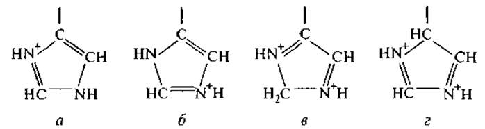
The relative contributions of these forms to the total energy of the ring state are estimated at 40% (a), 32% (b), 23% (c), and 5% (d) (Ching-Fa Wu et al., 1995). The resonance of electronic structures affects the interaction of histidyl with neighboring amino acid residues bearing aromatic side chains and determines the structural universality of the catalytic centers in many proteinases, particularly the so-called charge-Relay system block (D—H—S), which will be discussed in Section 1.2.3. Theoretical calculations of the maximum and minimum distances between the π- and τ-nitrogen atoms of the side chain and the polypeptide backbone have shown that histidine residues closely spaced along the chain tend to associate—i.e., to undergo spatial approximation—thereby stabilizing the active conformation of the peptide (Karlin et al., 1994). An example of high activity and tissue Specificity of peptides containing closely spaced histidine units is histatin, a basic peptide isolated from the human Cytology/practical/97.html">Parotid salivary gland. Its structure contains six histidine residues out of a total of 22 amino acids. The most active region of this peptide consists of eight residues, three of which are histidine residues:

This peptide from the histatin family induces the growth of periodontal fibroblasts at a concentration of 0.1 µg/mL and is recommended for the Treatment of periodontal disease (Takemura et al., 1994). In terms of its regulatory characteristics, it can be classified as a cytomedin, and its specific biological activity is determined by the combination of all PHYSICOCHEMICAL PROPERTIES OF the histidine units discussed in this section. It should merely be emphasized that two adjacent histidine residues significantly enhance the peptide's selectivity in chelation reactions with polyvalent metal ions, which appears to be important both for normal periodontal regeneration and for the execution of other regulatory functions.
Last update: 06/08/2026
Editorial and Educational Adaptation: This material has been compiled based on the primary/original source text. The project team performed an editorial review, corrected technical inaccuracies, structured sections, and adapted the content for an educational format.
What was processed:
- elimination of formatting defects (OCR errors, structural breaks, corrupted characters);
- editorial organization of content;
- standardization of terminology in accordance with academic sources;
- verification of factual statements against the original source text.
All mentions of the author, publication year, and origin of the primary text have been preserved in accordance with the source.