Peptide Self-Regulation of Living Systems (Facts and Hypotheses) - Shataeva L. K. 2003
Peptides in Aqueous Solutions
Physicochemical Characteristics of Peptides
Conformations of Peptides in Solution
The formation of a polypeptide chain is a special case of polymerization that results in a decrease in the total number of degrees of freedom of the monomers. In turn, this allows the macromolecule to acquire qualitatively new intramolecular degrees of freedom—specifically, conformational ones—which arise from the rotational isomerism of the polymer chain and are absent in simple molecules.
As the peptide chain elongates, the order within the system increases, much like a handful of scattered beads becomes organized when strung onto a thread. At the same time, the probability of individual beads contacting one another increases if the thread is twisted or coiled. Following this analogy, the joining of Amino Acids into a single chain molecule allows their side chains to collide and interact as the peptide chain bends and folds. The longer the chain, the larger the set of energetically accessible Conformations.
Although the relative arrangement of amino acid residues in a peptide molecule is fixed, it is precisely the rotation of atoms and groups around valence bonds—governed by a set of possible torsional angles—that gives rise to the peptide's numerous conformations. In solution, the main chain of a peptide never adopts the simple shape of an extended string. The most probable conformation of a synthetic homopolymer is a statistical (randomly coiled) globule with a maximum conformational Entropy. Natural peptides exhibit a more organized conformation, their chains folding into ordered blocks due to the twisting and compaction of the peptide backbone (Pauling, 1964). X-Ray Diffraction Analysis has established that a polypeptide chain composed of L-isomers is curved into a helix. A single turn of the α-Helix along the peptide axis measures 5.4 Å, with approximately 3.6 amino acid residues per turn. Each residue is linked to those of the preceding and succeeding turns via Hydrogen Bonds between the hydrogen atom of the amide group and the oxygen atom of the carbonyl group.
At this level of molecular Organization, a fundamentally new property of peptide molecules emerges: The ability to set aside a portion of space as intramolecular. This region within the α-helical Structure is inaccessible to Water molecules and low-molecular-weight electrolytes, whereas the rest of the space—occupied by the solvent, electrolytes, and other peptides—can be considered external. The amino acid side chains are directed toward the external space, hydrated in accordance with their hydrophobic properties, and arranged for maximum close packing based on their specific volumes (see Table 1). Conventional molecular models do not represent peptide chain conformations with sufficient accuracy because they are constructed without accounting for the Hydration of amino acid side chains.
It is well known that aqueous solutions of amphiphilic molecules, such as ionic Surfactants and Phospholipids, exhibit the ability to self-organize internal cavities. At certain concentrations in an aqueous medium, they form spherical or cylindrical micelles that preserve an internal space inaccessible to “external” molecules. However, these colloidal particles are unstable when the pH or Ionic strength of the solution changes, because the molecules forming them—unlike the units of a polypeptide chain—are not connected by covalent bonds. Furthermore, peptide structures possess a qualitative topological difference: they exhibit chirality, meaning they are fundamentally incompatible with their mirror image.
Not all amino acid residues participate in the Formation of the α-helix: Proline and Glycine act as units that disrupt helical order. Local disruption of the peptide's intramolecular hydrogen bonds by low-molecular-weight agents (such as ethanol, urea, or guanidine) also contributes to the unwinding of the helix. The side chains of the amino acid residues extend radially from the helix and therefore experience no steric hindrance when rotating around CH2—CH2 bonds. Nevertheless, the PHYSICOCHEMICAL PROPERTIES OF these groups (Hydrophobicity, ionization degree, and redox potential) dictate The Nature of their interactions with one another. These attractive and repulsive interactions can be quite intensive, serving either to stabilize the helical conformation of the main chain or to promote its destruction.
The preferred conformation of a macromolecule is determined by the intramolecular bonds of its side chains. It is precisely these bonds that maintain the conformational order of the macromolecule. This order reduces chain entropy, but its energetic stability depends on the strength of the intramolecular side-chain bonds. Alterations in these bonds lead to conformational changes within the chain. In living systems, all Conformational Changes in peptides are reversible, as the potential for conformational transitions is underpinned by the stability (robustness) of the peptide backbone.
The polypeptide backbone consists of a series of rigid planes with flexible hinge joints located at the asymmetric carbon atoms. The dipole moments of the peptide bonds are arranged collinearly from the N-terminus to the C-terminus, their vectors summing up, while the backbone STRUCTURE OF THE polypeptide with hydrophobic side chains becomes regular. Upon A change in the external electric field, such a polypeptide alters its spatial orientation in proportion to its own dipole moment while preserving its chain conformation (Birstein, 1975). Reversible changes in backbone conformation typically occur in Polypeptides containing ionogenic side groups, driven by variations in the ionization degree of these groups. In particular, The Emergence of α-Helical structures in copolymers of glutamic acid and Lysine depends on the pH of the external solution. Calculations have shown that these synthetic copolymers are characterized by a highly ordered chain structure with alternating patterns such as KEKEKEKE or KKEEKKEEKKEE (Volkenstein, Fishman, 1967). The first structural variant could be termed polyvilon.
Elongating a peptide with charged side chains broadens the range of its intermolecular interactions by expanding the ionization range of its side groups. This is readily illustrated by the decrease in the pKa of lysine amino groups when transitioning from a dimer to a pentamer (Stewart et al., 1962):
Peptide |
pKa |
Lysine |
8.95 |
Dilycine |
7.56 |
Trilysine |
7.26 |
Tetralysine |
7.15 |
Pentalysine |
7.07 |
A similar relationship is observed in polypeptides with carboxyl side groups. In other words, adjacent ionogenic side groups of a polypeptide carrying the same charge—much like those of any polyelectrolyte—exhibit an elevated mean pKa value (i.e., a lowered mean acidity) and an expanded range of ionization degrees.
Given that any change in the ionization degree of a polypeptide side group is invariably coupled with shifts in its local hydration and conformation, such a polypeptide can be expected to act simultaneously as a molecular pH sensor for its environment and as an actuator that responds to pH fluctuations by altering its hydration. An example of such a pH-sensitive sensor and hydration regulator is a composite hydrogel-polypeptide copolymer that modulates The rate of drug delivery into Cells (Akala et al., 1998). Living systems regulate metabolite transport with no less efficiency. As we will see below (see Section 2.2), the membrane protein responsible for water transport into The Cell operates on the exact same principle.
There is an immense body of literature dedicated to the principles of structural organization in natural Peptides and Proteins. These investigations rely on conformational analysis, wherein a few of the most accessible and energetically favorable conformations are singled out from the entire set of available states. A detailed analysis of such studies has been provided by E. M. Popov (1997). Synthesizing literature data with his own original research, the author arrived at specific Conclusions regarding the regularities governing conformational changes in polypeptide chains. Under physiological conditions, the Spatial Structure of a natural oligopeptide is described by a limited set of low-energy compact structures, the stability of which is maintained by the coordination of all intramolecular non-covalent interactions. When external conditions change (such as solvent, Temperature, acidity, or proximity to a neighboring receptor molecule), the natural peptide adapts by shifting its conformational state. The shift in equilibrium between the preferred peptide structures occurs through a successive transition of low-energy conformations in the form of an intramolecular excitation wave. In other words, this sequence of conformational changes is just as ordered as the Amino Acid Sequence of the peptide chain. The novel conformation is characterized by a new vibrational frequency spectrum and a corresponding change in the heat capacity of the peptide chain (Popov, 1997).
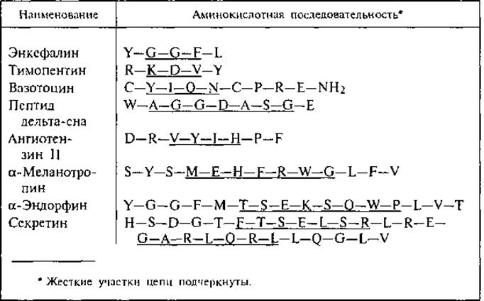
The results of theoretical calculations for specific natural peptides make it possible to identify structurally rigid regions within each molecule—whose conformations require significant energy inputs to change—as well as structurally flexible (labile) regions possessing high torsional mobility at physiological temperatures. Table 3 presents the structures of several regulatory peptides for which chain rigidity has been calculated (Popov, 1997). Notably, the rigid segments consist of either 4 or 7 amino acid residues, meaning they are multiples of the number of residues per turn of the α-helix. The distribution of hydrophilic and hydrophobic residues along the chain also influences the rigidity of these regions, as the α-helix enables hydrophobic amino acid residues to form intramolecular bonds. All peptide formulas presented in this table and throughout the text conform to the biochemical reference data (Oxford Dictionary..., 1997).
Class="center">Table 3. Rigidity of regulatory peptide chains (after Popov, 1997, p. 404)
When examining the relationship between the structure and BIOLOGICAL Functions OF polypeptides, one must bear in mind that every function relies on the high mobility of living cell components, which in turn is sustained by the Presence of water—one of the primary metabolites. Although Ribosomes and macromolecules of proteins and Nucleic Acids are packed densely within the Cytoplasm between Organelles and The Endoplasmic reticulum (ER), the presence of water allows them to undergo conformational transitions and diverse movements for which water serves less as an “environment” than as a lubricant. A hydrophobic microenvironment (such as adjacent hydrophobic ER surfaces or lipoprotein complexes) likewise enhances the molecular mobility of the hydrated polypeptide.
Three types of intracellular molecular motions can be distinguished: 1) macromolecular rotation; 2) relocation from one position to another (translational motion); 3) rapid oscillations (vibrations) of the atoms constituting the molecule. All these movements are essential for molecular surfaces to come into contact and engage in specific interactions to fulfill their biological functions.
The rates of molecular motions are investigated using various spectroscopic techniques. These Methods have established that polypeptide macromolecules in the cytoplasm constantly change conformation, bend, rotate, and collide with neighbors. The frequency of diffusive collisions between polypeptides and low-molecular-weight components is proportional to the concentration of the diffusing molecules. For instance, at a typical intracellular ATP concentration of around 1 mM, each peptide chain undergoes random collisions with ATP at a frequency of 106 per second (Alberts et al., 1994). Given that these molecular interactions and Chemical Reactions take place within a volume of 4×103 μm3—a scale vastly different from laboratory test-tube conditions—the high efficiency of enzymatic reactions in vivo becomes much easier to understand. The outcome of a “successful” collision with a binding site may be either the formation of an intermolecular complex or Enzymatic Hydrolysis, depending on the Nature of the binding site itself.
The catalytic center can be modeled as a sort of fractal surface. In this case, the chemical reaction rate ($v$) under diffusion-limited conditions is expressed by the equation
V = k • [А]X,
where [A] is the concentration of the interacting structures; $k$ is the rate constant; and $X$ is the exponent indicating the reaction concentration order:
X = 1 + 2 /DS,
where $D_s$ is the fractal dimension of the catalytic center (Kopelman, 1992).
In a model system, molecules A = IJ randomly collide with the binding site and immediately dissociate into two molecular fragments, I and J. Subsequently, they can migrate across the surface (or the surface can shift beneath them), resulting in the recombination of the fragments to form the product IJ, which immediately dissociates from the catalytic center. The maximum reaction rate of this kind is achieved when the binding site is not a surface, but a one-dimensional chain ($D_s$ = 1, $X$ = 3). In this scenario, due to reduced topological constraints, the chain can facilitate recombinations for reactions involving three colliding molecules (Kopelman, 1992).
This speculative model finds support in experimental studies investigating the relationship between the conformation of a peptide chain and its catalytic function. It has been established that when a specific conformation of the peptide chain brings low-reactivity side groups into close proximity, their reactivity is amplified manifold precisely due to this spatial arrangement. Such a conformation, which ensures the biochemically active proximity of the peptide's side groups, is termed native.
For instance, in the peptide chain of Chymotrypsin, the Nτ nitrogen atom of the Histidine residue His57 is spatially approximated to the carboxyl group of the aspartic acid residue Asp102, while its Nπ nitrogen atom is brought close to the side group of Serine Ser195. The carboxyl group of asparagine accepts a proton from the imidazole ring, shifting the electron density, and the Nπ nitrogen atom activates the serine hydroxyl group by abstracting a proton from it, enabling it to form a covalent bond with the carbonyl group of another peptide (substrate), thereby cleaving its peptide bond (Alberts et al., 1994). All Serine proteinases carry out peptide bond hydrolysis following this mechanism. The amphiphilic character of peptides and a specific alternation of hydrophilic and hydrophobic side groups in their structure give rise to additional conformational features of peptides in solution. These arise in accordance with THE PRINCIPLE OF minimizing the surface Free energy at the interface between the peptide and its aqueous environment. This Minimization is achieved through the spatial segregation of hydrophilic and hydrophobic side groups in the form of a ß-sheet or an a-helix (Kaiser, Kézdy, 1984). Schemes of such segregation are presented in Fig. 4, A. The features of a-helical structures with a characteristic arrangement of hydrophilic and hydrophobic groups on opposite sides of the a-helix were discovered as early as 1967 and have since been known as “Edmundson wheels,” or helical wheels (Schiffer, Edmundson, 1967). In a single a-helix, the side groups of the first and fourth amino acids lie close to each other on the outer cylindrical surface of the molecule. This alternation repeats with a period of 7 residues, corresponding to two turns of the a-helix. If amino acids with hydrophobic side groups occupy these positions, a hydrophobic “ridge” of the a-helix is formed. Hydrophobic interactions along this ridge further reinforce the hydrogen bonding system along the peptide backbone and the overall helical structure. Furthermore, if the hydrophobic side groups of the peptide fit the 1–4 rhythm, they exhibit increased partial volumes due to hydration peculiarities, and the hydrophobic ridge in this case acquires a longer pitch compared to a standard a-helical conformation. Fig. 4, B depicts, as a heptagonal cylinder, the twisted helix of the 16–32 sequence of the immunostimulant thymopoietin. Natural thymopoietin is a peptide consisting of 49 amino acid residues (see Appendix Table III) containing an “active center”—the peptide sequence 32–36. A chemically synthesized pentapeptide of identical structure exhibits The activity of the whole molecule and was therefore named thymopentin (Table 3, row 2). The regions preceding and following the “active center” (residues 16–32 and 37–49, respectively) are capable of forming helical “wheels.”
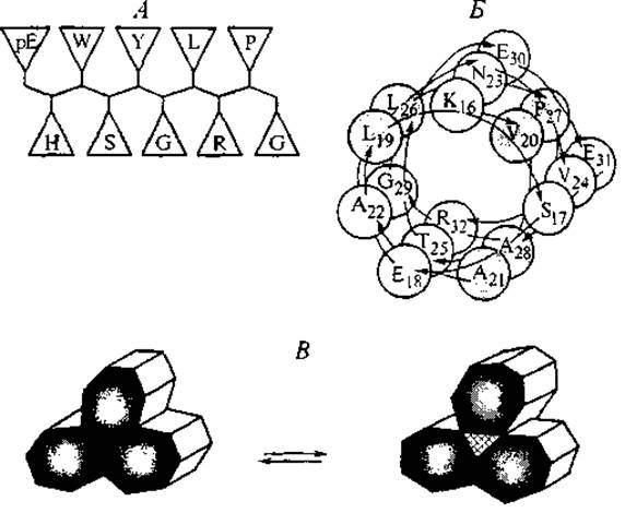
Fig. 4. Schemes of the ß-sheet structure of gonadoliberin (A) and a portion of the twisted a-helix (superhelix) of thymopoietin (B), demonstrating the spatial Separation of hydrophilic and hydrophobic (darkened) amino acid residues. Scheme C represents a cross-section of a bundle of associated superhelices and the conformational transition resulting in the formation of an internal channel between the superhelices.
Leucine residues are located at positions 19 and 26 within the presented region. It should be noted that leucine plays a special role in the formation and stabilization of helical structures. Structural studies have established METABOLISM/2.html">THE CONCEPT OF the “leucine zipper.” This consists of relatively short sequences with an average length of 24 amino acid residues, in which leucine residues are located at positions 2, 5, 7, 12, 16, 21, and 24, forming the hydrophobic faces of the helix. Such structures, characteristic of transmembrane domains of Membrane Proteins, participate in protein–protein and protein–DNA intermolecular interactions. In the latter case, the configuration of the leucine zipper determines the contact of the peptide with The surface of the DNA double helix (see Section 3.2).
Similar a-helical blocks have been discovered in the chains of several Peptide Hormones: Calcitonin, somatoliberin, corticoliberin, apolipoprotein A, and ß-endorphin (Kaiser, Kezdi, 1984). This principle of amino acid residue alternation along the backbone was utilized in the design and Chemical synthesis of natural hormone analogs (Somatostatin and Glucagon) with enhanced specific activity (Rivier et al., 1978).
A distinctive feature of twisted a-helical regions (superhelices) is their ability to assemble into asymmetrical bundles, as shown in Fig. 4, C. In this arrangement, the hydrophobic ridges of the helices can be oriented toward the interior of the bundle and pack tightly against one another—this represents one conformation. Upon altering the interactions at the hydrophilic ridges of the superhelix, the hydrophobic ridges change their orientation, and the interior of the bundle forms a hydrophilic channel (Alberts et al., 1994). In the next chapter, we will return to peptide superhelices when discussing the structures of Membrane Receptors.
The segregation of hydrophilic and hydrophobic groups on different faces of the heptagonal peptide helix confers additional functional properties upon the molecule (Zhou et al., 1992). Hydrophobic groups located on one ridge of the heptagon can serve as a recognition determinant for interacting with other peptides that possess hydrophobic patches (Immunostimulants, 1987). Meanwhile, hydrophilic, particularly ionogenic, side groups positioned sequentially along the perimeter of the heptagon cross-section with a specific alternation of positively and negatively charged groups act as determinant binding sites for many high-molecular-weight Antigens.
Table 4 Amino acid sequences of determinant regions of peptide antigens (after Hopp, Woods, 1981)
Protein antigen |
Determinant region of the peptide chain |
Plague hemagglutinin Cholera toxin a-chain E. coli enterotoxin Streptococcal N-protein Human interferon-I Human interferon-II |
—E—R—R—E—G—D— —E—A—K—V—E—K— —E—R—M—K—D—T— —R—K—A—D—L—E—K— —E—E—K—L—E—K—E—D— —E—R—L—R—R—K—E— |
Table 4 presents the amino acid sequences of hydrophilic epitopes from several antigens (Hopp, Woods, 1981). It is interesting to note the alternation of positively and negatively charged side groups along these chains, which enhances the thermodynamic favorability of their positioning on a shared ridge within the folded helix.
Attention is drawn to a distinct qualitative similarity between the determinant regions of certain microbial antigens and PARTS OF THE polypeptide chain of thymic peptides responsible for the body's Immunity. In terms of the local density of positively and negatively charged side groups and the pattern of their alternation (repetition of KE and EK blocks), these peptides bear a striking resemblance (see Appendix Table III).
A certain similarity is observed between thymopentin and the determinant of plague hemagglutinin, as well as between the cholera toxin determinant and interferon. It can be hypothesized that through a prolonged evolutionary process, pathogens of particularly dangerous infections have incorporated peptide regions into their antigenic determinants (epitopes) that mimic the amino acid sequences of Peptides of the human immune system.
In addition to recognition processes in the antigen–antibody system, a specific order of alternation of positively and negatively charged side groups is crucial for facilitating the Transmembrane Transport of the polypeptide chain. A statistical Analysis of the alternation of Lys and Asp, and Arg and Glu in The structure of several cell-secreted proteins revealed a consistent tendency for these residues to be spaced four amino acid residues apart (Heijne, 1980). This enables local intramolecular neutralization of electrostatic charges when the peptide folds into an amphiphilic superhelix.
A unique property of the amphiphilic peptide superhelix as a template for non-ribosomal Peptide Synthesis was demonstrated in the work of Lee et al. (1996). The authors experimentally proved that a twisted peptide chain consisting of 32 amino acid residues with a specific sequence, forming a heptagon with two hydrophobic ridges, exhibits the capacity for autocatalytic self-Replication—a property of macromolecules previously found exclusively in nucleic acids. The study utilized the method of autocatalytic peptide bond synthesis on a peptide template. The formation of a peptide bond between two oligopeptide fragments was achieved via the Condensation of peptide thioesters (Kent's method) (Dawson et al., 1994). The reaction mixture consisted of equal quantities of electrophilic and nucleophilic peptide fragments in the presence of a minor amount of the target template peptide. The autocatalytic nature of the reaction was confirmed by the linear dependence between the initial concentration of template molecules and the rate of synthesis. The reaction proved to be highly structure- and chemospecific.
It is known that polycondensation reactions of peptide thioesters occur in the cells of Bacteria and lower eukaryotes during the synthesis of peptides (particularly peptide Antibiotics) with the participation of specific peptide synthetases. Such non-ribosomal oligopeptide synthesis is considered the most evolutionarily ancient mode of peptide self-defense, since these peptides contain non-codable D-Amino Acids and exhibit enhanced affinity for the phospholipid structures of cell membranes. It is precisely these properties that allow them to actively disrupt membrane permeability and exert an antibiotic effect against competing cells (Biotechnology of Antibiotics, 1997).
Under physiological conditions, the conformations of individual Regions of the peptide chain can change As a result of the spatial mobility of side groups while maintaining an invariant sequence along the chain; however, these changes are reversible. As spectroscopic methods demonstrate, each conformation possesses a set of characteristic vibrational modes. Upon the emergence of non-physiological conditions, the conformation of a sufficiently long polypeptide can alter into a statistical coil—without breaking peptide bonds, but with a loss of biological activity. This conformational transition is termed Denaturation. In this state, the spectral CHARACTERISTICS OF THE macromolecule lose definition, and the conformational entropy of the coil reaches its maximum.
In A number of cases, a return to physiological conditions allows the polypeptide to revert to its native conformation, especially if it is stabilized by intrachain S–S bonds. However, such renaturation is possible only for single-chain peptides. For instance, the Insulin molecule, which consists of two peptide chains linked by S–S bridges, cannot return to its native conformation after the destruction of these bridges and subsequent denaturation. This is determined by the specifics of insulin Biosynthesis. The native conformation of insulin arises as a result of proinsulin hydrolysis. As shown in Fig. 5, proinsulin is synthesized as a single peptide chain containing two terminal regions—the A- and B-chains of future insulin—with six free Cysteine groups within them. The middle region of proinsulin (C; see Fig. 5) adopts a conformation that establishes a specific system of S–S bonds between the terminal segments of the chain. Following the excision of the middle region, an active two-chain insulin molecule is formed. Precisely because of the spatial predetermination of this structure by the middle region of proinsulin, spontaneous renaturation of insulin is impossible. In other words, the removal of a portion of the peptide chain is equivalent to a loss of molecular information for the remaining parts of insulin.
Recently, intensive research has been conducted on the post-translational modification of peptides, during which peptide bond synthesis is carried out with high selectivity, predetermined by the conformation of the joining oligopeptides. This modification involves two coordinated proteolytic cleavages of the peptide and the subsequent ligation of the ends of the two resulting fragments. An example of such combinatorics is the joining and cyclization of two truncated chains during The biosynthesis of the macrocyclic peptide antibiotic q-defensin, produced by monocytes and neutrophils (Tang et al., 1999). Such processes are regarded as peptide splicing, occurring analogously to RNA splicing (Cooper, Stevens, 1995).
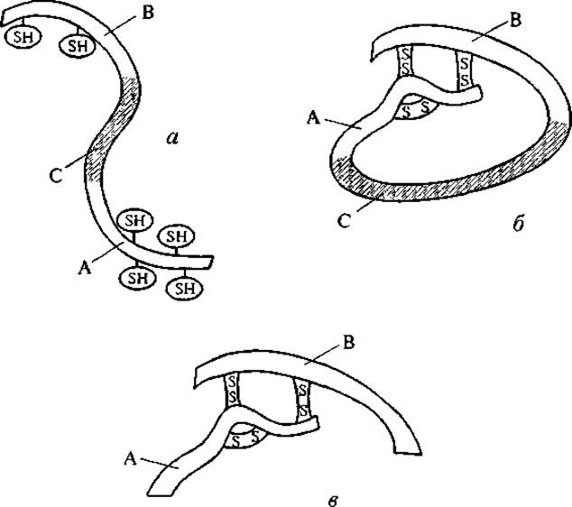
Fig. 5. Formation of the insulin molecule from proinsulin. a — structure of the proinsulin peptide chain: A and B are terminal regions, C is the middle region; b — conformational change of proinsulin leading to intramolecular S–S cross-linking of its terminal regions; c — structure of the native insulin molecule.
The compact conformation of the polypeptide, stabilized by intramolecular Hydrophobic bonds, determines the topographical arrangement of hydrophilic and hydrophobic side groups On the surface of the macromolecule, i.e., its mosaic structure. The relative positioning of charged side groups of amino acid residues on the surface of the peptide globule is heterogeneous: there are regions with charges of varying magnitude and sign, which can be visualized on an Electrostatic Surface potential map.
Direct calculations of this potential based on X-ray diffraction data for a number of polypeptides and nucleic acids have demonstrated not only spatial complementarity, but also electrostatic surface potential (ESP) complementarity between the binding sites of Enzymes and substrates, Antibodies and antigens, and enzymes and inhibitors (Wainer et al., 1982). Using the Examples of ESP complementarity between Ribonuclease and RNA, Trypsin and its inhibitor, and thyroxine and prealbumin, it has been shown that the regions of these molecules not involved in the electrostatic attraction of ligands and binding sites provide mutual local repulsion between the interacting entities (including via electrostatic repulsion of like charges), so that the energy barriers for the reverse dissociation reaction of the complex are not excessively high. In other words, intermolecular complementarity does not shift the equilibrium of peptide interaction exclusively toward binding; adjacent regions of the peptide chain ensure the reversibility of this process and the dissociation of the complex. Such dynamic equilibria underlie the intermolecular association of biologically active polypeptides both in Enzymatic Catalysis and in the peptide Regulation of Specific cellular functions.
Electrostatic complementarity between interacting peptides, when not counterbalanced by repulsive forces, leads to irreversible phase transformations. An example of such transformations is the spontaneous and irreversible self-association of peptides consisting of alternating tetrapeptide blocks that are complementary to each other in charge sign:
(Ala–Glu–Ala–Glu–Ala–Lys–Ala–Lys–)2.
In aqueous solutions, these peptides form intermolecular associates with a ß-sheet structure and, in the presence of salts, precipitate as macroscopic membranes and fibers (Zhang et al., 1993). Complex formation leads to a sharp decrease in peptide hydration. Electron Microscopy reveals their microstructure: intertwined filaments 10—20 nm in thick. They are completely insoluble—neither in acids nor in alkalis, neither upon The addition of urea or guanidine, nor under the action of Proteolytic Enzymes. There is every reason to believe that this type of intermolecular peptide association underlies the formation of amyloid deposits in various tissues (Tomas et al., 1996). In particular, the serum protein serum amyloid A, with a molecular mass of 13.4 kDa, is produced by the Liver under The Influence of cytokines and deposits as self-associates in various Organs and tissues, damaging them and causing amyloidosis; the ß-amyloid glycoprotein, with a molecular mass of 86.8 kDa, deposits primarily in Nervous Tissue and is one of the causes of Alzheimer's disease. Similar Brain lesions are caused by Prions—membrane proteins with a molecular mass of 33—35 kDa, which apparently lose their native a-helical structure and acquire a ß-sheet structure upon losing their glycophospholipid moiety. In all the aforementioned examples, the cause of amyloid deposits is the irreversible, electrostatically anchored self-association of complementary regions of peptide chains adopting a ß-configuration.
Significant changes in polypeptide conformations are observed upon enzymatic modification of their side chains, such as methylation, decarboxylation, and deamidation. The most pronounced changes in polypeptide hydrophilicity and conformation occur during the phosphorylation of serine, Threonine, or Tyrosine side chains (mediated by phosphokinases) or during polypeptide glycosylation. These processes belong to another domain of the body's self-regulation—the realm of coordinated enzyme systems that ensure metabolism and the interaction of peptides with other classes of molecules, such as CARBOHYDRATES, polyphosphates, and Lipids.
As already mentioned, all conformational transitions and intermolecular interactions in living systems occur in an aqueous environment, although this water is organized in the form of hydration shells (bound water) surrounding the macromolecule.
Obviously, a change in the Introduction/10.html">Peptide Conformation is accompanied by alterations in its hydration shells and the hydrogen-bonding system, which possesses a well-defined dynamic structure within the hydration water. The side chains of the peptide are arranged in such a way that their hydrogen bonds with other groups easily change orientation and transform into hydrogen bonds with surrounding water molecules. The frequency of “Hydrogen bond partner” exchange depends on the Chemical Nature of the side chain and its position within the given peptide conformation.
The cause of a peptide chain conformation change can be not only its intermolecular interactions, but also alterations in the intramolecular electrostatic characteristics of the molecule associated with Changes in the ionization degree of its side chains. The orientation of the local dipole vector (from the center of gravity of the negative charge to the center of gravity of the positive charge) changes as the side chains become deionized (see Fig. 3) due to shifts in the dielectric permittivity or pH of the external environment. The resulting change in the electrostatic component of the free energy typically leads to a conformational change of the entire molecule. Since each conformation represents an ordered structure, a conformational shift is coupled with changes in order not only within the Peptide Structure but also within the coordinated H-bond network of the aqueous environment (see Fig. 4, C — transition of a-helical conformations into a superhelix). Against the Background of thermal fluctuations, these ordered changes are perceived by bulk (free) water not as disturbances, but as orderly molecular signals indicating a change in peptide conformation. In this capacity, they can be transmitted through the aqueous medium to other macromolecules and cellular receptors.
The proposed simplified model of information action at a distance highlights only one feature of molecular signals: their transmission does not involve The transfer of mass or energy. It is based on the alternation of reversible conformational transitions between the native peptide and its associated water. No special code is required to transmit the peptide's signals regarding its own conformational state, since they originally arise in the form of localized changes and shifts in the water's hydrogen bond network and are transmitted and perceived in this very same form.
Solving the specific problem—determining what form of signal is characteristic of a given peptide and what response this signal can elicit in a recipient peptide—requires a preliminary systematic comparison of the amino acid sequences of peptides that perform specific regulatory (informational) functions. As a rule, their activity manifests at the level of specialized functional systems of the body and is often defined as tissue-specific. We will examine this issue in more detail in the next section.
Last update: 06/08/2026
Editorial and Educational Adaptation: This material has been compiled based on the primary/original source text. The project team performed an editorial review, corrected technical inaccuracies, structured sections, and adapted the content for an educational format.
What was processed:
- elimination of formatting defects (OCR errors, structural breaks, corrupted characters);
- editorial organization of content;
- standardization of terminology in accordance with academic sources;
- verification of factual statements against the original source text.
All mentions of the author, publication year, and origin of the primary text have been preserved in accordance with the source.