Peptide Self-Regulation of Living Systems (Facts and Hypotheses) - Shataeva L. K. 2003
Peptides in Aqueous Solutions
Physicochemical Characteristics of Peptides
Hydrophilic-Hydrophobic Properties of Oligopeptides
As noted earlier, the dissociation of zwitterions, particularly Peptides, proceeds as a complex multi-step process characterized by multiple ionization constants. Altering the charge of a single ionogenic group not only affects its interaction with inorganic counterions (Na+, K+, Ca2+), the polarizability, and the overall dipole moment of the peptide, but also, as mentioned above, changes the orientation of surrounding Water molecules and shifts the balance between Hydration and clathrate hydration of the entire molecule. Thus, the range of biological activity of dipeptides is broadened due to their higher sensitivity to fluctuations in environmental Ionic strength and acidity: their biological effects manifest at lower molar concentrations yet with greater selectivity compared to individual Amino Acids.
By way of example, we can examine the Structure and regulatory properties of three dipeptides: carnosine, thymogen, and vilon, which are thoroughly described in the literature (Khavinson, Zhukov, 1992; Kuznik et al., 1998; Hipkiss et al., 1998; Morozov et al., 2000b).
Carnosine
The dipeptide carnosine (β-alanyl-Histidine) was first isolated from Muscle tissue and subsequently found in all innervated tissues at concentrations up to 20 mM. Its molecular structure is illustrated in Fig. 3; its defining feature is the combination of the histidine imidazole ring at the C-terminus of the molecule with a β-amino group at the opposite end. This β-position amino group lies sufficiently far from the plane of the peptide bond, thereby retaining greater conformational mobility with free rotation around the C—C and N—C bonds than an α-position amino group. The distinct Properties of the histidine imidazole ring are worthy of note. For free histidine, the dissociation constant of this side chain is pKa = 6.0, meaning this group associates a proton less strongly than water (pKa = 7.0) (Chang, 1980). However, the two nitrogen atoms (Nπ and Nτ) differ in their distance from the main peptide backbone and in their electron-donating properties. Consequently, A change in the degree of dissociation of one of them is accompanied by a shift of electron density across the ring, enabling histidine to participate in Ligand and redox interactions when its α-amino group is incorporated into the peptide bond. This may explain why carnosine positively influences Glycolysis and Oxidative Phosphorylation, increasing the yield of ATP. Furthermore, carnosine enhances the efficiency of active K+ and Na+ Transport Across the cytoplasmic membrane.
Class="center">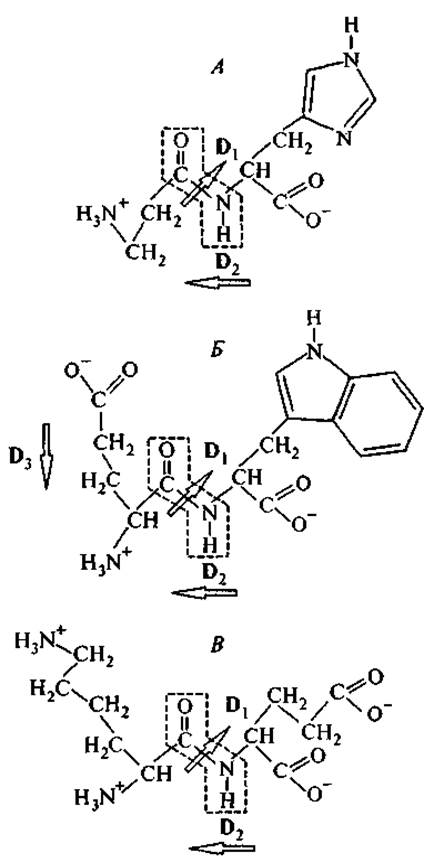
Fig. 3. Structural formulas of carnosine (A), thymogen (B), and vilon (C).
The frame encloses the plane of the peptide bond, which lies in the plane of the page. Arrows indicate the dipole moments D of the peptide bond and side chains. The local dipole moment vector is directed from the negative charge to the positive charge.
Carnosinase, an enzyme that cleaves carnosine into β-Alanine and histidine, is found in the Liver, Adrenal Glands, Spleen, and the mucous secretions of the Small Intestine. Carnosine is known to inhibit Lipid Peroxidation, promote the Repair of Damaged tissues, and prove effective in treating senile cataracts. Currently, carnosine is manufactured in eye drop formulations for this very purpose.
Relatively recently, it was demonstrated that carnosine also protects Brain tissue from The formation of amyloid protein deposits. Amyloidosis is a systemic disorder characterized by the deposition of protein-carbohydrate complexes within the intercellular spaces of Nervous Tissue. Besides the association of peptide fragments, an important role in this process is played by the cross-linking of peptide fibrils via aldehyde groups from reducing sugars and malondialdehyde, a harmful end product of lipid peroxidation. The protective effect of carnosine against amyloid tissue degeneration is presumably attributable to the electron-accepting activity of its imidazole ring, which counteracts lipid peroxidation. However, this activity manifests only after histidine is incorporated into the dipeptide, which possesses a significant dipole moment and spatial stabilization of the ring relative to this dipole. Free histidine lacks such activity (Alberts et al., 1994). A certain role in the destabilization of amyloid deposits may also be played by the β-structure of alanine (Iverson, 1997).
Thymogen
The dipeptide thymogen (L-glutamyl-L-Tryptophan) was first isolated from oligopeptide fractions of Thymus preparations. Following the discovery of its T-lymphocyte-stimulating activity, this dipeptide was successfully produced via chemical synthesis and is now widely used in medical practice (Morozov, Khavinson, 1996; Kuznik et al., 1998; Morozov et al., 2000b). Its structure is shown in Fig. 3.
Carnosine and thymogen combine hydrophilic and hydrophobic moieties within their structures, which in aqueous solution reside on opposite sides of the peptide bond plane. The hydrophilic groups of these dipeptides (the left-hand portions of the molecules depicted in Fig. 3) differ in charge sign: in carnosine, it is a positively charged amino group, whereas in thymogen, it is a negatively charged carboxyl group. When the glutamic acid residue of thymogen is not in the pyroform, its molecule possesses two local, oppositely oriented dipole moments: one between the C- and N-termini of the dipeptide (D2), and another between the α-amino and carboxyl groups of the glutamic acid residue (D3). Thus, the total permanent dipole moment of thymogen is lower than that of the carnosine molecule.
A distinctive feature of the thymogen molecule is that it combines the most hydrophilic (glutamyl) and the most hydrophobic (tryptophan) amino acid residues. The presence of tryptophan sets thymogen apart from all known peptide regulators of the thymic system, bringing it closer to Neuropeptides of the neuromedin and peptide-releasing hormone classes. At the same time, thymogen exhibits all the immunomodulatory properties of thymalin while significantly surpassing the latter in specific activity (Khavinson, Zhukov, 1992). Studies have shown that thymogen in vitro promotes the regeneration of specific T- and B-lymphocyte receptors and activates the mononuclear phagocyte system (Morozov, Khavinson, 1997; Kiseleva et al., 1999), whereas in vivo in patients with secondary immunodeficiency, it normalizes the levels of T-helper Cells, T-suppressor cells, and B-lymphocytes (Kuznik et al., 1999; Morozov et al., 2000b).
Apparently, the combination of a bulky hydrophobic tryptophan side chain and compensated dipolarity facilitates the interaction of thymogen with the hydrophobic regions of Cell membranes. Specifically, it has been demonstrated that after attaching the EW dipeptide to the N-terminus of the α-helical polypeptide (LSSLLSL)3, the extended polypeptide binds to The Cell membrane surface, penetrates its bilayer phospholipid structure, forms Ion Channels within the membrane, and thereby alters the selectivity of ion transport (Lear et al., 1997). This is one of several Examples showing that a tryptophan residue near the N-terminus of a polypeptide chain enhances the selectivity of polypeptide interaction with the outer surface of the cell membrane.
Vilon
The dipeptide vilon (L-Lys—L-Glu) was designed based on statistical Analysis of the Amino Acid Composition of the drug Thymalin (Morozov et al., 2000b; Khavinson, 2001b). Data from Table III of the Appendix indicate that this dipeptide serves as a structural building block in numerous thymic Hormones. As Fig. 3 shows, a defining characteristic of this dipeptide is the distinct segregation of electrostatic charges between its two ends: positively charged amino groups belong to the Lysine residue, while negative charges are concentrated on the glutamic acid residue. Such a structure points to vilon's capacity to actively participate in electrostatic (ion-ion and ion-dipole) interactions.
Investigating the effects of thymogen and vilon on The Development of organotypic cultures of the thymus and spleen revealed an identical stimulatory effect on thymus explants, yet a divergent effect on spleen explants harvested from animals of different ages: thymogen stimulated tissue growth in both old and young animals, whereas vilon stimulated it only in old ones (Khavinson et al., 1999; Chalisova et al., 1999). Subsequent studies demonstrated that vilon exerts a pronounced stabilizing and regenerative influence on the morphofunctional characteristics of organotypic spleen culture and exerts a stimulatory effect on the stromal cellular microenvironment in aged and surviving cultures. In various models of premature Aging induced by gamma irradiation, vilon was found to modulate animal immune Homeostasis, stimulate reparative processes in the Thymus gland, and increase the proliferative activity of thymocytes, thereby accelerating the compensation of microcirculatory disorders in the thymus and spleen (Khavinson et al., 2001d; Knyazkin, Polyakova, 2002; Knyazkin et al., 2002). Other authors established that vilon is the shortest
regulatory fragment that facilitates the delivery of METABOLISM/31.html">Transcription factors into The Nucleus or integrates into The structure of functionally active transcription factor complexes required for activating interleukin-2 Gene transcription in lymphocytes (Khavinson et al., 2000c). Further research revealed that vilon is capable of enhancing neutral sphingomyelinase activity, either by modulating the passage of known biologically active signals (specifically IL-1β) along the sphingomyelin pathway or by directly transmitting its regulatory information into thymocytes via this signal Transduction pathway (Khavinson et al., 2002b).
An investigation of vilon's biological activity conducted on CBA mice revealed a substantial increase in the maximum lifespan of these animals following administration of the preparation. Moreover, vilon Treatment exerted an inhibitory effect on the development of malignant tumors and neoplasms in female CBA mice (Khavinson, Anisimov, 2000; Anisimov V. N. et al., 2002a). Studying vilon's influence on Gene Expression demonstrated that genes whose expression levels changed under the peptide's action belong to A wide variety of cellular systems. However, genes associated with Cell Division and cellular/organismal defense systems are the most prominently represented. Specifically, noteworthy changes were observed in the expression of genes related to Cell Cycle regulation and membrane transport, as well as genes implicated in oncogenesis and calcium metabolism (Anisimov S. V. et al., 2002).
Thus, the regulatory and immunomodulatory action of vilon on mechanisms of regeneration and carcinogenesis is mediated by microenvironmental stromal cells (macrophages, fibroblasts, mast cells, endothelial, and reticular cells) and realized through the microvasculature. Data obtained from experimental studies of vilon's biological effects warrant recommending its further investigation as a geroprotective agent. The application of vilon in geriatrics is purposeful for correcting age-related disorders of angio- and immunogenesis, as well as for modulating cellular proliferation and inhibiting carcinogenesis.
The values of total side-chain Hydrophobicity for carnosine, vilon, and thymogen, calculated from the data in Table 1 (1.5, 5.1, and 15.2 kJ/mol, respectively), indicate that the carnosine molecule possesses the lowest hydrophobicity, i.e., the highest hydrophilicity. Presumably, this hydrophilicity plays a definite role in destabilizing hydrophobic amyloid β-structures (see Section 1.2.3).
Alternating amino acid residues with positively and negatively charged side chains are characteristic of most hydrophilic dipeptides exhibiting biological activity. For example, Lys—Asp and Glu—Arg are regarded as the simplest regulators and stimulators of physiological Functions (Belokrylov et al., 1998). At the same time, THE SPECTRUM OF functions influenced by these peptides is very broad and displays no tissue Specificity. One may hypothesize that these molecules not only exhibit intrinsic regulatory activity but can also act as agonists for other endogenous regulators (including non-peptide ones), heightening the sensitivity of cellular receptors to their action.
The biologically active dipeptides discussed above possess a relatively simple structure while remaining capable of engaging in various types of intermolecular interactions. Lengthening the peptide chain enhances the polyfunctionality of the molecule and broadens the spectrum of its regulatory functions. The synthetic tetrapeptide Epitalon (Anisimov et al., 2001) serves as a prime example.
Last update: 06/08/2026
Editorial and Educational Adaptation: This material has been compiled based on the primary/original source text. The project team performed an editorial review, corrected technical inaccuracies, structured sections, and adapted the content for an educational format.
What was processed:
- elimination of formatting defects (OCR errors, structural breaks, corrupted characters);
- editorial organization of content;
- standardization of terminology in accordance with academic sources;
- verification of factual statements against the original source text.
All mentions of the author, publication year, and origin of the primary text have been preserved in accordance with the source.