Peptide Self-Regulation of Living Systems (Facts and Hypotheses) - Shataeva L. K. 2003
Peptides in Aqueous Solutions
Structural Features of Amino Acids and Oligopeptides
As is well known, Proteins are composed of a-aminocarboxylic acids, commonly referred to as Amino Acids. Their structures can be found in any biochemistry textbook or reference guide. To date, more than 100 amino acids of various structures have been discovered in the PLANT AND ANIMAL kingdoms, yet only 20 of them are encoded by METABOLISM/28.html">The Genetic Code. That is, for each of these 20 amino acids, There is a specific combination of nucleotide units (codons) that determines The amino acid's position in the polypeptide chain during ribosomal synthesis. In this monograph, we will focus exclusively on the encoded amino acids, which form the foundation of biological processes on Earth. Their structures exhibit distinct spatial and electrochemical characteristics.
1. All amino acids, with the exception of Glycine, exhibit chirality—meaning they exist in two mirror-symmetrical, spatially incompatible forms, L and D (Fig. 1, A). Amino acids incorporated into natural Peptides and Proteins are predominantly in the L-form; exceptions are found in peptide Antibiotics of animal origin (Kreil, 1997).
2. The amino group exhibits a pronounced basic character, while the carboxyl group is acidic: for glycine, the acid dissociation constant values (pKa) are 9.6 and 2.3, respectively. In an aqueous solution under physiological conditions, the proton of the carboxyl group transfers to the amino group (Fig. 1, B), converting the molecule into a zwitterion (Pauling, 1964). Due to the spatially fixed arrangement of their positive and negative charges, amino acids possess a permanent dipole moment in Water.
3. The R side chains of amino acids (Fig. 1, A) display a wide range of hydrophilic and hydrophobic properties (Table 1), which govern the intermolecular interactions of amino acids. The side chains of Lysine and Arginine act as strong bases (pKa values are 10.5 and 12.5, respectively); the side chains of aspartic and glutamic acids exhibit strong acidic properties (pKa values are 3.6 and 4.2, respectively).
Class="center">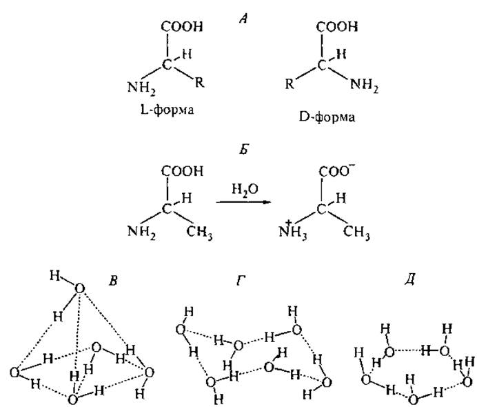
Fig. 1. Spatial structures of Amino Acids and their aqueous environment. A — stereoisomers of a-carboxylic amino acids, where R is the side chain; B — zwitterionic Structure of L-Alanine in aqueous solution at pH 7.0. C–E — coordination of water molecules via Hydrogen Bonds, determining the Hydration of the hydrophilic and hydrophobic parts of Amino Acids and Proteins: in bulk water (C), in the ice structure (D), and in clathrates (E).
4. The combination of proton-donor and proton-acceptor groups in the Structure of Amino acids enables them to participate in hydrogen bonding with other molecules, particularly with solvent molecules.
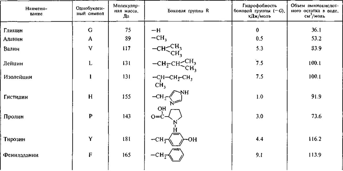
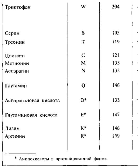
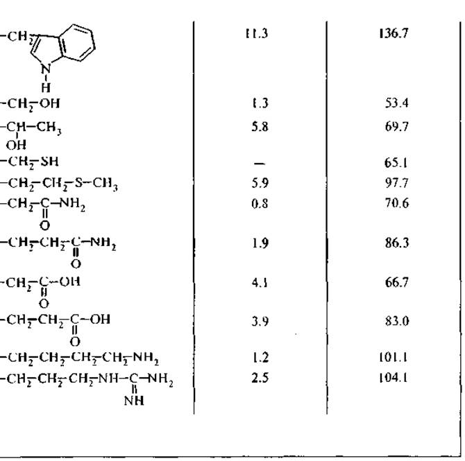
Hydrophobicity is a property of nonpolar molecules and chemical groups, reflecting their tendency to dissolve in nonpolar Solvents. At the same time, these nonpolar molecules are practically insoluble in water and immiscible with it. Among inorganic substances, noble gases are considered hydrophobic, while among organic substances, Hydrocarbons and their derivatives fall into this category. The partition coefficient between an organic solvent and water is conventionally used as a measure of a substance's hydrophobicity; Tanford utilized ethanol as this organic solvent, whereas McGregor used 2-butanol. This Classification divided amino acid residues into three categories: hydrophilic, hydrophobic, and those distributed equally between the organic and aqueous phases. The most comprehensive systematization of hydrophilic-hydrophobic characteristics was developed by Hansch, who employed octanol as the nonpolar solvent (Leo et al., 1971). Using data from his review on the partition coefficients of amino acids between octanol and water, we calculated the Standard Free energy of transfer (G) of an amino acid from water to octanol. Based on this, the most hydrophilic amino acid is glycine—a zwitterion lacking a side chain, with an intrinsic dipole moment of 20 D.3 Transferring it from water to octanol requires an input of 17.4 kJ/mol. All Other Amino Acids are more hydrophobic and transfer into a nonpolar environment with a lower energy cost. Taking the hydrophobicity of the glycine residue as 0, one can estimate the hydrophobicity of the side chains of other amino acids. Table 1 presents the calculated values for the Free energy of transfer of an amino acid side chain from water to octanol, i.e., from a polar to a nonpolar solvent. This scale of amino acid residue hydrophobicity reveals that all of them are hydrophobic to some degree, although for most of them the energy of transfer into a nonpolar environment is small and comparable to thermal energy: RT ~ 2.58 kJ/mol at 37 °C (where R is the gas constant and T is the Temperature). In the following discussions, we will use this hydrophobicity scale to evaluate intermolecular interactions between peptides and with other cellular components—Phospholipids and Nucleic Acids—although theoretical calculations of optimal protein Conformations most frequently employ the simplified Tanford scale.
Amino acid residues differ significantly in their partial molar volumes in the hydrated state (Table 1). The highest molar volume values belong to the most hydrophobic amino acids—Tyrosine, phenylalanine, and Tryptophan—surpassing the molar volumes of the hydrophilic glutamic and aspartic acid residues, as well as lysine and arginine, which carry electrical charges on their side chains (Zamyatnin, 1973).
3 Debye (D) is a non-SI unit of molecular dipole moment: 1 D = 3.3 — 10-30 C ∙ m.
Table 1 Amino acids encoded by DNA (after: Leo et al., 1971; Zamyatnin, 1973)
The measurement of the hydrophobicity of Organic compounds has a long history. As early as the beginning of the 20th century, pharmacologists noted a correlation between hydrophobicity and the physiological activity of toxins, neuroleptics, and narcotic substances. It was hypothesized that the lipid structures of Cell membranes and the myelin structures of nerve fibers represent a non-polar phase into which hydrophobic molecules partition with high selectivity. Only later did L. Pauling suggest that The structure of hydration water plays a decisive role in mediating physiological intermolecular interactions (Pauling, 1961).
It is now experimentally established that water is an essential participant in all biological processes on Earth. The structure and mobility of water form the basis for the mobility of all other components in living systems. The average weight percentage of water in mammalian Cells is 70%, with proteins and phospholipids accounting for 18% and 3%, respectively. All Other components of a living cell make up only 9% of its weight.
The water molecule has a nearly isosceles triangular geometry. The distance between the oxygen atom Nucleus and the protons is 0.96 Å, and the crystallographic diameter of a water molecule is 2.8 Å. In liquid water, each molecule is bonded to two neighboring ones, as shown in Fig. 1, B–E. The central oxygen atom participates in four hydrogen bonds: acting as an electron donor in two and an acceptor in the other two. The angle in the O...H—O system is close to 180°. This structure determines the high polarity of the water molecule, with an intrinsic dipole moment of 1.84 D.
The STRUCTURE OF THE water molecule and its intermolecular hydrogen bonds play a pivotal role in the hydration of solutes, particularly peptides and proteins. Various types of hydration shells form around polar and non-polar groups in water. Around non-polar (hydrophobic) groups, which cannot participate in hydrogen bonds, an openwork cage of water molecules is constructed, bound exclusively to one another, as illustrated in Fig. 1, G and E. Six-membered curved rings and unstrained planar pentagons with 108° angles form spatial polyhedra consisting of triplet and more voluminous structures with cubic Symmetry—cages inside which the hydrophobic molecule is accommodated (Adamson, 1984). Structures with this type of water molecule coordination are termed "clathrates," or cellular hydrates (Gabuda, 1982), and are classified as inclusion compounds. These loose, openwork hydrate structures have an average density of 0.79 g/cm3, whereas the density of ordinary ice is 0.92 g/cm3. Clathrates are stable only when molecules or atoms incapable of participating in Hydrogen bonds are present in their cavities, meaning that The amount of clathrate water reaches a maximum for hydrates of hydrophobic substances (Zamyatnin, 1973). Not all clathrate water structures are electroneutral: in five-membered rings, the dipole moments of the water molecules point outward relative to the molecule enclosed within the cage (Fig. 1, E).
For each type of such clathrate compound, there is a critical temperature above which they break down, essentially melting, and this "phase" transition is observed as an endogenous thermal effect during differential thermal analysis.
In the late 1950s, Pauling demonstrated that water can form complex compounds with hydrocarbons (e.g., СН4 ∙ 6Н2О) and with the hydrocarbon moieties of biologically active molecules, particularly analgesics. He proposed the hypothesis that the combination of a polar (or electrically charged) group with the hydrophobic parts of a molecule underlies the interaction of analgesics and neurotoxins with cell membranes (Pauling, 1961).
If an amino acid residue carries an electrostatic charge or interacts with water molecules as a Hydrogen bond donor or acceptor, a denser layer of hydration water with an ice-like structure forms around it. The dipole moments of the water molecules in this layer are oriented under the Influence of the local electrostatic field of the hydrated group (Fig. 1, E). The ionization of an amino acid carboxyl or amino group causes a shift from clathrate to hydrate-type hydration, which is reflected in the hydrogen bond network of the immediate environment. Calculation of the association energy of water molecules with charged centers has demonstrated that the sign of the charge
of a particle affects the polarization vector of the aqueous environment (Rusanov, 1978). The solvation radius r also depends on the sign and density of the electrical charge of the hydrated group in the series: r+ < r- < r2 < r2+. For example, during water vapor Condensation, the condensing activity of negative ions is an order of magnitude higher than that of positive ions. For this reason, raindrops carry a negative excess surface polarization vector, while the atmospheric potential remains positive.
Studying the dynamics of hydration water around molecular regions of varying polarity makes it possible to define the boundaries of hydration shell domains that differ in the orientation and packing density of water molecules (Okouchi et al., 2002).
The ionization of a simple organic acid (such as acetic acid) involves a single deprotonation step, resulting in a negative charge on the carboxyl group:

The equilibrium of such a reaction is determined by a single acid dissociation constant:

Acid dissociation (deprotonation) of zwitterions is a more complex, multistep process, in which the equilibrium constants at each stage depend on the dissociation pathway of the entire molecule. The dissociation of the simplest amino acid, glycine, can be represented by the following scheme (Chang, 1980):
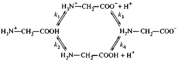
where k1, k2, k3, and k4 are microscopic dissociation constants related by the thermodynamic equation:
k1k3 = k2k4.
However, only two ionization constants can be determined experimentally: K1 = k1 + k2 for carboxyl groups; K2 = k3k4/k3 + k4 for amino groups.
Calculations based on glycine potentiometric titration data demonstrate that depending on the ionization pathway of the molecule (varying the pH from acidic to alkaline values or vice versa), the microscopic dissociation constants of the amino group (k2 and k1) differ by two orders of magnitude. Changes in the ionization state of a particular group affect its interaction with the immediate microenvironment, altering The ratio of clathrate to hydrate structures in bound water.
The physicochemical properties and multifunctional nature of Amino acids are essential for executing numerous regulatory Functions in living organisms. Individual amino acids, their derivatives (peptides), and metabolic products (ammonia, urea, aromatic amines) serve notably as nerve terminal Neurotransmitters, environmental signaling molecules, inhibitors of specific biochemical reactions, adaptogens, and precursors for the synthesis of Neuropeptides and Hormones (Krichevskaya et al., 1983). Blood Plasma acts as their reservoir, ensuring the Transport of Amino acids to specific target Organs.
The free amino acid pool in the cells of living organisms exhibits evolutionary, organ-specific, and tissue-specific patterns. For instance, the amino acid profile of the Brain differs significantly from that of other organs and Tissues due to an excess of dicarboxylic acids and their amides, which account for two-thirds of the total amino acid content in the brains of all animal species. Glutamine, asparagine, and their residues within peptide chains undergo non-Enzymatic Hydrolysis in the Organism to yield the corresponding dicarboxylic acids. In this context, it is noteworthy that the proteins of young cells exhibit a significantly higher degree of amidation than those of Aging cells (Pushkina, 1977).
Four aromatic amino acids—Histidine, tyrosine, phenylalanine, and tryptophan—possess enhanced chemical reactivity in their side chains. This reactivity is governed by a system of conjugated bonds and delocalized electrons, as well as the capacity of these groups to participate in nucleophilic and electrophilic substitution reactions. Aromatic amino acids form the foundation of numerous biologically active derivatives, including hormones, neurotransmitters, and Coenzymes.

Fig. 2. PATHWAYS OF AMINO acid transformation into molecular regulators of biological functions.
A — Tryptophan conversion: serotonin is a neurotransmitter that excites postganglionic nerve fibers; melatonin is a pineal hormone; mexamine is a radioprotector. B — Histidine conversion into histamine, a biologically active amine with hormonal and neurotransmitter functions.
Figure 2 illustrates Examples of such transformations for tryptophan and histidine.
Tryptophan is an essential amino acid synthesized naturally by microorganisms. The daily human requirement for tryptophan is 250 mg, and tryptophan deficiency is tolerated much less easily than starvation. Tryptophan itself exhibits a wide spectrum of physiological activity, exerting a beneficial effect on Lipid Metabolism and hepatic Protein Synthesis. At low doses, tryptophan displays a hypoglycemic effect, whereas high doses reverse this outcome. For instance, the diabetogenic effect of tryptophan was observed in rats fed a dose of 2.5 g per kg of body weight, which exceeds the normal daily requirement by 700 times (Rudzit, 1981). As we will discuss later, several gastrointestinal (GI) Peptide Hormones that stimulate Insulin secretion contain tryptophan within their active determinants (Appendix, Table II). It can be hypothesized that free tryptophan at ultra-high doses acts as a competitive antagonist of these hormones, inhibiting specific receptors On the surface of insulin-secreting cells.
Histidine is likewise an essential amino acid. Its regulatory functions are determined by The chemical properties of its imidazole side chain. Specifically, this group participates in redox reactions and is capable of forming coordination bonds with transition metals. Free histidine is present in tissues at very low concentrations. At the same time, owing to the donor-acceptor properties of its imidazole group, it constitutes an integral part of the catalytic (active) sites of numerous Enzymes (Ribonuclease, Chymotrypsin, convertase) and regulatory peptides (carnosine, histatin, neurokinins). The decarboxylation of histidine yields histamine, a neurotransmitter that regulates vascular tone, capillary permeability, and allergic reactions. As a neurotransmitter, histamine acts through three distinct classes of cellular receptors, including those in the brain.
In other words, the diverse regulatory repertoire of amino acids and their derivatives is underpinned by their physicochemical multifunctionality and participation in reversible biochemical reactions.
As noted previously, in an aqueous environment amino acids exist as zwitterions, making The formation of a peptide bond In aqueous solutions thermodynamically unfavorable. Peptide Synthesis is therefore conventionally carried out in organic solvents following the temporary protection of functional groups that do not participate in peptide bond formation. Depending on the length and compositional profile of the polypeptide, the optimal synthetic strategy is selected—whether solution-phase, solid-phase, or recombinant (Andersson et al., 2000).
Peptide bond formation can be represented by the following scheme:
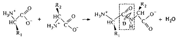
In the resulting dipeptide, the lone electron pair of the nitrogen atom is conjugated with the double bond of the carbonyl group, imparting partial double-bond character to the C—N bond and restricting rotation around it. The peptide bond possesses a constant dipole moment D. The side chains R1 and R2 dictate the combination of physicochemical properties characteristic of the constituent individual amino acids. As is well established, barring any bond Cleavage at the asymmetric carbon atom, the molecular configuration remains intact during these transformations. Consequently, the Fundamental properties of the parent amino acids are preserved upon peptide bond formation: 1) optical activity, arising from the chirality of the amino acid residues; and 2) the capacity to participate in intermolecular hydrogen bonding.
Nevertheless, when amino acids assemble into a peptide molecule, each unique Amino Acid Sequence gives rise to a specific hydrophilic-hydrophobic balance of the peptide side chains (Alberts et al., 1994).
The spatial distribution of opposing electrical charges, combined with a peptide bond dipole moment of 3.5 D, endows the peptide with a permanent dipole moment and high polarizability. Owing to these acquired properties, dipeptides exhibit a broader spectrum of energy states than individual amino acids, although this is offset by a reduction in the spatial degrees of freedom of the system. In solution, each simple molecule possesses 6 degrees of motion: 3 rotational and 3 translational. Two non-interacting amino acids thus possess 12 degrees of freedom; upon their linkage into a single dipeptide, the number of degrees of freedom decreases to 6. From the standpoint of statistical Thermodynamics, this is equivalent to an increase in system order and a corresponding decrease in Entropy.
It is worth dwelling separately on the fundamentally novel quality that amino acids acquire upon assembling into a polypeptide chain: the complementarity of dynamic conformations.
The General concept of complementarity traces its roots back to early natural philosophy. During The Development of alchemy, two descriptive principles emerged: “like tends to like” and “opposites attract.” The former was grounded in the practice of substance Separation and purification, whereas the latter derived from chemical transformations in which elements with contrasting qualities interact and Complement one another. In modern structural science, THE PRINCIPLE OF “like tends to like” is embodied in The Theory of hydrophilic-hydrophobic interactions, whereas complementarity is exemplified by the attraction of positive and negative charges (in electrostatics), the alignment of convex-concave surfaces, and the mutual correspondence between an object and its mirror reflection (matrix complementarity in early printing technology). In all these instances, the “opposites” are treated as invariant entities.
Biological macromolecules, particularly peptides, maintain the complementarity of intermolecular interactions across a fairly wide range of conformations. This ability underpins the catalytic activity of enzymes, and the “lock-and-key” model of complementary interaction was first applied at the dawn of enzymology. Later, this same model was used to discuss the Specificity of antigen–antibody binding and the selectivity of receptor–Ligand interactions.
In The Study of nucleic acids, the model of template nucleotide-pair complementarity was first employed. It proved that these macromolecules govern the precise Replication of their own structure, since the initial macromolecule serves as a template for the Formation of the next one.
The principle of template complementarity is remarkably simple and elegant. It dictates that every element of an ordered molecular construct (an electrostatic charge, a proton-donor or hydrophobic group) is copied in the exact same sequence onto another macromolecule, whose elements are complementary to those of the first.
Upon closer examination, template complementarity was also discovered in peptide synthesis, the formation of phospholipid bilayer membranes, and the self-assembly of polymers (Conjugated oligomers..., 1998).
Last update: 06/08/2026
Editorial and Educational Adaptation: This material has been compiled based on the primary/original source text. The project team performed an editorial review, corrected technical inaccuracies, structured sections, and adapted the content for an educational format.
What was processed:
- elimination of formatting defects (OCR errors, structural breaks, corrupted characters);
- editorial organization of content;
- standardization of terminology in accordance with academic sources;
- verification of factual statements against the original source text.
All mentions of the author, publication year, and origin of the primary text have been preserved in accordance with the source.