LEHNINGER PRINCIPLES OF BIOCHEMISTRY - VOL. 2. BIOENERGETICS AND METABOLISM - 2014
PART II. BIOENERGETICS AND METABOLISM
13. BIOENERGETICS AND BIOCHEMICAL REACTION TYPES
13.3. Phosphoryl Group Transfers and ATP
Having examined some of the fundamental principles of energy changes in chemical systems, we can now look at the cellular energy cycle and the special role of ATP as the energy currency linking Catabolic and anabolic processes (see Figs. 1–28, Vol. 1). Heterotrophic Cells obtain Free energy in chemical form through the Catabolism of nutrient molecules and use this energy to synthesize ATP from ADP and inorganic phosphate (Pi). ATP then donates some of its chemical energy in endergonic processes, such as the synthesis of metabolic intermediates and macromolecules from smaller precursors, the active Transport of substances across membranes against concentration gradients, and molecular motility. This energy transfer from ATP ultimately results in The breakdown of ATP to ADP and Pi, or in some cases to AMP and 2Pi. Here we discuss the chemical basis for the large free-energy changes associated with the Hydrolysis of ATP and other high-energy phosphate compounds, and we will show that most ATP-driven energy-transducing reactions involve group transfers rather than simple ATP hydrolysis. To illustrate the energy conversions powered by ATP, we will consider the synthesis of informational macromolecules, transmembrane solute transport, and Muscle contraction.
The Free-Energy Change for ATP Hydrolysis Is a Large Negative Number
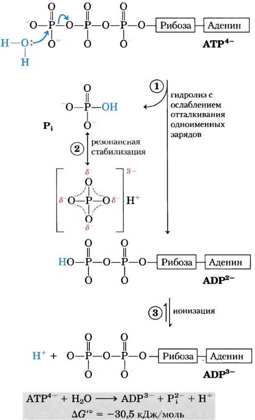
Why is the Standard Free Energy of ATP hydrolysis so negative? Let us examine the chemical basis of this process using Figure 13.11. Hydrolytic Cleavage of the terminal phosphate group, breaking a phosphoanhydride bond in ATP, relieves some of the electrostatic repulsion among the four negative charges on the ATP molecule. The released Pi (HPO42-) is stabilized by The formation of Resonance structures that are not possible in ATP. ADP2-, the other direct product of ATP hydrolysis, immediately ionizes, releasing hydrogen ions into the medium at a very low concentration, [H+] ≈ 10-7 M. The concentrations of the direct products of ATP hydrolysis within The Cell are much lower than their concentrations in an equilibrium system (Table 13-5); thus, According to the mass action ratio, cellular conditions strongly favor the hydrolysis of ATP.
Class="center">Figure 13-11. Chemical basis for the large free-energy change accompanying ATP hydrolysis. (1) Cleavage of the terminal phosphoanhydride bond during hydrolysis relieves electrostatic strain in the tetravalent ATP molecule with its four negative charges. (2) The reaction product, inorganic phosphate (Pi), is stabilized by the formation of a hybrid resonance Structure in which each of the four phosphorus-oxygen bonds acquires a degree of double-bond character, and no single oxygen atom is permanently bound to a hydrogen ion. (Resonance stabilization also occurs in phosphates involved in ester or anhydride linkages, but fewer resonance forms are available to them than to Pi.) (3) The ADP2- product immediately ionizes, releasing protons into the medium at a very low concentration (pH 7). A fourth factor (not shown in the figure) that favors ATP hydrolysis is the greater degree of solvation (in this case, Hydration) of the Pi and ADP products compared with the reactant ATP, which renders the products more stable than the reactants.
The standard free-energy change for ATP hydrolysis is -30.5 kJ/mol, but in living cells this parameter differs substantially from this value because cellular concentrations of ATP, ADP, and Pi are not equal to each other and are far lower than the standard concentration of 1.0 M (Table 13-5). Moreover, in the Cytosol, Mg2+ ions associate with ATP and ADP (Fig. 13-12), and in most enzymatic reactions where ATP acts as a phosphoryl group donor, the active species is actually the magnesium-ATP complex, MgATP2-. Therefore, it is more accurate to refer to the ∆G'° of MgATP2- hydrolysis. Box 13-1 details the calculation of ∆G for ATP hydrolysis in intact erythrocytes using the data from Table 13-5. In intact cells, the ∆G of ATP hydrolysis is commonly denoted as ∆Gp and is referred to as the phosphorylation potential.
Table 13-5. Concentrations of Adenine NUCLEOTIDES, Inorganic Phosphates, and Creatine phosphate in Some Cells
Cells |
Concentration (mM) |
||||||||||
ATP |
ADP6 |
AMP |
Pi |
PCr |
|||||||
Rat hepatocytes |
3.38 |
1.32 |
0.29 |
4.8 |
0 |
||||||
Rat myocytes |
8.05 |
0.93 |
0.04 |
8.05 |
28 |
||||||
Rat Neurons |
2.59 |
0.73 |
0.06 |
2.72 |
4.7 |
||||||
Human erythrocytes |
2.25 |
0.25 |
0.02 |
1.65 |
0 |
||||||
E. coli cells |
7.90 |
1.04 |
0.82 |
7.9 |
0 |
||||||
a For erythrocytes, cytosolic concentrations are given (human erythrocytes lack both nuclei and Mitochondria). In all other cases, total cellular content is meant; cytosolic ADP concentration differs significantly. PCr = phosphocreatine (discussed on p. 33).
b Total ADP concentration is given; the concentration of free ADP may be considerably lower (see pp. 30–31).
Because the concentrations of ATP, ADP, and Pi vary among different cell types (see Table 13-5), The values of ∆Gp for ATP hydrolysis also differ. Furthermore, within any given cell, ∆Gp can fluctuate over time depending on metabolic conditions influenced by the concentrations of ATP, ADP, Pi, and H+ (pH). We can calculate ∆Gp for any metabolic reaction in the cell provided that the concentrations of all reactants and products are known, along with other parameters such as pH, Temperature, and Mg2+ ion concentration.
These calculations are complicated by the fact that the total concentrations of ATP, ADP, Pi, and H+ can be significantly higher than the corresponding free concentrations that enter into thermodynamic calculations, as a fraction of ATP, ADP, and Pi is bound to cellular Proteins. For instance, according to various estimates, the concentration of free ADP molecules in relaxed muscle ranges from 1 to 37 µM. In Example 13-2, using a concentration of 25 µM yields ∆Gp = -64 kJ/mol. While this particular calculation of ∆Gp may not be entirely definitive, we must remember our overarching Conclusion regarding free-energy changes: in vivo, The energy released by ATP hydrolysis exceeds the standard free-energy change ∆G'°.
Example 13-2. Calculation of ∆Gp
Calculate the Free energy of ATP hydrolysis (∆Gp) in human erythrocytes, given that the standard free energy of ATP hydrolysis is -30.5 kJ/mol, the intracellular concentrations of ATP, ADP, and Pi in erythrocytes are as listed in Table 13-5, the pH is 7.0, and the body temperature is 37 °C. What can be inferred about The amount of energy required to synthesize ATP under these conditions?
Solution. In human erythrocytes, the concentrations of ATP, ADP, and Pi are 2.25 mM, 0.25 mM, and 1.65 mM, respectively. The free energy of ATP hydrolysis in erythrocytes under standard conditions (see Equation 13-4)
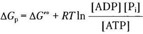
Substituting the numerical values, we obtain

(Note that the answer is rounded—52.5 was rounded down to 52 following the rule of rounding the digit 5 to the nearest lower rather than higher integer to avoid overestimating the values.) Thus, in intact erythrocytes, the ∆Gp of ATP hydrolysis (-52 kJ/mol) is substantially more negative than ∆G'° = -30.5 kJ/mol.
Consequently, the synthesis of ATP from ADP and Pi in erythrocytes requires 52 kJ/mol of energy.
In subsequent discussions of ATP hydrolysis, we will use the standard value ∆G'°, as this allows for comparison of energetics across different cellular reactions. However, one should bear in mind that in the living cell, the actual ∆G of ATP hydrolysis—as well as that of many other reactions—may deviate significantly from ∆G'°.
An important point should be made here regarding cellular ATP content. We have shown (and will discuss further below) which Chemical properties of ATP make this molecule a convenient energy currency for the cell. However, ATP drives metabolic reactions and other energy-demanding processes not merely through its chemical properties. More importantly, evolution has shaped regulatory mechanisms that maintain intracellular ATP concentrations significantly higher than the equilibrium concentrations of its hydrolysis reactions. When ATP levels drop, not only does the fuel supply decrease, but the fuel itself loses its driving force: the hydrolysis ∆G of this molecule drops (i.e., the phosphorylation potential, ∆Gр). As we will see when examining metabolic processes involving ATP synthesis and consumption, living cells have developed sophisticated mechanisms (which might at first glance seem inefficient and counterintuitive) to maintain high ATP concentrations.
The standard free energies of other phosphorylated compounds and thioesters are also large
Phosphoenolpyruvate (PEP; Fig. 13-13) contains a phosphoester bond that undergoes hydrolysis to yield the enol form of Pyruvate, and this direct reaction product can immediately tautomerize into the more stable keto form. Because the starting PEP exists in only one form
(the enol form), whereas the product (pyruvate) can exist in two forms (enol and keto), the reaction product is more stable than the reactant. This is the primary reason for the large standard free energy of hydrolysis of phosphoenolpyruvate: ∆G′° = -61.9 kJ/mol.
Fig. 13-13. Hydrolysis of phosphoenolpyruvate (PEP). The reaction is catalyzed by pyruvate kinase and is followed by the tautomerization of pyruvate. Tautomerization of PEP is impossible, making the hydrolysis products more stable than the reactants. As shown in Fig. 13-11, resonance stabilization of inorganic phosphate also contributes

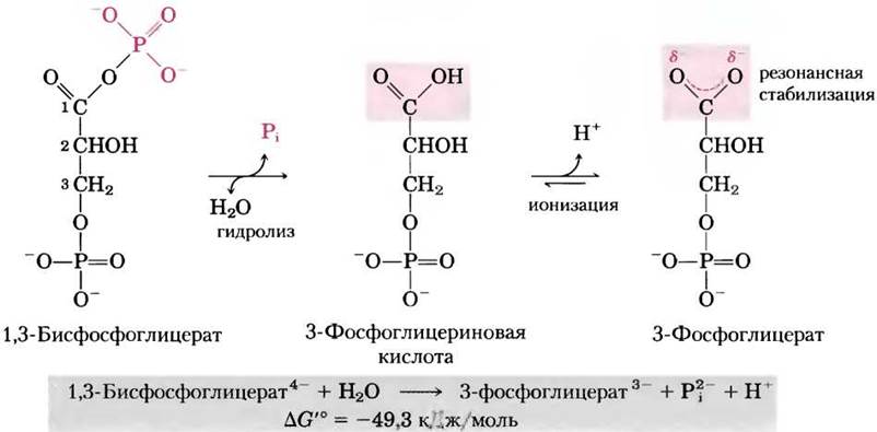
Another 3-carbon compound, 1,3-bisphosphoglycerate (Fig. 13-14), contains an anhydride bond between the C-1 carboxyl group and a phosphate group. The hydrolysis of this acyl phosphate is accompanied by a large negative change in standard free energy (∆G′° = -49.3 kJ/mol), which can be explained by the Structural Features of the reactant and products. When Water attacks the anhydride bond of 1,3-bisphosphoglycerate, one of the direct products, 3-phosphoglyceric acid, can rapidly lose a proton to form 3-phosphoglycerate (a carboxylate ion) with two equivalent resonance structures (Fig. 13-14). The forward reaction is favored by the removal of the direct reaction product (3-phosphoglyceric acid) and the formation of a resonance-stabilized ion.
Fig. 13-14. Hydrolysis of 1,3-bisphosphoglycerate. The direct product of hydrolysis is 3-phosphoglyceric acid, which contains an un ionized carboxyl group that, however, dissociates immediately. Due to ionization and resonance stabilization, the reaction product is more stable than the reactants. In addition, the negative free-energy change is enhanced by the resonance stabilization of Pi.
In creatine phosphate (Fig. 13-15), the P—N bond can undergo hydrolysis to yield free creatine and Pi. The release of Pi and the resonance stabilization of creatine drive the reaction forward. The standard free-energy change for creatine phosphate hydrolysis is likewise large (-43.0 kJ/mol).
Fig. 13-15. Hydrolysis of creatine phosphate. Cleavage of the P-N bond in creatine phosphate yields creatine, which is stabilized by a hybrid resonance structure. The other reaction product, Pi, is also resonance-stabilized.

In all these reactions involving phosphate release, the formation of multiple resonance forms of Pi increases the Stability of the product compared to the reactant, which is a major factor contributing to the large negative free-energy change. Table 13-6 lists the standard free energies of hydrolysis for several phosphorylated compounds.
Table 13-6. Standard Free Energies of Hydrolysis of Some Phosphorylated Compounds and Acetyl-CoA (a Thioester)
∆G′° |
||
kJ/mol |
kcal/mol |
|
Phosphoenolpyruvate |
-61.9 |
14.8 |
1,3-Bisphosphoglycerate (—> 3-phosphoglycerate + Pi) |
-49.3 |
-11.8 |
Creatine phosphate |
-43.0 |
-10.3 |
ADP (—> AMP + Pi) |
-32.8 |
-7.8 |
ATP (—> ADP + Pi) |
-30.5 |
-7.3 |
ATP (—> AMP + PPi) |
-45.6 |
-10.9 |
AMP (—> adenosine + Pi) |
-14.2 |
-3.4 |
PPi (—> 2 Pi) |
-19.2 |
-10 |
Glucose 1-phosphate |
-20.9 |
-5.0 |
Fructose 6-phosphate |
-15.9 |
-3.8 |
Glucose 6-phosphate |
-13.8 |
-3.3 |
Glycerol 1-phosphate |
-9.2 |
-2.2 |
Acetyl-CoA |
-31.4 |
-7.5 |
Thioesters, in which sulfur replaces the usual oxygen in the ester linkage, also exhibit a large negative standard free energy of hydrolysis. Acetyl-coenzyme A, or acetyl-CoA (Fig. 13-16), is one such thioester playing a vital role in METABOLISM. The acyl group in these compounds is activated in transacylation, Condensation, or oxidation-reduction reactions. Thioesters undergo significantly less resonance stabilization than oxygen esters. Consequently, the difference in free energy between reactants and resonance-stabilized products is even greater for thioesters than for analogous oxygen esters (Fig. 13-17). In both cases, ester hydrolysis yields a carboxylic acid, which can ionize and form multiple resonance structures. Together, these two factors account for the large negative ∆G′° (-31.4 kJ/mol) for acetyl-CoA hydrolysis.
Fig. 13-16. Hydrolysis of acetyl-coenzyme A. The hydrolysis of the thioester acetyl-CoA is characterized by a large negative standard free energy. In thioesters, sulfur takes THE PLACE OF oxygen in the ester bond. The complete structural formula of coenzyme A (CoA or CoASH) is given in Fig. 8-41, Vol. 1.
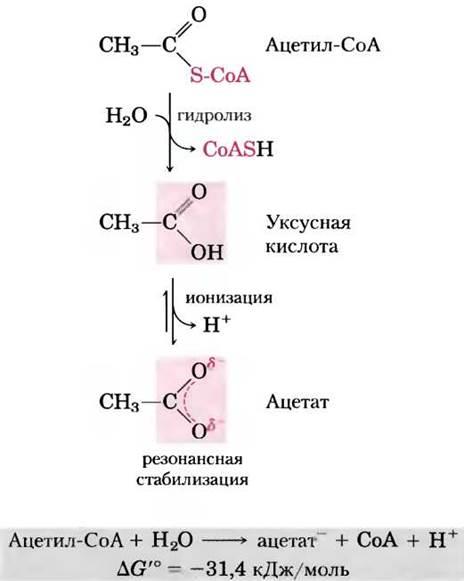
Fig. 13-17. Free energy of hydrolysis of esters and thioesters. The hydrolysis products of these esters have similar free energies (G), but the free energy of a thioester is higher than that of an oxygen ester. Partial orbital overlap between O and C provides resonance stabilization in oxygen esters, whereas orbital overlap between S and C is poor, resulting in negligible resonance stabilization in thioesters.

To summarize, hydrolysis reactions characterized by large negative standard free-energy changes yield products that are more stable than the reactants. This is due to one or more of the following factors: (1) relief of electrostatic repulsion upon Charge Separation in the reactants, as in the case of ATP; (2) resonance stabilization of the reaction products through ionization, as seen with ATP, Acyl phosphates, and thioesters; (3) stabilization of products via isomerization (tautomerization), as in phosphoenolpyruvate; and/or (4) stabilization of products due to resonance structures, such as in creatine released from phosphocreatine, carboxylate ions (from acyl phosphates and thioesters), and phosphate Pi (from anhydride and ester compounds).
ATP drives energy-requiring processes through group transfers, not simple hydrolysis
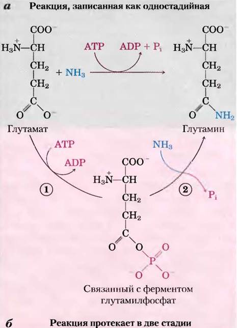
Throughout this book, we will encounter reactions or processes that utilize ATP energy, and ATP involvement in these reactions is usually depicted, as in Fig. 13-18a, by a curved arrow showing The conversion of ATP to ADP and Pi (or in some cases ATP to AMP and pyrophosphate PPi). Such representations can make the reactions appear to be simple hydrolyses in which water displaces Pi (or PPi), tempting one to say that ATP-dependent reactions "are driven by ATP hydrolysis." In reality, this is not the case. ATP hydrolysis typically results in nothing more than heat dissipation, which cannot drive a chemical process in an isothermal system. The single arrow in the reaction schemes of Fig. 13-18a and elsewhere almost always denotes a two-stage process (Fig. 13-18b) in which a portion of the ATP molecule—specifically a phosphoryl, pyrophosphoryl, or adenylate group (AMP)—is first transferred to a substrate molecule or an amino acid residue within an enzyme, forming a covalent linkage and raising its free energy. In the second stage, this phosphate-containing moiety is replaced to yield Pi, PPi, or AMP. Thus, ATP participates covalently in the enzymatic reaction by supplying free energy.
Fig. 13-18. Two stages of ATP hydrolysis. (a) ATP participation in reactions is often depicted as a single step, but the process is almost invariably two-stage, (b) Reaction catalyzed by ATP-dependent Glutamine Synthetase. (1) A phosphoryl group from ATP is transferred to glutamate, after which (2) the phosphoryl group is replaced by NH3 and released as Pi.
However, some processes indeed involve the direct hydrolysis of ATP (or GTP). For instance, the non-covalent binding of ATP (or GTP) followed by hydrolysis to ADP (or GDP) and Pi can provide the energy required for the conformational changes of certain proteins involved in mechanical movement. This occurs During muscle contraction, as well as when Enzymes translocate along DNA or Messenger RNA shifts relative to the ribosome. The direct hydrolysis of phosphoanhydride bonds also powers Reactions Catalyzed by helicases, RecA proteins, and certain topoisomerases (Chapter 25). GTP-binding proteins acting in signaling pathways directly hydrolyze GTP, triggering conformational changes that terminate signals initiated by Hormones or other extracellular agents (Chapter 12).
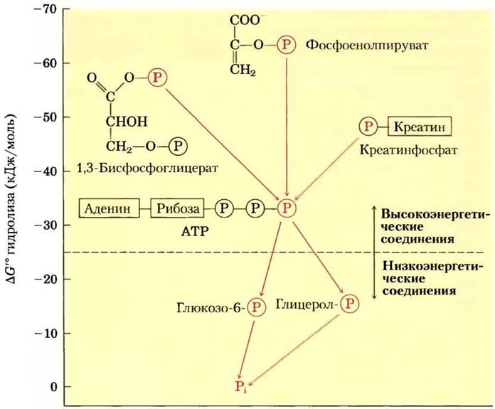
Based on the magnitude of the standard free energy of hydrolysis, phosphate compounds found in living organisms can be arbitrarily divided into two groups (Fig. 13-19). "High-energy" compounds have a hydrolysis $\Delta G'^0$ of less than -25 kJ/mol; "low-energy" compounds have a higher $\Delta G'^0$. By this criterion, ATP (hydrolysis $\Delta G'^0 = -30.5$ kJ/mol $=-7.3$ kcal/mol) is a high-energy compound, whereas glucose 6-phosphate ($\Delta G'^0 = -13.8$ kJ/mol $=-3.3$ kcal/mol) is a low-energy compound.
Fig. 13-19. Evaluation of biological phosphate compounds based on their standard free energy of hydrolysis. The transfer of phosphoryl groups, designated by  from high-energy phosphoryl Donors via ATP to acceptor molecules (such as glucose and glycerol) to form their low-energy phosphate derivatives is shown. Under intracellular conditions, this enzyme-catalyzed phosphoryl transfer mediated by Kinases proceeds with a net loss of free energy at each step. The hydrolysis of low-energy phosphate compounds releases Pi, which has an even lower phosphoryl group transfer potential (see text).
The term "high-energy phosphate bond" has long been used by biochemists to describe the P–O bond cleaved during hydrolysis; however, this is somewhat misleading, as it may incorrectly imply that the energy resides within the bond itself. In reality, breaking any chemical bond requires an input of energy. The free energy released during the hydrolysis of phosphate compounds stems not from the rupture of a specific bond, but from the fact that the reaction products possess a lower free energy than the reactants. For simplicity, we will occasionally use the term "high-energy phosphate compound" when referring to ATP or other phosphate compounds with a large negative standard free energy of hydrolysis.
Because the Free Energy Changes of consecutive reactions are additive, any phosphorylated compound can be synthesized by coupling its formation to the breakdown of another phosphorylated compound that has a lower negative free energy of hydrolysis. For example, because the cleavage of Pi from phosphoenolpyruvate (PEP) releases more energy than is required to drive the condensation of Pi with ADP, a direct phosphoryl transfer from PEP to ADP is thermodynamically feasible:
$\Delta G'^0$, (kJ/mol)
(1) PEP + H2O —> pyruvate + Pi -61.9
(2) ADP + Pi —> ATP + H2O +30.5
Net: PEP + ADP —> pyruvate + ATP -31.4
Note that although the overall reaction is presented as the algebraic sum of the first two reactions, it is essentially a third, distinct reaction that does not involve free Pi; PEP transfers its phosphoryl group directly to ADP. Based on their standard free energies of hydrolysis, we can describe phosphorylated compounds as having either a high or low phosphoryl group transfer potential (Table 13-6). Phosphoenolpyruvate has a very high phosphoryl group transfer potential, ATP has a high potential, and glucose 6-phosphate has a low potential (Fig. 13-19).
Although many catabolic processes are directed toward the synthesis of high-energy phosphate compounds, their formation is not an end in itself. These compounds merely serve as a means to activate A wide variety of substances for subsequent chemical transformations. Phosphoryl group transfer effectively delivers the free energy required to drive downstream metabolic conversions. As described above, the Synthesis of glucose 6-phosphate involves phosphoryl transfer from ATP. The following chapter will demonstrate how the phosphorylation of glucose activates the molecule—effectively "fueling" it for the catabolic reactions that take place in every living cell. Occupying an intermediate position on the group transfer potential scale, ATP can channel energy from high-energy phosphate compounds generated during catabolism to molecules like glucose, converting them into more reactive forms. Thus, ATP acts as the universal energy currency in all living cells.
ATP has another feature that is crucial for its metabolic role: although thermodynamically unstable In aqueous solutions—making it an effective phosphoryl group donor—it is kinetically stable. Due to the very high activation energy (ranging from 200 to 400 kJ/mol) required for the non-Enzymatic cleavage of its phosphoanhydride bonds, ATP cannot spontaneously transfer phosphoryl groups to water or to the hundreds of other potential acceptors present within the cell. Phosphoryl group transfer from ATP occurs exclusively in the presence of specific enzymes that lower this activation energy. Consequently, the cell is able to control the distribution of energy carried by ATP molecules by regulating the enzymes that act upon them.
ATP donates phosphoryl, pyrophosphoryl, and adenylyl groups
Reactions involving ATP are typically SN2 Nucleophilic substitution reactions (see Section 13-2), in which the nucleophile may be, for example, the oxygen atom of an alcohol or carboxylate, or the nitrogen atom of creatine, an Arginine side chain, or Histidine. Each of the three phosphorus atoms of ATP is susceptible to nucleophilic attack (Fig. 13-20), yielding different reaction products.
Nucleophilic attack by an alcohol on the y-phosphate (Fig. 13-20, a) displaces ADP and results in the formation of a new phosphoester. Experiments with 18O-labeled reactants have demonstrated that the oxygen atom in the ester linkage of the new compound originates from the alcohol rather than from ATP; consequently, ATP transfers a phosphoryl group (-PO2-3) rather than a phosphate group (-OPO2-3). During the transfer of a phosphoryl group from ATP to glutamate (Fig. 13-18) or glucose (Vol. 1, p. 308), the attack occurs at the y-position of the ATP molecule.
Fig. 13-20. Nucleophilic substitution reactions of ATP. Any of the three phosphorus atoms (α, β, or y) can serve as an electrophilic target for nucleophilic attack—in this case, by a labeled nucleophile R-18O. Potential nucleophiles include an alcohol (ROH), a carboxyl group (RCOO-), or a phosphoanhydride (e.g., a nucleoside monophosphate or nucleoside diphosphate). a — when the oxygen of the nucleophile attacks the y-position, the labeled oxygen ends up in the ester bond of the product. This indicates that ATP transfers a phosphoryl group (-PO2-3) rather than a phosphate group (-OPO2-3). b — attack at the β-position displaces AMP and leads to the transfer of a pyrophosphoryl (not pyrophosphate) group to the nucleophile. c — attack at the α-position displaces PPi and transfers an adenylyl group to the nucleophile. The three sites on the ATP molecule for nucleophilic attack by R18O
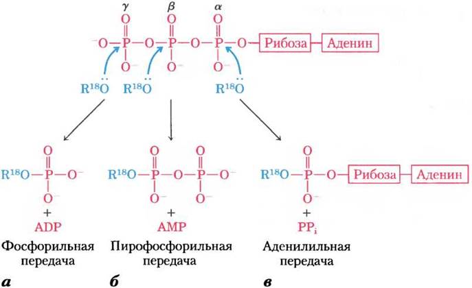
Attack on the β-phosphate of ATP results in the displacement of AMP and the transfer of a pyrophosphoryl (rather than pyrophosphate) group to the attacking nucleophile (Fig. 13-20, b). For example, the formation of 5'-phosphoribosyl-1-pyrophosphate (p. 861), a key intermediate in nucleotide synthesis, occurs via the attack of the ribose -OH group on the β-phosphate.
Nucleophilic attack at the α-position of the ATP molecule causes the displacement of PPi and the transfer of adenylate (5'-AMP) as an adenylyl group (Fig. 13-20, c). This reaction is known as adenylylation (arguably the clumsiest term in biochemical nomenclature). Note that the hydrolysis of the α-β-phosphoanhydride bond releases considerably more energy (~ 46 kJ/mol) than the hydrolysis of the β-γ-bond (-31 kJ/mol) (Table 13-6). Furthermore, the byproduct of adenylylation, inorganic pyrophosphate PPi, is subsequently hydrolyzed by the ubiquitous enzyme inorganic pyrophosphatase to yield two molecules of inorganic orthophosphate Pi. This releases 19 kJ/mol of energy, providing an additional thermodynamic push for the adenylylation reaction. As a result of this overall reaction, both phosphoanhydride bonds of ATP are cleaved. Therefore, adenylylation reactions are thermodynamically very favorable. Whenever ATP Energy is required to drive particularly unfavorable metabolic reactions, adenylylation is frequently employed as an energy-coupling mechanism. Fatty acid activation serves as an excellent example of this type of energy coupling.
The First stage in the Activation of a fatty acid—necessary either for energy-yielding oxidation or for the synthesis of more Complex Lipids—is the formation of a thioester (see Fig. 17-5). Direct condensation of a fatty acid with coenzyme A is an endergonic reaction; however, through the stepwise removal of two phosphoryl groups from ATP, the formation of a fatty acyl-CoA derivative becomes an exergonic process. First, adenylate (AMP) is transferred from ATP to the carboxyl group of the fatty acid to form a mixed anhydride (a fatty acid adenylate derivative) with the release of PPi. Then, the thiol group of coenzyme A displaces the adenylate group to form a thioester with the fatty acid. The sum of these two reactions is energetically equivalent to the exergonic hydrolysis of ATP to AMP and PPi (ΔG'° = -45.6 kJ/mol) and the endergonic Formation of the fatty acyl-CoA derivative (ΔG'° = 31.4 kJ/mol). The formation of the fatty acyl-CoA derivative is rendered thermodynamically favorable owing to the hydrolysis of PPi by inorganic pyrophosphatase. Thus, fatty acid activation results in the cleavage of both phosphoanhydride bonds of ATP. Consequently, ΔG'° is the sum of the ΔG'° values for the cleavage of these bonds, i.e., -45.6 kJ/mol + (-19.2) kJ/mol:
ATP + 2H2O —> AMP + 2Pi
ΔG'° = -64.8 kJ/mol
Box 13-1. ATP Provides Energy for Fireflies
Bioluminescence requires substantial amounts of energy. Fireflies utilize ATP in a series of reactions that convert chemical energy into light. In the 1950s, William McElroy and his colleagues at Johns Hopkins University isolated the key players in these energy-yielding processes from several thousand fireflies collected by children in and around Baltimore: the carboxylic acid luciferin and the enzyme luciferase. Generating a flash of light requires the activation of luciferin, which involves the pyrophosphate cleavage of ATP to form luciferyl adenylate (Fig. 1). In the presence of molecular oxygen and luciferase, luciferin undergoes a multi-step oxidative decarboxylation to (hydr)oxyluciferin, accompanied by the emission of light. The color of the light flashes varies among different firefly species, presumably due to structural differences in the luciferase enzyme. Luciferin is regenerated from hydroxyluciferin through a subsequent series of reactions.
Fig. 1. Key Components of the firefly bioluminescence cycle

In the laboratory, purified firefly luciferin and luciferase are used to measure extremely small amounts of ATP by monitoring the intensity of the generated light flash; quantities of ATP down to a few picomoles (10-12 mol) can thus be detected. In more advanced studies on luciferase, the corresponding Gene has been successfully cloned in tobacco plants. When such plants are watered with a solution containing luciferin, they glow in the dark (see Fig. 9-29).
The activation of Amino Acids prior to their polymerization into proteins (see Fig. 27-19) proceeds via an analogous set of reactions, except that a Transfer RNA molecule replaces coenzyme A. An interesting utilization of ATP cleavage to AMP and PPi occurs in fireflies, which employ ATP as an energy source to generate light flashes (Box 13-1).
Energy is Required for the Assembly of Information Macromolecules
During the assembly of macromolecules such as DNA, RNA, and proteins (i.e., Biopolymers) from simple precursor molecules, as discussed in detail in Volume 3, energy is required both for the condensation of monomeric units and for the creation of ordered sequences. Nucleoside triphosphates serve as precursors for DNA and RNA in Biosynthesis; this polymerization reaction is accompanied by the cleavage of the phosphoanhydride bond between the α- and β-phosphates, releasing PPi (Fig. 13-21). In RNA Synthesis, the residues transferred to the growing biopolymer are adenylate (AMP), guanylate (GMP), cytidylate (CMP), or uridylate (UMP), whereas in DNA Synthesis, their deoxy analogues are used (with TMP replacing UMP). As noted above, the Amino Acid Activation required for Protein Synthesis involves the transfer of adenylate groups from ATP; furthermore, as we will see in Chapter 27, these several Stages of Protein Synthesis are also accompanied by GTP hydrolysis. In all these cases, the exergonic breakdown of a nucleoside triphosphate is coupled with the endergonic synthesis of a sequence-specific biopolymer.
ATP Provides Energy for Active Transport and Muscle Contraction
ATP can supply energy for The transport of an ion (or molecule) across a membrane into another aqueous compartment where its concentration is higher (see Fig. 11-38). Processes associated with substance transport are major energy consumers. For example, in the human Kidneys and Brain, two-thirds of the resting energy consumption is devoted to pumping Na+ and K+ across Plasma Membranes via the Na+/K+-ATPase. The transport of Na+ and K+ is coupled with the cyclic phosphorylation and dephosphorylation of the transport protein, with ATP acting as the phosphoryl group donor (see Fig. 11-37). Na+-dependent phosphorylation of the Na+/K+-ATPase induces a conformational change in the protein, whereas K+-dependent dephosphorylation restores it to its initial state. Each cycle of the transport process is accompanied by the cleavage of ATP to ADP and Pi, wherein The change in the free energy of ATP hydrolysis drives cyclic Conformational Changes in the protein, resulting in the electrogenic pumping of Na+ and K+ ions. Note that in this case, the phosphoryl group from ATP is transferred to the enzyme molecule rather than to a substrate.
In the Cells of the Skeletal Muscle contractile system, Myosin and Actin are specialized for converting the chemical energy of ATP into mechanical energy (motion) (see Fig. 5-31). ATP binds tightly (via noncovalent bonds) to one of the Conformations of myosin, holding the protein in that state. When myosin catalyzes the hydrolysis of the bound ATP, ADP and Pi are released from the protein; this once again triggers a conformational change in the protein molecule, which persists until another ATP molecule binds. The binding and subsequent hydrolysis of ATP (mediated by myosin ATPase) provides the energy that drives cyclic conformational Changes in the myosin HEAD. Alterations in the conformation of many individual myosin molecules cause myosin fibrils to slide along actin filaments (see Fig. 5-30), which ultimately produces the macroscopic contraction of the muscle fiber.
As noted earlier, mechanical movement driven by ATP consumption is one of the few instances where the chemical energy in the coupled process derives directly from the ATP hydrolysis reaction itself rather than from group transfer from ATP.
Transphosphorylation Between Nucleotides Occurs in All Cell Types
Although we have focused on ATP as the primary cellular energy carrier and phosphate group donor, all other nucleoside triphosphates (GTP, UTP, CTP) and deoxyribonucleoside triphosphates (dATP, dGTP, dUTP, dCTP) are energetically equivalent to ATP. The change in free energy upon the hydrolysis of the phosphoanhydride bonds in these compounds is nearly identical to the values for ATP listed in Table 13-6. To fulfill various biological Functions, these nucleotides are synthesized and maintained in their nucleoside triphosphate (NTP) forms through phosphoryl group transfer to the corresponding nucleoside diphosphates (NDPs) and monophosphates (NMPs).
ATP is a crucial high-energy phosphate compound generated during catabolic processes such as Glycolysis, Oxidative Phosphorylation, and, in photosynthetic cells, Photophosphorylation. Subsequently, specific enzymes facilitate the transfer of phosphoryl groups from ATP to other nucleotides. Nucleoside diphosphate kinases, found in all cells, catalyze the following reaction:

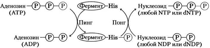
Although this reaction is reversible, the relatively high [ATP]/[ADP] ratio within normal cells drives the reaction forward from left to right, producing NTPs and dNTPs. In fact, the enzyme catalyzes a two-stage phosphoryl transfer process—a classical double-displacement mechanism (the "ping-pong" mechanism; Fig. 13-21, see also Fig. 6-13, b). First, a phosphoryl group is transferred from ATP to an active-site histidine residue of the enzyme, forming a phosphorylated enzyme intermediate; then, the phosphoryl group is transferred from this intermediate to the NDP acceptor. Because the enzyme is nonspecific for the base in the NDP and functions equally well with both dNDPs and NDPs, it can synthesize all NTPs and dNTPs from their corresponding NDPs in the presence of ATP.
Fig. 13-21. The "ping-pong" mechanism of nucleoside diphosphate kinase action. First, the enzyme binds to its substrate (ATP in this example), and a phosphoryl group from ATP is transferred to the side chain of a histidine residue. ADP is then released and replaced by another nucleoside diphosphate (or deoxynucleoside diphosphate), which is converted into the corresponding triphosphate through phosphoryl transfer from the phosphohistidine residue.
Phosphoryl group transfers from ATP lead to the accumulation of ADP. For instance, during vigorous muscle contraction, ADP accumulates, which hinders ATP-dependent contraction. During periods of acute cellular ATP demand, ADP concentrations drop while adenylate kinase activity causes ATP to accumulate.

This reaction is reversible; therefore, once the acute need for ATP subsides, the enzyme can reconvert AMP into ADP, which can subsequently be phosphorylated to ATP in the mitochondria. An analogous enzyme, guanylate kinase, converts GMP into GDP at the expense of ATP. In this manner, the energy stored during catabolic ATP generation is utilized to supply the cell with necessary amounts of NTPs and dNTPs.
Creatine phosphate serves as a readily available source of phosphoryl groups for the rapid synthesis of ATP from ADP (Fig. 13-15). The concentration of creatine phosphate (PCr) in skeletal muscle is approximately 30 mM, which is nearly 10 times higher than that of ATP. In other Tissues, such as smooth muscle, brain, and Kidney, PCr concentrations range from 5 to 10 mM. The reversible phosphoryl transfer reaction from creatine phosphate to ADP is catalyzed by creatine kinase

Creatine phosphate acts as a reservoir of phosphoryl groups whenever sudden energy demands deplete ATP reserves, allowing ATP to be regenerated much faster than via catabolic synthesis. When energy demands decrease, ATP molecules produced through catabolism are used to replenish PCr stores via the reverse creatine kinase reaction. In phylogenetically lower organisms, other PCr-like molecules, collectively termed phosphagens, serve as phosphoryl group reservoirs.
Inorganic Polyphosphates as Potential Phosphoryl Group Donors
Inorganic polyphosphates, polyP (or (polyP)n, where n is the number of orthophosphate groups), are linear polymers consisting of many tens or hundreds of Pi residues linked together by phosphoanhydride bonds. Present in all organisms, these substances can accumulate in large quantities in certain cells. For example, in Yeast, the concentration of polyP accumulated in the vacuole would amount to 20 mM if distributed evenly throughout the entire cell. (Compare with the concentrations of other phosphoryl group donors, see Table 13-5.)
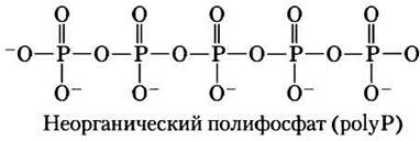
The potential role of polyP is that it can act as a phosphagen—a reservoir of phosphoryl groups for ATP production, much like creatine phosphagen functions in Muscle tissue. PolyP has a phosphoryl group transfer potential (energy) roughly comparable to that of PPi. The shortest polyphosphate, PPi (n = 2), serves as an energy source for active transport in plant vacuoles. In plants, pyrophosphate PPi acts as a phosphate group donor for at least one enzyme, Phosphofructokinase, thus playing the same role that ATP does in animals and microorganisms (p. 73). The discovery of high concentrations of polyP in volcanic condensates and steam emissions suggests that it may have served as an energy source in prebiotic and early cellular evolution.
In Bacteria, the enzyme polyphosphate kinase-1 (PPK-1) catalyzes the reversible reaction

via a mechanism involving an enzyme-bound phosphohistidine intermediate (recall The Mechanism of nucleoside diphosphate kinase described above). Another enzyme, polyphosphate kinase-2 (PPK-2), catalyzes the reversible synthesis of GTP (or ATP) from polyphosphate and GDP (or ADP)

It is believed that PPK-2 functions primarily in the direction of GTP and ATP synthesis, whereas PPK-1 acts mainly toward polyphosphate synthesis. PPK-1 and PPK-2 kinases are present in the cells of various bacteria, including many pathogenic species.
As has been demonstrated in bacteria, elevated levels of polyP stimulate the expression of several genes responsible for adapting the Organism to starvation and other life-threatening conditions. For instance, in Escherichia coli, polyP accumulates when cells experience a shortage of amino acids or Pi, and this reserve helps them survive these adverse conditions. Deletion of polyphosphate kinase genes reduces the ability of certain pathogenic bacteria to infect animal tissues. Consequently, this enzyme could serve as a vulnerable target for The Development of novel antimicrobial therapeutics.
Yeast lack the gene encoding a PPK-like protein, yet they possess four genes (unrelated to bacterial PPK genes) that are required for polyphosphate synthesis. This indicates that the mechanism of polyphosphate synthesis in eukaryotes is entirely different from that in prokaryotes.
Summary of Section 13.3 Phosphoryl Group Transfer and ATP
■ Adenosine triphosphate (ATP) links catabolic and anabolic processes. This chemical compound serves as the energy "currency" of the living cell. The exergonic conversion of ATP to ADP and Pi, or to AMP and PPi, is coupled to many endergonic reactions.
■ ATP hydrolysis provides the energy for conformational changes that result in muscle contraction. However, energy coupling between ATP breakdown and endergonic substrate conversions is typically achieved not by the hydrolysis of the ATP molecule itself, but by the transfer of a phosphoryl, pyrophosphoryl, or adenylyl group from ATP to a substrate or enzyme molecule.
■ Through these group-transfer reactions, ATP supplies energy for anabolic processes, including the synthesis of informational molecules, as well as the Transport of Molecules and ions across membranes against concentration and electrical potential gradients.
■ To maintain its capacity for phosphate group transfer, the concentration of ATP must be held far above its equilibrium concentration, a state sustained by energy-yielding catabolic reactions.
■ Cells contain other metabolites with large negative standard free energies of hydrolysis, including phosphoenolpyruvate, 1,3-bisphosphoglycerate, and creatine phosphate. Like ATP, these high-energy compounds with a high phosphoryl group transfer potential are effective phosphoryl group donors. Thioesters are also characterized by high free energies of hydrolysis.
■ Inorganic polyphosphates, present in all cells and possessing a high group transfer potential, can function as a reservoir of phosphoryl groups.
Last update: 06/08/2026
Editorial and Educational Adaptation: This material has been compiled based on the primary/original source text. The project team performed an editorial review, corrected technical inaccuracies, structured sections, and adapted the content for an educational format.
What was processed:
- elimination of formatting defects (OCR errors, structural breaks, corrupted characters);
- editorial organization of content;
- standardization of terminology in accordance with academic sources;
- verification of factual statements against the original source text.
All mentions of the author, publication year, and origin of the primary text have been preserved in accordance with the source.