Biochemistry - The Chemical Reactions of Living Cells Volume 3 - D. Metzler 1980
Light in Biology
Other Types of Light Reactions
Bioluminescence
The emission of visible light by living organisms is one of the most striking and remarkable phenomena in nature. Luminescent Bacteria and Protozoa responsible for ocean glow, glowing Mollusks, fantastically illuminated worms, and breathtaking fireflies all continually capture the attention of biochemists [162–164]. The most fascinating aspect is the chemical basis of the phenomenon. The light emitted by fireflies has a wavelength of ~560 nm (17,900 cm-1) and an energy of 214 kJ∙Einstein-1. This raises a natural question: what chemical reaction releases such a massive amount of energy? After all, it far exceeds the energy yield of ATP Hydrolysis. Even The oxidation of NADH by oxygen can hardly supply the required energy.
A key to this puzzle is provided by chemiluminescence, frequently observed when O2 is used as an oxidant in non-enzymatic processes. The slow oxidation of alcohols, aldehydes, and many nitrogenous compounds is accompanied by the emission of visible light. Chemiluminescence is most pronounced in reactions thought to involve the generation of free radicals. The recombination of these radicals releases sufficient energy to drive luminescence in the visible spectrum.
In light of all these findings, the ability of many organisms to convert The energy released during oxygenation reactions into light energy no longer seems so surprising.
Attempts to isolate light-emitting material from organisms date back to the last century, when the French physiologist R. Dubois, in 1887, obtained two extracts from luminous mollusks using cold Water for one and hot water for the other [162]. He was able to demonstrate that adding a thermostable compound extracted with hot water to the cold-water extract—which he termed luciferase—resulted in light emission. Dubois named the thermostable compound luciferin. These terms have endured and become standard. Luciferins comprise a family of compounds whose structures have now been elucidated for many Organism species (Fig. 13-30).
Firefly luciferin is a carboxylic acid that is activated in an ATP-dependent reaction, being converted to luciferyl adenylate (Fig. 13-30). In the presence of O2 and luciferase, this compound emits light. As seen in the figure, the carboxyl group is cleaved off as CO2, and the ring is converted to its oxidized form. Concurrently, the acyl-adenylate bond is cleaved. In Renilla reniformis (a coelenterate), luciferin has a completely different Structure [165]; however, much like in fireflies, light emission results from a reaction with O2 that yields CO2 and an oxidized product. In Renilla, luciferin exists as luciferyl sulfate; its putative structure is shown in Fig. 13-30. The conversion of this compound into active luciferin is accomplished by transferring a sulfuryl group to adenosine-3',5'-diphosphate to yield 3'-phosphoadenosine-5'-phosphosulfate—that is, via the reverse of step g in equation (11-4).
Another intriguing feature of bioluminescence in Renilla is the presence of a distinct green fluorescent protein in the light-emitting Cells. In the absence of this protein, blue light is emitted with vmax = 20,500 cm-1 (488 nm), whereas in its presence, light is emitted in a narrow spectral band with vmax = 19,600 cm-1 (509 nm). Apparently, this involves efficient energy transfer between the two chromophores [164].
1) The reader should bear in mind that the scientists who first investigated luciferins had very limited amounts of material at their disposal, which is why A number of errors can be found in the older literature.
Class="center">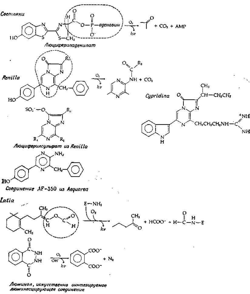
FIG. 13-30. Structure of luciferins from several luminous organisms. Formulas are given for the "activated" molecules "ready" for reaction with O2. However, the compound AF-350 is a Cleavage product of the photoprotein aequorin, which is activated by Ca2+ ions.
The luciferin of the hermit crab Cypridina is very similar in structure to that of Renilla. In Cypridina, luciferin and luciferase are synthesized in separate glands and secreted into the surrounding aqueous environment, where they mix and emit light. The light-emitting reaction is utilized quite differently by the limpet Latia. In this case, the luciferin is an unusual terpene derivative lacking any light-emitting chromophore [166]. Evidently, the oxidation of this luciferin induces electronic excitation in some other molecule—most likely a "purple protein" that is also required for luminescence. It is believed that the complex of luciferin and the purple protein reacts with luciferase (abbreviated as E—NH2 in Fig. 13-30), releasing a formyl group that was previously involved in forming an enol-ether bond. The resulting aldehyde group interacts with an amino group of the enzyme, and the resulting Schiff base further reacts with oxygen [scheme (13-40)]:

In describing the MECHANISM OF ACTION of most luciferases, it is generally assumed that O2 reacts with a carbon atom, which is subsequently incorporated into the carbonyl group of the product. For Renilla luciferin, such a process is easy to envision as the result of an Electron transfer from the pyrazine nitrogen (Fig. 13-30, bottom) to O2. In the case of firefly luciferin, a proton is apparently removed from a carbon atom prior to interaction with O2, driven by the electron-withdrawing Properties of the adjacent ring system. This reaction can be compared to those catalyzed by oxygenases [e.g., the reaction described by equation (10-50)]. According to one hypothesis, the resulting peroxide group adds to the carbonyl (or to RC = N— in Latia luciferin), generating a four-membered dioxetane ring [schemes (13-40) to (13-41)].

This ring subsequently opens via a concerted process (as indicated by the arrows) to yield the final products.
The validity of this proposed theory was tested in experiments using 18O2. In the case of Cypridina luciferin, the incorporation of a single 18O atom into the CO2 molecule was indeed observed; however, when examining firefly and Renilla luciferins, no 18O incorporation into CO2 occurred. Thus, in the latter two cases, The Mechanism of luciferase action is different. It is possible that a hydroxide ion adds to the carbonyl group to form a hydroperoxide intermediate, the concerted breakdown of which into products is likewise accompanied by light emission. This idea is supported by the incorporation of two 18O atoms from H182O into CO2. The exchange of both oxygen atoms in CO2 can take place via reaction (13-42) between the adduct and the solvent.

Much attention has been drawn to the bioluminescent systems of the jellyfish Aequorea and related Coelenterates [167]. Aequorea contains a photoprotein that emits light in the presence of Calcium Ions. Because the intensity of emitted light can be measured with high sensitivity (modern photomultipliers allow the counting of light quanta), the protein aequorin and related photoproteins are currently employed as sensitive indicators of calcium ion concentration. (Similarly, the firefly luciferin–luciferase system, which requires ATP for activation, is widely used as a sensitive method for estimating ATP content.)
To identify the chromophore of aequorin, more than 4,000 kg of jellyfish had to be processed, yielding 125 mg of electrophoretically pure photoprotein [169]. From this quantity, 1 mg of the AF-350 chromophore was isolated, the formula of which is shown in Fig. 13-30. The resemblance of this chromophore to the luciferins of Renilla and Cypridina is readily apparent. It is hypothesized that in intact aequorin, an imidazole ring is "fused" to AF-350. It has been postulated that aequorin and other photoproteins contain a stabilized oxygen-containing intermediate, meaning that completion of reaction (13-41) does not require additional oxygen, but merely requires Ca2+ to induce a conformational change in the protein [170]. Energy transfer to other fluorescent Proteins is also observed in the coelenterates in question [171].
Entirely different light-emitting reactions take place in luminous bacteria. In this case, reduced riboflavin 5'-phosphate is oxidized by oxygen, requiring the presence of a long-chain aldehyde (such as palmitaldehyde). Apparently, a significant portion of the energy subsequently emitted as light is supplied by the oxidation of the aldehyde to a carboxylic acid [equation (13-43); FH2 denotes riboflavin 5'-phosphate here]:
FH2 + R - СНО + О2 → F + Н2О + R — СООН (13-43)
The bacterial luminescence spectrum matches the fluorescence spectrum of the oxidized flavin ring. Evidence has been obtained indicating the bacterial formation of an enzyme-bound reduced-flavin hydroperoxide [as in equation (10-50)]. This hydroperoxide breaks down into flavin and H2O2, and in addition, it can oxidize the aldehyde in a process accompanied by light emission [172, 173]. The presence of a yet-unidentified novel flavin in certain luciferases has also been reported [174].
1. Define the following concepts: photon, energy quantum, wavenumber, Einstein, circularly polarized light, action spectrum, fluorescence, phosphorescence.
2. What is the Franck–Condon principle?
3. A 0.1 mL aliquot of an adenosine solution in distilled water was diluted with neutral phosphate buffer (pH 7.0) to a final volume of 25 mL. The absorbance at 259 nm was found to be 0.77. The molar extinction coefficient of adenosine at 259 nm is 1.54∙104 M-1∙cm-1. What is the concentration of the initial adenosine solution? What is the transmittance of the diluted solution at 259 nm?
4. The difference spectrum was recorded for a protein solution with an absorbance of 2.0 at 280 nm and an identical solution that additionally contained an allosteric modifier at a specified concentration. A series of positive and negative bands appeared in the 260–300 nm spectral region. When the experiment was repeated with a protein solution having an absorbance of 3.0 at 280 nm, to which 1.5 times more of the allosteric modifier had been added accordingly, the heights of the maxima and the depths of the minima in the difference spectrum increased significantly less than 1.5-fold. Explain why.
5. The titration of Tyrosine residues in a protein can be performed spectrophotometrically. a. Explain how this can be done. b. At what wavelength (or wavelengths) would you carry out the measurements? c. Show that for a compound with a single dissociating group and molar extinction coefficients εHA and εA for the undissociated and dissociated forms, respectively, the following relation holds (where ε is the apparent molar extinction coefficient at a given pH):

6. Anthracene crystals absorb ultraviolet light at 339 nm. Calculate the energy of the excited level relative to the ground state, expressing it in kcal∙mol-1, in kJ∙mol-1, and in cm-1.
7. Calculate the energy of one Einstein of quanta for the wavelengths corresponding to the absorption maxima of chlorophyll, i.e., 430 and 660 nm.
Why do molecules that fluoresce at room Temperature typically phosphoresce only at very low temperatures (e.g., at —180°C)?
9. Chlorophyll molecules dimerize under certain conditions. How does this dimerization affect the electronic spectrum?
10. What is the ultimate fate of an absorbed photon (based on the Discussion in the text)? Which of the pathways is most probable in functioning METABOLISM/14.html">Chloroplasts?
11. What is meant by the "light" and "dark" reactions of Photosynthesis?
12. What serves as the primary source of electrons required for the reduction of NADP in green plants during photosynthesis?
13. Why is the "Emerson enhancement effect" (Sec. D, 1) not observed in photosynthetic bacteria?
14. In what other ways do the photosynthetic processes of bacteria differ from those of blue-green Algae or eukaryotic plants?
15. What are the similarities between Cell/35.html">Mitochondria and chloroplasts?
16. The CO2-concentrating mechanism (Fig. 13-26 operating in C4 plants can be described by the following equation:
(СО2 + NADPH)M + ATP → (СО2 + NADPH)о + 2Рi + АМР,
where the subscripts "m" and "b" correspond to mesophyll and bundle-sheath cells, respectively. Assuming the ratio [AMP][Pi]2/[ATP] is equal to 10-3 and the NADPH concentration is the same in both cell types, what will be the equilibrium concentration ratio [CO2]b/[CO2]m?
17. Into which positions will 14C be incorporated a few seconds after the initiation of photosynthesis in the presence of 14CO2 in the following molecules: a) 3-phosphoglycerate, b) fructose-6-phosphate, c) Serine, d) oxaloacetate?
18. What is the minimum crop area required to meet your personal energy needs through photosynthesis?
Last update: 06/08/2026
Editorial and Educational Adaptation: This material has been compiled based on the primary/original source text. The project team performed an editorial review, corrected technical inaccuracies, structured sections, and adapted the content for an educational format.
What was processed:
- elimination of formatting defects (OCR errors, structural breaks, corrupted characters);
- editorial organization of content;
- standardization of terminology in accordance with academic sources;
- verification of factual statements against the original source text.
All mentions of the author, publication year, and origin of the primary text have been preserved in accordance with the source.