Practical Course in Zoology: Study Guide - T. A. Dauda 2014
Multicellular Animals
Coelenterates
Class Hydrozoan Polyps
Freshwater hydra (Hydra sp.).
Materials and Equipment. Freshwater hydras are easily found in ponds, lakes, and slow-flowing rivers on aquatic plants, stones, logs, etc. To find hydras, collect small sprigs of aquatic plants (such as Elodea), place them in a jar with clean Water, and examine them against the light. Their size ranges from 2–3 mm to 1.0–1.5 cm (excluding tentacle length), depending on the species and physiological state.
Hydras thrive and reproduce in aquariums with aquatic plants if fed regularly. Daphnia is the best food for them. If Daphnia is unavailable, finely chopped mosquito larvae (Chironomus) or earthworms can be used. If the food is motionless, it should be brought directly to the hydra's tentacles using fine forceps.
Under natural conditions in our region, hydras can be found from June until late September. In summer, they reproduce exclusively asexually by budding. Gamete formation is typically observed in autumn. Following sexual reproduction, the hydras die off, and in spring, new individuals develop from fertilized, overwintered eggs.
Hydras inhabiting freshwaters belong to various genera and species. Commonly encountered species include the brown hydra (Hydra oligactis), common hydra (H. vulgaris), and green hydra (H. viridis). The Morphology and Biological features of hydras can be studied using both live and fixed specimens.
Required tools and supplies: Microscope; dissecting loupe; watch glasses and Glass slides; depression slides; dissecting needles; pipettes; filter paper; acetic acid (1–2%); microscope slides — a whole mount of a hydra and a Cytology/practical/72.html">Cross section of a hydra.
Assignment. Observe a live hydra, noting its body shape, gastrovascular cavity, and tentacles. Study The Structure of the hydra using a whole mount and a cross section; examine the STRUCTURE OF THE body wall.
Observations of a Live Hydra. Examine a hydra under a loupe or low microscope magnification, placed in a watch glass with a small amount of water or in the depression of a glass slide filled with water.
The hydra is the simplest structured polyp. Note that its body resembles a cylindrical sac about 1 cm in length (Fig. 26). Locate the Mouth at the anterior end, surrounded by tentacles (ranging from 6 to 12). The mouth serves both for food intake and the elimination of undigested waste. Observe that the hydra attaches to the substrate by its opposite end, known as the basal disc (or FOOT). Like all Cnidarians, the hydra is a diploblastic animal. Examine its two body layers: the outer transparent Cell layer, the ectoderm, and the inner dark cell layer, the endoderm.
Class="center">
Fig. 26 Hydra (Hydra oligactis):
A — young hydra; B — budding hydra; 1 — digestive (gastric) cavity; 2 — basal disc; 3 — tentacles; 4 — young hydras.
Note the single cavity, which Functions simultaneously as both the body cavity and the gut cavity (gastric cavity). It is visible as a dark space running through the hydra's body and extending into the tentacles.
Observe the hydra for several minutes. It is evident that its body and tentacles possess a high degree of contractility. Lightly Touch the hydra's body with a needle — the animal contracts into a small ball, while the tentacles become short and thick. After a short time, the hydra's body expands, and the tentacles lengthen and become thin.
Examine The surface of the tentacles under low microscope magnification to observe bumpy swellings — clusters of cnidocytes, or stinging Cells, which are characteristic of most cnidarians. They serve as a unique mechanism for defense and offense, functionally analogous to the trichocysts of Ciliates.
Upon mechanical stimulation (such as when a small animal brushes against the hydra) or chemical irritation, the cnidocytes discharge fine, elastic threads (see Fig. 27). The sting of these threads is venomous and paralyzes planktonic crustaceans and other small prey.
Reproduction in hydras occurs both asexually and sexually. Asexual reproduction involves The formation of daughter hydras on the parent Organism via budding. A small bump forms on the hydra's body slightly above the basal disc, with both cell layers participating in its formation. An extension of the gastric cavity enters the interior of the bud. As the bud grows, a mouth breaks through at its apex, tentacle primordia develop, and a young hydra is formed. It eventually detaches from the parent body and begins an independent existence. When food is abundant, several daughter hydras may often be observed budding simultaneously on a single parent.
Make a drawing of the hydra.
Study of the Whole Mount. The prepared whole mount contains a fixed and stained hydra embedded in Canada balsam. Under low and high microscope magnification, examine and draw the overall body shape, the mouth, the tentacles with batteries of stinging cells, the basal disc, and the gastrovascular cavity.

Fig. 27 Cnidocytes and discharge of cnidae capsules:
A–G — stenotele capsule: A — resting capsule, ready for discharge; 1 — operculum of the cnida capsule, with powerful bristles located on the expanded Base of the stinging thread (4) and pressed against nested stylets (5); 2 — cnidocil; 3 — Nucleus of the cnidocyte; B–G — successive stages of capsule discharge: B — operculum popped open, base of the thread everted, projecting the stylet apparatus forward; C — stylets diverge, enlarging the wound in soft tissue; D — tip of the stinging thread fully ejected; D [Note: letter corresponds to original D/E sequence in Russian, keeping original structure] — agglutinant capsules; G and Z — volvent capsules.
Study of the Hydra Cross Section. Locate the stained cross section of the hydra on the prepared slide under low microscope magnification and examine it. Study structural details under high microscope magnification. The body wall, enclosing the gastrovascular cavity on all sides, is clearly visible (Fig. 28). It is formed by two cell layers: the outer layer represented by the ectoderm, and the inner layer by the endoderm. Between them lies a thin, acellular layer — the mesoglea — which is secreted by the Cells of the ectoderm and endoderm and serves a supportive function.
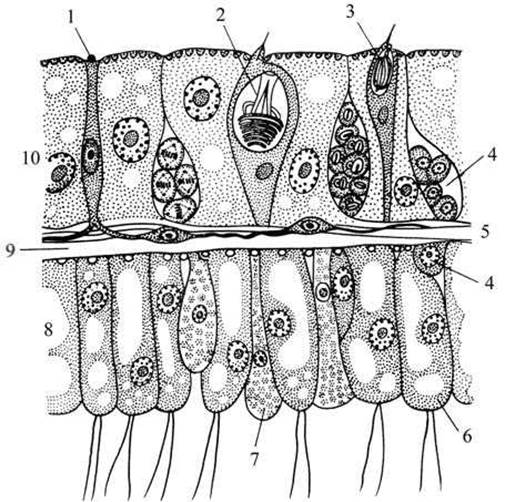
Fig. 28 Fragment of the hydra body wall:
1 — nerve cell; 2, 3 — cnidocytes with Different types of capsules; 4 — interstitial cells; 5 — nerve cell; 6 — epitheliomuscular cell; 7 — gland cell; 8 — endoderm; 9 — mesoglea (supporting lamella); 10 — ectoderm.
The ectoderm and endoderm are composed of several cell types that differ in Structure and function. Under high magnification, examine the ectoderm, the large endoderm cells, and the thin supporting lamella between them in cross-section. The bulk of the cells in both the ectoderm and endoderm consist of epithelial (covering) cells, which are cubic or cylindrical in shape. In the endoderm, these cells are larger, feature a flagellum, and are capable of extending pseudopodia.
In coelenterates, food is only partially digested within the gastrovascular cavity (extracellular Digestion); smaller food particles are engulfed by the pseudopodia of the endodermal epithelial cells and digested within the Cytoplasm (intracellular digestion).
In some slide preparations, remnants of digested food can be observed within the gastral cavity of the hydra. Make a drawing of the hydra cross-section (Fig. 29).
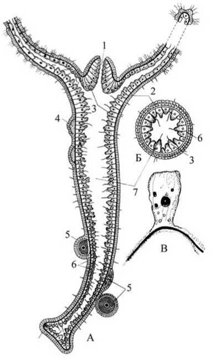
Fig. 29 Structure of Hydra:
A — longitudinal section; B — cross-section of Hydra; C — epitheliomuscular cell (high magnification): 1 — mouth; 2 — ectoderm; 3 — endoderm; 4 — Testis; 5 — egg cells; 6 — supporting lamella; 7 — gastrovascular cavity.
Upon careful examination of the slide preparation, darker-stained cnidocytes can be found in the ectoderm, resembling small, transparent flasks or round ampoules; they are particularly abundant in the tentacles and around the oral opening. Cnidocytes that have discharged their thread rather quickly perish and are replaced by new ones generated from interstitial cells.
Last update: 13/08/2026
Editorial and Educational Adaptation: This material has been compiled based on the primary/original source text. The project team performed an editorial review, corrected technical inaccuracies, structured sections, and adapted the content for an educational format.
What was processed:
- elimination of formatting defects (OCR errors, structural breaks, corrupted characters);
- editorial organization of content;
- standardization of terminology in accordance with academic sources;
- verification of factual statements against the original source text.
All mentions of the author, publication year, and origin of the primary text have been preserved in accordance with the source.