Review of Medical Physiology - William F. Ganong 2002
Physiology of Nerve and Muscle Cells
Synaptic and Junctional Transmission
Synaptic Transmission - Major Neurotransmitter Systems
Synaptic physiology is a complex and rapidly evolving branch of neurophysiology; therefore, it cannot be covered in exhaustive detail within this book. Nevertheless, summarizing the key information regarding major Neurotransmitters and their receptors remains highly appropriate.
Acetylcholine
The relatively simple Structure of acetylcholine, which is an acetic acid ester of Choline, is shown in Fig. 4-16. Acetylcholine is found in high concentrations primarily within the small, clear synaptic vesicles of terminal buttons in Neurons that synthesize acetylcholine (cholinergic neurons).
Synthesis of Acetylcholine
Acetylcholine is synthesized through the reaction of choline with acetate. Choline is an essential amine that also serves as a precursor for membrane Phospholipids—phosphatidylcholine and sphingomyelin—as well as the signaling phospholipids platelet-activating factor and sphingosylphosphorylcholine. Choline is actively taken up by cholinergic neurons via a transporter (Fig. 4-17) and is also synthesized within neurons. Acetate is activated by coupling acetate groups with reduced coenzyme A. The reaction between activated acetate (acetyl-coenzyme A, acetyl-CoA) and choline is catalyzed by the enzyme choline acetyltransferase. This enzyme is present in high concentrations in the Cytoplasm of cholinergic nerve terminals. Its localization is so specific that a high concentration of this enzyme in a particular neural region indicates that the synapses in that area are cholinergic. Acetylcholine enters synaptic vesicles via a vesicular transporter known as VAChT.
Cholinesterases
Upon repolarization, acetylcholine must be rapidly cleared from the synaptic cleft. This occurs via the Hydrolysis of acetylcholine into choline and acetate—a reaction catalyzed by the enzyme acetylcholinesterase (also known as true or specific cholinesterase). Acetylcholinesterase exhibits the highest affinity for acetylcholine, though it also hydrolyizes other choline esters.
The body contains numerous esterases. One of these, found in plasma, is also capable of hydrolyzing acetylcholine, although it differs from acetylcholinesterase. This enzyme is designated as pseudocholinesterase or nonspecific cholinesterase. Plasma pseudocholinesterase is partially regulated humorally and is also influenced by functional Changes in the Liver. Conversely, molecules of specific cholinesterase form clusters in the postsynaptic membrane of cholinergic synapses. The esterase is encoded by a single Gene; however, through Alternative Splicing of various mRNAs, two catalytic units are formed: one is anchored to The Cell membrane via Glycolipids, while the other possesses a Collagen tail. The hydrolysis of acetylcholine occurs rapidly, which accounts for the changes in Na+ conductivity and the electrical activity phenomena observed during synaptic transmission.
Class="center">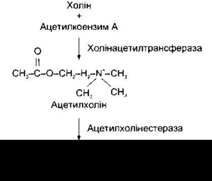
Fig. 4-16. Biosynthesis AND Catabolism of acetylcholine.
Acetylcholine Receptors
Traditionally, acetylcholine receptors are divided into two major types based on their pharmacological properties. Muscarine is a toxic alkaloid found in poisonous mushrooms: it has only a minor effect on receptors in autonomic ganglia, yet it fully mimics the stimulatory action of acetylcholine on smooth Muscles and glands. Consequently, this action of acetylcholine is termed the muscarinic effect, and the corresponding receptors are called muscarinic cholinergic receptors. This effect is prevented by atropine, which blocks muscarinic receptors. In sympathetic ganglia, small amounts of acetylcholine stimulate postganglionic neurons, whereas large amounts block the passage of impulses from pre- to postganglionic neurons. This type of effect is abolished by atropine but replicated by nicotine. This action of acetylcholine is termed the nicotinic effect, and the corresponding receptors are called nicotinic cholinergic receptors. Nicotinic receptors are subdivided into those located at neuromuscular junctions and those found in autonomic ganglia and the Central Nervous system. Both muscarinic and nicotinic receptors are abundant in the Brain.

Fig. 4-17. Biochemical transformations in cholinergic terminals. ACh - acetylcholine; AChE - acetylcholinesterase; X - receptor. Compare with Figs. 4-21 and 4-24.
Nicotinic cholinergic receptors belong to the superfamily of Ligand-Gated Ion Channels, which also includes GABAA receptors, Glycine receptors, and certain glutamate receptors. They are formed by multiple subunits encoded by different genes. Each nicotinic cholinergic receptor consists of five subunits that form a central channel, which upon receptor activation conducts Na+ or other cations. These five subunits are drawn from a pool of 16 known subunits: a1-a9, ß2-ß5, y, δ, and є, encoded by 16 distinct genes. Some receptors are homomeric, meaning they contain, for example, five a7 subunits, but most are heteromeric. The Muscle-type nicotinic receptor found in the fetus is composed of two a1 subunits, a ß1 subunit, a y subunit, and a δ subunit (Fig. 4-18). In adult mammals, the δ subunit is replaced by an є subunit, which decreases the channel open time while increasing its conductance. Nicotinic cholinergic receptors of autonomic ganglia are heteromers containing predominantly a3 subunits combined with other subunits, whereas brain nicotinic receptors are formed by numerous diverse subunits. A large population of nicotinic cholinergic receptors in the brain is located presynaptically on axon terminals that produce glutamate (see below), where they enhance the release of this transmitter. Others, however, occupy a postsynaptic position. Some are found in non-neuronal structures, and certain ones likely even reside in the interstitial fluid, thus occupying a perisynaptic position.
Each a subunit contains an acetylcholine-binding site, and when an acetylcholine molecule binds to each of these sites, it induces a conformational change in the protein, causing the channel to open. This results in the conductance of Na+ and other cations, and the influx of Na+ triggers a depolarization potential. An important feature of neuronal nicotinic cholinergic receptors is their high permeability to Ca2+, which may indicate their involvement in synaptic excitation and learning processes (see below).
Muscarinic cholinergic receptors differ profoundly from nicotinic ones. Five types of these receptors have been cloned, encoded by five separate genes. The exact Properties of the M5 receptor remain unclear. The other four are serpentine receptors coupled via G Proteins to adenylate cyclase, K+ channels, or phospholipase C (see Table 4-2). The nomenclature for these receptors is not yet standardized. The receptor designated in Table 4-2 is abundant in the brain. The M2 receptor is found in The Heart (see Chapter 28), and the M4 receptor is located in pancreatic acinar and islet tissue, where it mediates an increase in the secretion of pancreatic Enzymes and Insulin. M1 and M4 receptors are present in smooth muscles.
Norepinephrine and Epinephrine
The chemical transmitter at most sympathetic postganglionic terminals is norepinephrine. It is stored in synaptic vesicles of neurons that synthesize the transmitter and accumulate it in characteristic small vesicles containing a dense core (granular vesicles; see above). Norepinephrine and its methyl derivative, epinephrine, are produced by The adrenal medulla (see Chapter 20), although epinephrine is not a transmitter at sympathetic postganglionic terminals. The endings of sympathetic postganglionic neurons on smooth muscles are described below; each neuron features numerous varicosities along its axons, each of which apparently serves as a site where norepinephrine is released. The brain also contains norepinephrine-, dopamine-, and epinephrine-producing neurons (see Chapter 15). Neurons that synthesize norepinephrine are accordingly termed noradrenergic neurons, although the term adrenergic neurons is also used. Obviously, the latter term is more appropriate for epinephrine-producing neurons. Dopamine-synthesizing neurons are referred to as dopaminergic neurons.

Fig. 4-18. Fetal nicotinic Acetylcholine Receptor; lateral view (top) and view from above (bottom). a1, ß1, y, δ are receptor subunits (from McCarthy MP et al: Molecular biology of the acetylcholine receptor. Annu Rev Neurosci 1986;9:383. Reprinted with permission from Annual Review of Neuroscience, vol. 9, 1986).
Biosynthesis and Release of Catecholamines
The main catecholamines of the body—norepinephrine, epinephrine, and dopamine—are formed through the hydroxylation and decarboxylation of The amino acid Tyrosine (Fig. 4-19). Part of the tyrosine is derived from phenylalanine, but the majority is of dietary origin. Phenylalanine hydroxylase is located principally in the liver. Tyrosine is transported into catecholamine-synthesizing neurons as well as adrenal medullary Cells via a concentration mechanism. In the cytoplasm of the cells, tyrosine is converted to dihydroxyphenylalanine (DOPA), after which tyrosine hydroxylase and DOPA decarboxylase convert it to dopamine. Decarboxylase, also known as aromatic L-amino acid decarboxylase, is very similar to, yet apparently not identical with, 5-hydroxytryptophan decarboxylase. Dopamine then enters granular vesicles inside which dopamine ß-hydroxylase converts it to norepinephrine. In this reaction, the starting substance is L-DOPA, yet the resulting norepinephrine possesses the D-configuration. This is true despite the fact that it rotates polarized light to the left (-). Dextrorotatory (+) norepinephrine is far less active. The rate-limiting step in this synthesis is The conversion of tyrosine to DOPA. Tyrosine hydroxylase, which catalyzes this step, is inhibited by dopamine and norepinephrine, thereby achieving intrinsic Regulation of the synthesis process via feedback. The cofactor for tyrosine hydroxylase is tetrahydrobiopterin, which is converted to dihydrobiopterin during the transformation of tyrosine into DOPA.
Some neurons and adrenal medullary cells contain cytoplasmic phenylethanolamine-N-methyltransferase (PNMT), which catalyzes the conversion of norepinephrine to epinephrine. In these cells, norepinephrine presumably exits the vesicles, is converted to epinephrine, and then enters other vesicles where it is stored.
Within the granular vesicles, norepinephrine and epinephrine bind to ATP and associate with chromogranin A, a protein of unknown function. Large granular vesicles of certain noradrenergic neurons also contain neuropeptide Y (see below). Chromogranin A is a protein with a Molecular Weight of 49 kDa found in numerous endocrine and neuroendocrine cells, and it may play an important role in hormone secretion and storage. A similar protein, chromogranin B, is produced in certain Tissues. Plasma levels of chromogranin A are elevated in patients with various endocrine tumors.
The transport of catecholamines into granular vesicles is carried out by two vesicular transporters (see above), and this process is inhibited by reserpine.
Catecholamines are released from autonomic neurons and the Cells of the adrenal medulla via exocytosis (see Chapter 1). Since granular vesicles also contain non-membrane-bound ATP, chromogranin A, and dopamine $\beta$-hydroxylase, these substances are released from the vesicles along with noradrenaline and adrenaline. The half-life of circulating dopamine $\beta$-hydroxylase is much longer than that of catecholamines; the level of this circulating enzyme is influenced by genetic and other factors, and furthermore, it depends on the activity level of the sympathetic nervous system. Evidently, the level of circulating chromogranin A serves as a better indicator of sympathetic nervous system activity.
Phenylketonuria, or phenylpyruvic oligophrenia, is a disorder characterized by severe intellectual disability and the accumulation of large amounts of phenylalanine and its keto acid derivatives in the Blood, tissues, and urine. This is caused by a congenital deficiency of phenylalanine hydroxylase (see Fig. 4-19). The gene for this enzyme is located on the long arm of chromosome 12. Numerous Mutations in this gene that cause phenylketonuria have been described. Catecholamines are continuously synthesized from tyrosine, and the intellectual disability is primarily caused by the accumulation of phenylalanine and its derivatives in the blood. Therefore, the management of phenylketonuria requires dietary therapy involving restricted phenylalanine intake.

Fig. 4-19. Biosynthesis of Catecholamines. Dashed lines indicate the inhibition of tyrosine hydroxylase by noradrenaline and dopamine. Main Cofactors are highlighted in italics.
The disorder may also be caused by tetrahydrobiopterin deficiency. Since tetrahydrobiopterin is a cofactor for tyrosine hydroxylase, Tryptophan hydroxylase (see above), and phenylalanine hydroxylase, its deficiency leads to a shortage of catecholamines and serotonin, combined with hyperphenylalaninemia. This results in hypotonia, reduced activity, and developmental abnormalities. Treatment involves the administration of tetrahydrobiopterin, levodopa, 5-hydroxytryptophan, as well as a low-phenylalanine diet.
Catabolism of catecholamines
Noradrenaline, like other amine and amino acid Transmitters, is cleared from the synaptic cleft by binding to post- or presynaptic receptors (see Fig. 4-15), by reuptake into presynaptic neurons, or through catabolism (Fig. 4-20). The primary mechanism for noradrenaline clearance is its reuptake, which partly explains the supersensitivity of structures deprived of sympathetic influence due to denervation.
Following the transection of noradrenergic neurons, their terminals degenerate and, consequently, reuptake becomes impossible. Therefore, a significant amount of noradrenaline originating from other sources is able to stimulate the receptors of autonomic effector Organs.
Adrenaline and noradrenaline are metabolized via oxidation and methylation to form biologically inactive products. Oxidation is catalyzed by monoamine oxidase (MAO), and methylation by catechol-O-methyltransferase (COMT) (see Fig. 4-20). MAO is located on the outer surface of Mitochondria.
There are two isoforms of MAO: MAO-A and MAO-B, which differ in substrate Specificity and pharmacological sensitivity. Both isoforms are present in neurons. MAO is widely distributed, with particularly high concentrations found in nerve terminals that produce catecholamines. COMT is also widely distributed, especially in the liver, Kidneys, and smooth muscles. In the brain, it is localized in glial cells, with only minor amounts found in postsynaptic neurons, whereas COMT is completely absent in presynaptic noradrenergic neurons. Thus, there are two distinct pathways for Catecholamine METABOLISM.
Extracellular adrenaline and noradrenaline are predominantly O-methylated, and measuring the concentration of O-methylated derivatives (normetanephrine and metanephrine in urine) serves as an important indicator of norepinephrine and epinephrine secretion rates. Rather than being excreted directly, these O-methylated derivatives are primarily oxidized; consequently, the largest fraction of catecholamine metabolites in the urine appears as 3-methoxy-4-hydroxymandelic acid (vanillylmandelic acid, VMA) (see Fig. 4-20). Small amounts of O-methylated derivatives are also conjugated with sulfates and glucuronides.
In noradrenergic nerve terminals (Fig. 4-21), on the other hand, a portion of noradrenaline is continuously converted by MAO into physiologically inactive deaminated derivatives: 3,4-dihydroxymandelic acid (DOMA) and its corresponding glycol (DOPEG). These are subsequently converted into their respective O-methylated derivatives, VMA and MOPEG (see Fig. 4-20).
Alpha and beta receptors
Adrenaline and noradrenaline act on $\alpha$- and $\beta$-receptors, respectively, as noradrenaline has a higher affinity for $\alpha$-adrenergic receptors, and adrenaline for $\beta$-adrenergic receptors. As noted above, $\alpha$- and $\beta$-receptors are typical G protein-coupled serpentine receptors and exist in various complex forms. They are closely related to cloned dopamine and serotonin receptors, as well as to muscarinic cholinergic receptors.
Dopamine
In small intensely fluorescent (SIF) cells of autonomic ganglia (see Chapter 13) and in certain areas of the brain (see Chapter 15), catecholamine synthesis stops at The formation of dopamine (see Fig. 4-19), and this catecholamine acts as a synaptic transmitter. Dopamine undergoes active reuptake via a Na+- and Cl--dependent transporter (see above). It is metabolized by MAO and COMT to yield inactive components (Fig. 4-22) in a manner analogous to the inactivation of noradrenaline. DOPAC and HVA are also conjugated predominantly with sulfates.
Five distinct dopamine receptors have been cloned, some of which exist in multiple complex forms. All of them are coupled via G proteins to seven transmembrane domains. D1 and D5 receptors cause an increase in cAMP concentration (see Table 4-2), although their distribution in the brain differs. D2, D3, and D4 (presumably D4 intended here) receptors mediate a decrease in cAMP levels and also show somewhat distinct distributions. The D4 receptor has a higher affinity than other dopamine receptors for the "atypical" antipsychotic drug clozapine, which is effective in Schizophrenia yet produces fewer extrapyramidal effects than most other tranquilizers. Furthermore, as noted, the density of D4 receptors in the brain is increased sixfold in schizophrenia.
Serotonin
Serotonin (5-hydroxytryptamine, 5-HT) is found in high concentrations in Blood Platelets and the gastrointestinal tract, where it resides in enterochromaffin cells and the myenteric plexus (see Chapter 26). Lower amounts of serotonin are detected in the brain and retina. Serotonin is synthesized in the body via the hydroxylation and decarboxylation of the essential amino acid tryptophan (Figs. 4-23 and 4-24). Normally, hydroxylase is not saturated; consequently, an increased Dietary intake of tryptophan can lead to elevated serotonin levels in the brain. Following release from serotonergic neurons, the majority of serotonin is reclaimed via active reuptake (see Fig. 4-24), while the remainder is inactivated by MAO (see Fig. 4-23) to form 5-hydroxyindoleacetic acid (5-HIAA). This substance represents the primary urinary metabolite of serotonin; therefore, measuring its urinary excretion serves as an index of the body's serotonin turnover rate. In the Pineal Gland, serotonin is converted into melatonin (see Chapter 24).

Fig. 4-20. Top: Catabolism of extracellular adrenaline and noradrenaline. Catabolism occurs primarily in the liver. Catabolites are predominantly conjugated with glucuronides and sulfates. 3-Methoxy-4-hydroxyphenylglycol (MOPEG) is also conjugated. Bottom: Catabolism of noradrenaline within noradrenergic nerve terminals. The acid and glycol produced via MAO actions diffuse into the extracellular fluid and subsequently undergo O-methylation to yield 3-methoxy-4-hydroxymandelic acid (VMA) and MOPEG. Adrenaline in nerve terminals is evidently catabolized via the same pathway.
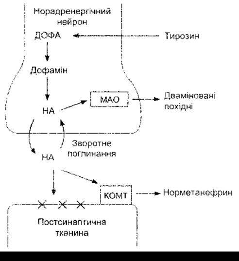
Fig. 4-21. Biochemical transformations at noradrenergic terminals. NA – noradrenaline; COMT – catechol-O-methyltransferase; MAO – monoamine oxidase; X – receptor. Presynaptic receptors are omitted for simplicity. Note that MAO is localized intracellularly; therefore, noradrenaline is continually deaminated within nerve terminals. COMT acts primarily on released noradrenaline. Compare with Figs. 4-17 and 4-24.
The number of cloned and characterized serotonin receptors is steadily increasing. The known types include 5-HT1, 5-HT2, 5-HT3, 5-HT4, 5-HT5, 5-HT6, and 5-HT7 receptors. Within the 5-HT1 group, several subtypes are distinguished: 5-HT1A, 5-HT1B, 5-HT1D, 5-HT1E, and 5-HT1F. The 5-HT2 group comprises 5-HT2A, 5-HT2B, and 5-HT2C (formerly known as 5-HT1C) subtypes. There are two 5-HT5 subtypes: 5-HT5A and 5-HT5B. Most of these receptors are G protein-coupled and affect adenylate cyclase and phospholipase C (see Table 4-2). Like nicotinic cholinergic receptors, 5-HT3 receptors function as ligand-gated ion channels. Some serotonin receptors are presynaptic, whereas others are postsynaptic. The 5-HT2A receptors mediate platelet aggregation and smooth Muscle contraction. Mice with a knockout of the 5-HT2A receptor gene gain weight due to increased food intake, despite responding normally to leptin (see Chapter 14), and reach an impressive size. 5-HT3 receptors are located in the gastrointestinal tract and the area postrema, where they are involved in the vomiting reflex (see Chapter 14). 5-HT4 receptors are also found in the gastrointestinal tract—where they enhance secretion and peristalsis—as well as in the brain. In the brain, 5-HT6 and 5-HT7 receptors are localized within the limbic system, with 5-HT6 exhibiting a high affinity for antidepressants.

Fig. 4-22. Dopamine catabolism. As in other MAO-catalyzed Oxidative Deamination reactions, aldehydes are initially formed; they are subsequently oxidized in the presence of aldehyde dehydrogenase to yield the corresponding acids (DOPAC and HVA). Aldehydes are also reduced to 3,4-dihydroxyphenylethanol and 3-methoxy-4-hydroxyphenylethanol. DOPAC and HVA form sulfate conjugates.
Histamine
The cell bodies of histaminergic neurons are located in the tuberomammillary Nucleus of the posterior hypothalamic region (see Fig. 15-6), and their axons project to all Regions of the brain, including the Cerebral Cortex AND Spinal Cord. Thus, the histaminergic system resembles the noradrenergic, adrenergic, dopaminergic, and serotonergic systems in that a relatively small number of cells establish connections with all PARTS OF THE CNS.
Histamine is also present in the cells of the gastric mucosa (see Chapter 6) and in heparin-containing cells known as mast cells, which are particularly abundant in the anterior and posterior lobes of the Pituitary Gland.
Histamine is synthesized by the decarboxylation of the amino acid Histidine (Fig. 4-25). The enzyme catalyzing this step differs from the L-aromatic amino acid Decarboxylases that decarboxylate 5-hydroxytryptophan and L-DOPA. Histamine is metabolized to methylhistamine or, alternatively, to imidazoleacetate. The latter pathway is quantitatively less significant in humans. Diamine oxidase (histaminase) is more suitable for this reaction than MAO, although MAO catalyzes The oxidation of methylhistamine to methylimidazoleacetate.

Fig. 4-23. Biosynthesis and catabolism of serotonin (5-hydroxytryptamine). The enzyme catalyzing the decarboxylation of 5-hydroxytryptophan is very similar, though presumably not identical, to the enzyme that catalyzes the decarboxylation of DOPA. Tetrahydrobiopterin serves as a cofactor for tryptophan hydroxylase. The formation of melatonin is shown in detail in Fig. 24-11.
Three types of histamine receptors are known: H1, H2, and H3, all of which are found in both peripheral tissues and the brain. Most, if not all, H3 receptors are presynaptic and mediate the inhibition of histamine and other neurotransmitter release via G proteins. H1 receptors activate phospholipase C, whereas H2 receptors increase intracellular cAMP concentrations. The function of brain histaminergic systems remains unclear; however, histamine is known to be involved in processes related to arousal, motivation, Sexual Behavior, The regulation of certain Anterior Pituitary Hormones, blood pressure, fluid intake, and the pain threshold.

Fig. 4-24. Biochemical transformations in serotonergic synapses. Compare with Figs. 4-17 and 4-21. 5-HTP – 5-hydroxytryptophan; 5-HT – 5-hydroxytryptamine (serotonin); 5-HIAA – 5-hydroxyindoleacetic acid; X – serotonin receptor. Presynaptic receptors are omitted from the diagram for simplicity.
Excitatory Amino Acids: glutamate and aspartate
Glutamate and aspartate depolarize many different mammalian neurons by directly affecting their cell membranes via ionophoresis. Glutamate is the principal excitatory neurotransmitter in the BRAIN AND SPINAL cord, accounting for an estimated 75% of all excitatory transmission within the brain. Aspartate is likely the transmitter in pyramidal and spiny stellate cells of the visual cortex, although this has not yet been thoroughly investigated. Glutamate is synthesized through the reductive amination of the Krebs cycle intermediate α-ketoglutarate (Fig. 4-26), whereas aspartate is formed via Transamination of the Krebs cycle intermediate oxaloacetate (see Chapter 17). Both reactions are reversible, with further metabolism taking place in The Citric Acid Cycle. As noted above, glial cells and presynaptic neurons reuptake glutamate using at least three distinct transporters.
There are Two Types of glutamate receptors: metabotropic and ionotropic. Metabotropic receptors are G protein-coupled serpentine receptors that lead to an increase in intracellular IP3 and DAG, or a decrease in intracellular cAMP. Eleven different subunits of these receptors have been identified (see Table 4-2). Widely distributed throughout the brain, they appear to play a key role in synaptic plasticity, particularly in the hippocampus and Cerebellum. Knockout of the gene for one of these receptors (a form of mGluR1) results in severe impairments in motor coordination and spatial learning.
Ionotropic receptors are ligand-gated ion channels structurally related to nicotinic cholinergic receptors (see above), as well as to GABA and glycine receptors (see below). There are three Major Types of ionotropic receptors, named after their related compounds: kainate receptors (kainate being an acid isolated from sea Algae), AMPA receptors (for α-amino-3-hydroxy-5-methyl-4-isoxazolepropionate), and NMDA receptors (for N-methyl-D-aspartate). Like nicotinic, GABA, and glycine ionotropic receptors, these are multi-subunit complexes. Four subunits of AMPA receptors, five subunits of kainate receptors, and six subunits of NMDA receptors have been identified, all encoded by different genes. While these receptors are believed to have a pentameric structure, some may be tetramers, and their exact stoichiometry remains to be fully elucidated.
Kainate and AMPA receptors are simple ion channels that, upon opening, allow an influx of Na+ and an efflux of K+. The NMDA receptor is also a cation channel, but it is unique in permitting the influx of relatively large amounts of Ca2+. First, binding of glycine enhances its function, and glycine appears to be required for the receptor to respond normally to glutamate (Fig. 4-27). Second, while glutamate binding opens the channel, at resting membrane potentials the channel is blocked by Mg2+ ions. This block is relieved only when the receptor-bearing neuron is partially depolarized through the activation of AMPA channels or other channels mediating rapid depolarization via other synaptic circuits. Third, phencyclidine and ketamine—agents that induce amnesia and a sense of environmental dissociation—bind to separate sites within the channel. Most glutamatergic neurons possess both AMPA and NMDA receptors. Kainate receptors are located presentatively on GABA-producing nerve terminals and postsynaptically at various sites throughout the brain. Kainate and AMPA receptors are found in both Glia and neurons, whereas NMDA receptors are believed to be restricted to neurons.
A high concentration of NMDA receptors is found in the hippocampus. Blocking these receptors prevents long-term potentiation—a sustained enhancement of synaptic transmission that follows a brief period of high-frequency stimulation (see below). Consequently, these receptors are likely crucial for memory and learning processes. A distinctive feature of glutamate and some of its synthetic analogues is that, by acting on nerve cell bodies, they can trigger an influx of Ca2+ massive enough to cause cell death. This property allows these excitatory substances to be used via microinjections to selectively destroy nerve cell bodies while sparing adjacent axons.

Fig. 4-25. Synthesis and catabolism of histamine.

Fig. 4-26. Formation and metabolism of glutamate and GABA.
There is substantial evidence that excitatory amino acids play a major role in brain damage associated with stroke (see Chapter 32). Clearance of glutamate from the extracellular fluid of the brain is mediated primarily by Na+-dependent uptake systems in neurons and glia. When a cerebral artery is occluded, cells in the acutely ischemic core die, whereas cells in the surrounding penumbra may survive initially but lose the capacity to maintain the transmembrane Na+ gradient required for glutamate uptake. As a result, glutamate accumulates in the extracellular fluid to levels that trigger excitotoxic injury and cell death in the peri-infarct zone (the ischemic penumbra). The pharmacological modulation of these processes in stroke therapy is discussed in Chapter 33.
Inhibitory amino acids: gamma-aminobutyric acid
Gamma-aminobutyric acid (GABA) is the principal inhibitory neurotransmitter in the brain, operating at approximately 20% of all CNS synapses. GABA is also found in the retina and is the neurotransmitter mediating presynaptic inhibition (see above).
GABA, which exists in Body Fluids as y-aminobutyrate, is formed by the decarboxylation of glutamate (see Fig. 4-26). This reaction is catalyzed by glutamate decarboxylase (GAD), an enzyme that immunocytochemical Methods have shown to be localized in nerve terminals throughout many regions of the brain. (GABA is metabolized primarily by transamination to succinic semialdehyde and is subsequently converted to succinate in The Citric Acid cycle [see Chapter 17].) Transamination is catalyzed by GABA transaminase (GABA-T). Pyridoxal phosphate, a derivative of pyridoxine (vitamin B6), serves as a cofactor for both GAD and GABA-T. GABA is actively reuptaken via the GABA transporter (see above). The vesicular GABA transporter mediates GABA uptake into secretory vesicles. This transporter contains 10 transmembrane domains, whereas the vesicular monoamine transporter has 12 (see above).

Fig. 4-27. Schematic representation of the NMDA receptor. Upon binding of glycine and glutamate, the previously closed ion channel (left) opens, but at the resting Membrane Potential, the channel is blocked by Mg2+ (right). This block is removed by partial depolarization resulting from other inputs to the receptor-bearing neuron, allowing Ca2+ and Na+ to enter the neuron. The block can also be induced by dizocilpine maleate (MK-801).
An autoimmune response against GAD and the resulting GABA deficiency lead to stiff-man syndrome (SMS), which is characterized by fluctuating yet progressive muscle rigidity and painful muscle spasms.
Interestingly, GAD is also present in synaptic vesicle-like structures within insulin-producing B-cells of the pancreatic islets, where GABA may act as a paracrine transmitter (see Chapter 19). Insulin-dependent Diabetes Mellitus (type 1 diabetes) is an autoimmune disease characterized by the destruction of B-cells, with the majority of autoantibodies being directed against GAD. However, SMS is a rare condition whereas type 1 diabetes is quite common, and not all patients with SMS develop type 1 diabetes. Consequently, the exact relationship between these two disorders remains unclear.
Three types of GABA receptors have been described: GABAA, GABAB, and GABAC. GABAA and GABAB receptors are widespread in the central nervous system, whereas in adult vertebrates, GABAC receptors are found almost exclusively in the retina. Both GABAA and GABAC receptors are ligand-gated ion channels composed of five subunits surrounding a central pore, which is characteristic of nicotinic acetylcholine receptors as well as many glutamate receptors. In this specific case, the permeant ion is Cl- (Fig. 4-28).
GABAB receptors are metabotropic receptors coupled to heterotrimeric G proteins that increase K+ channel conductance while inhibiting adenylate cyclase activity and Ca2+ influx. The increased Cl- influx, increased K+ efflux, and decreased Ca2+ influx lead to hyperpolarization in neurons, resulting in an IPSP. The G-protein-mediated signaling of the GABAB receptor is unique in this context because it predominantly involves a G-protein heterodimer rather than individual monomers.
GABAC receptors have a relatively simple structure. They are pentamers composed of three p-subunits combined in various arrangements. GABAA receptors are also pentamers, but they are assembled from various combinations of six a, four ß, four y, one δ, and one ε subunits, which accounts for their remarkable functional diversity across different anatomical locations.
The Cl- influx mediated by GABAA receptors is enhanced by benzodiazepines, compounds known for their pronounced sedative properties as well as their effectiveness as muscle relaxants and anticonvulsants. Benzodiazepines bind to a-subunits. Diazepam and other benzodiazepines are widely used worldwide. Barbiturates and alcohol also promote Cl- flux through Cl- channels, at least in part. Steroid hormone metabolites—progesterone and deoxycorticosterone—bind to GABAA receptors and enhance Cl- conductance. It has been known for many years that high doses of progesterone and deoxycorticosterone exert hypnotic and anesthetic effects, which are mediated by their action on GABAA receptors.
Another class of benzodiazepine receptors has been identified in steroidogenic Endocrine glands and other peripheral tissues, leading to their designation as peripheral benzodiazepine receptors. They are thought to participate in steroid biosynthesis by fulfilling a function similar to that of the StAR protein (see Chapter 20), namely facilitating the translocation of Steroids into mitochondria. Another potential role of peripheral benzodiazepine receptors involves the Regulation of Cell proliferation. Peripheral benzodiazepine receptors have also been detected in astrocytes and Brain Tumors.
Glycine
By acting on NMDA receptors, glycine exerts an excitatory effect in the brain. However, it also mediates direct inhibition, predominantly within the Brainstem and spinal cord. Like GABA, glycine acts by increasing Cl- conductance. Its antagonist in this regard is strychnine. The Clinical presentation of convulsions and hyperexcitability induced by strychnine underscores the critical importance of postsynaptic inhibition for normal nervous system function. Inhibitory glycine receptors are Cl- channels. They are pentamers consisting of two types of subunits: a ligand-binding a-subunit and a structural ß-subunit. Recently, strong evidence has emerged for the existence of three populations of neurons involved in direct inhibition in the spinal cord: glycinergic neurons, GABAergic neurons, and neurons that release both glycine and GABA. Presumably, strictly glycinergic neurons contain the glycine transporter GLYT2, strictly GABAergic neurons contain GAD, and dual-action neurons contain both substances. The third category of neurons is of particular interest, as glycine and GABA appear to be packaged within the same synaptic vesicles.
Some individuals exhibit an exaggerated startle reflex (hyperekplexia), and at least in certain cases, this condition has been linked to single Amino Acid Substitutions within glycine receptor subunits.
Anesthesia
The MECHANISM OF ACTION of major anesthetics remained a mystery until recently. However, accumulating evidence indicates that not only alcohol and barbiturates, but also volatile inhalational anesthetics act on ion channels, notably GABAA and glycine receptors, thereby enhancing Cl- conductance.
Substance P and Other Tachykinins
Substance P is an 11-amino acid polypeptide found in the intestine, various peripheral nerves, and numerous regions of the central nervous system. Its structure is presented in Table 26-2. Substance P is one of six members of a mammalian polypeptide family known as tachykinins. These Peptides differ at their NH2-termini but share a conserved carboxy-terminal sequence, Phe-X-Gly-Leu-Met-NH2, where X represents Val, His, Lys, or Phe. The members of this family are listed in Table 4-3. Many similar tachykinins are also found in other vertebrates and invertebrates.

Fig. 4-28. Schematic representation of GABAA and GABAB receptors and their primary Mechanisms of action. The G protein mediating GABAB receptor effects is a heterodimer (reproduced with permission from Bowery NG, Brown DA: The cloning of GABAB receptors. Nature 1997;386:223).
Mammalian tachykinins are encoded by two genes. The neurokinin B gene encodes a single polypeptide, neurokinin B, whereas the substance P (neurokinin A) gene encodes the remaining five Polypeptides. Three of these are generated by alternative splicing of the primary RNA transcript, and the other two by post-translational Processing.
There are three neurokinin receptors. Two of them, the substance P receptor and the neurokinin K receptor, have been cloned and identified as serpentine, G-protein-coupled receptors. Activation of the substance P receptor triggers phospholipase C activation, leading to increased production of IP3 and DAG.
Substance P is present in high concentrations in the terminals of primary afferent neurons in the spinal cord and is believed to serve as the neurotransmitter at the first synapses of slow pain pathways (see Chapter 7). In addition, this peptide is found in the nigrostriatal system, where its concentration parallels that of dopamine, as well as in the Hypothalamus, where it may play a role in neuroendocrine regulation. When administered intradermally, substance P causes erythema and edema, likely acting as a neurotransmitter released by nerve fibers during an axon reflex (see Chapter 32). Substance P is also involved in the regulation of intestinal peristalsis. It has recently been demonstrated that centrally active NK-1 receptor antagonists exhibit antidepressant activity in humans. Similar to conventional monoamine-based antidepressants (see Chapter 15), this antidepressant effect requires a certain latency period to develop. However, NK-1 inhibitors failed to induce changes in brain monoamine metabolism in animal experiments. The physiological Functions of other tachykinins remain elusive.
Opioid Peptides
Receptors that bind morphine are located throughout the brain and gastrointestinal tract. Investigations into the endogenous ligands for these receptors led to the discovery of two related pentapeptides, termed enkephalins (Table 4-4), which bind to these opioid receptors. One contains Methionine (Met-enkephalin) and the other leucine (Leu-enkephalin). These and other opioid-receptor-binding polypeptides are collectively referred to as opioid peptides. Enkephalins are present in nerve terminals within the gastrointestinal tract and across various brain regions, where they apparently function as synaptic transmitters. They are found in the substantia gelatinosa, and when microinjected into the brainstem, they produce analgesia. Enkephalins also slow intestinal motility (see Chapter 26).
Table 4-3. Mammalian Tachykinins
Gene |
Polypeptide substances |
Receptors |
SP/NKA |
Substance P |
Substance P (NK-1) |
К+ |
Neurokinin A |
|
Н2O |
Neuropeptide K |
Neuropeptide K (NK-2) |
Са2+ |
Neuropeptide gamma |
|
Na+ |
Neurokinin A (3-10) |
|
NKB |
Neurokinin B |
Neurokinin B (NK-3) |
Like other small peptides, opioid peptides are synthesized as part of a large precursor molecule (see Chapter 1). Over 20 active opioid peptides have been identified. Unlike other peptides, opioids have multiple distinct precursors. Each of these exists as a preproprotein and a proprotein from which the signal peptide is cleaved. Three investigated precursors, along with the opioid peptides derived from them, are listed in Table 4-4. Proenkephalin was first discovered in the adrenal medulla (see Chapter 20) and also serves as the precursor for met-enkephalin and leu-enkephalin in the brain. Each proenkephalin molecule contains four met-enkephalins, one leu-enkephalin, one octapeptide, and one heptapeptide. Proopiomelanocortin—a large precursor molecule found in the anterior and intermediate lobes of the pituitary gland and in the brain—contains beta-endorphin, a 31-amino-acid polypeptide that features met-enkephalin at its NH2 terminus (see Chapter 22). Other, shorter endorphins are also formed.
The precursor molecule also gives rise to ACTH and melanocyte-stimulating hormone (MSH). In the brain, there are separate enkephalin- and beta-endorphin-producing neuronal systems (see Chapter 15). Beta-endorphin is released into the bloodstream by the pituitary gland. The third precursor molecule is prodynorphin, a protein containing three leu-enkephalin residues linked to dynorphin and neoendorphin. Dynorphin 1-17 is found in the duodenum, whereas dynorphin 1-8 is located in the posterior pituitary and the hypothalamus. The hypothalamus also contains alpha- and beta-neoendorphins. The physiological rationale for the existence of multiple precursor molecules for opioid peptides, as well as the presence of these peptides in both the bloodstream and the central nervous and gastrointestinal systems, remains incompletely understood today.
Enkephalins are primarily metabolized by two peptidases: enkephalinase A, which cleaves the Gly-Phe bond, and enkephalinase B, which cleaves the Gly-Gly bond. An aminopeptidase that cleaves the Tyr-Gly bond also participates in enkephalin metabolism.
Opioid receptors have been studied in detail. Mu (μ)-, kappa (κ)-, and delta (δ)-receptors have been identified, differing in their pharmacological properties, localization in the brain and other organs, and affinity for various opioid peptides. All three are serpentine receptors coupled to G-proteins, and all of them inhibit adenylate cyclase activity. There are likely subtypes of μ- and κ-receptors. Activation of μ-receptors leads to an increase in K+ conductance via hyperpolarization of central and primary afferent neurons. Activation of κ- and δ-receptors results in the closure of Ca2+ channels.
A recent Study of the natural ligands of the μ-receptor—the receptor targeted by morphine—led to the discovery of two additional opioid peptides: endomorphin 1 (Tyr-Pro-Trp-Phe-NH2) and endomorphin 2 (Tyr-Pro-Phe-Phe-NH2). The precursor molecule for these two μ-agonists remains unknown. The affinity of natural opioid peptides for κ-, μ-, and δ-receptors is illustrated in Fig. 4-29. Although the physiological effects resulting from receptor stimulation overlap significantly, the major outcomes of this stimulation can be defined and are summarized in Table 4-5.
Other Polypeptides
Numerous other polypeptides have been identified in the brain. Trophic pituitary hormones are present in various parts of The Nervous System, and most, if not all, appear to function as both neurotransmitters and hormones. Preprosomatostatin is the precursor of two polypeptides: Somatostatin-14 (see Fig. 14-19) and somatostatin-28 (Fig. 4-30). They occur together in tissues. Somatostatin is found in various regions of the brain, where it likely acts as a neurotransmitter, influencing sensory input, motor activity, and cognitive processes. In the hypothalamus, it is released into the hypophyseal portal vessels and acts as a Growth Hormone-inhibiting hormone (see Chapter 14). In the endocrine Pancreas, it inhibits the secretion of insulin and other pancreatic hormones (see Chapter 19), and it also serves as an important inhibitory hormone in the gastrointestinal tract (see Chapter 26). Both polypeptides are biologically active, but somatostatin-28 is more potent than somatostatin-14 in inhibiting insulin secretion. The somatostatin receptor family comprises five distinct G protein-coupled members (SSTR1 through SSTR5). All of them inhibit adenylate cyclase activity and affect intracellular messenger systems. It is likely that SSTR2 mediates cognitive processes and inhibits growth hormone secretion, whereas SSTR5 is responsible for suppressing insulin secretion.
Table 4-4. Opioid Peptides and Their Precursors
Precursor |
Opioid peptides |
Structure |
Proenkephalin |
Met-enkephalin |
Tyr-Gly-Gly-Phe-Met5 |
(see Chapter 20) |
Leu-enkephalin |
Tyr-Gly-Gly-Phe-Leu5 |
Octapeptide |
Tyr-Gly-Gly-Phe-Met-Arg-Gly-Leu8 |
|
Heptapeptide |
Tyr-Gly-Gly-Phe-Met-Arg-Phe7 |
|
Proopiomelanocortin |
ß-Endorphin |
See Chapter 22 |
(see Chapter 22) |
Other endorphins |
See Chapter 22 |
Prodynorphin |
Dynorphin 1-8 |
Tyr-Gly-Gly-Phe-Leu-Arg-Arg-Ile8 |
Dynorphin 1-17 |
Tyr-Gly-Gly-Phe-Leu-Arg-Arg-Ile-Arg-Pro-Lys-Leu-Lys-Trp-Asp-Asn-Gln17 |
|
alpha-Neoendorphin |
Tyr-Gly-Gly-Phe-Leu-Arg-Lys-Tyr-Pro-Lys10 |
|
beta-Neoendorphin |
Tyr-Gly-Gly-Phe-Leu-Arg-Lys-Tyr-Pro9 |
Vasopressin and Oxytocin are not only released into the blood but are also found in neurons whose axons project to the brainstem and spinal cord. The brain contains bradykinin, angiotensin II, and endothelin (see Chapters 24 and 31), the Gastrointestinal Hormones VIP, CCK-4, and CCK-8 (see Chapter 26), as well as two types of CCK receptors: CCK-A and CCK-B. CCK-8 acts on the binding sites of both receptors, whereas CCK-4 acts exclusively on CCK-B receptor sites (see Chapters 14 and 26). Gastrin, neurotensin, galanin, and gastrin-releasing peptide are also present in both the gastrointestinal tract and the brain. Neurotensin and VIP receptors have been cloned and shown to be serpentine receptors. The hypothalamus contains gastrin-17 and gastrin-34 (see Chapter 26). VIP causes vasodilation and is present in vasomotor nerve fibers. The functions of these peptides in the nervous system remain unclear.
Calcitonin gene-related peptide (CGRP) is a peptide that exists in two forms in rats and humans: CGRPa and CGRPß. In humans, these two forms differ by only three amino acid residues and are encoded by different genes. In rats and presumably in humans, CGRPß is found in the gastrointestinal tract, whereas CGRPa is located in primary afferent neurons, neurons transmitting taste signals to the thalamus, and neurons of the medial Forebrain bundle. Together with substance P, it is also present in the terminals of primary afferent neurons ending on Blood Vessels. CGRP-like immunoreactivity is detectable in the blood, and injection of CGRP causes vasodilation. CGRPa and calcitonin—a calcium-lowering hormone (see Chapter 21)—are encoded by the calcitonin gene. However, in The Thyroid Gland, mRNA encoding calcitonin is produced via splicing, whereas in the brain, alternative splicing forms mRNA encoding CGRPa. CGRP has little effect on Ca2+ metabolism, while calcitonin exhibits only a weak vasodilatory effect. Neuropeptide Y is a 36-amino-acid polypeptide related to the Pancreatic Polypeptide (see Chapter 19). It is found in numerous regions of the brain and in the Autonomic nervous system. Outside the brain, as well as in the autonomic nervous system, it is predominantly located in noradrenergic neurons, from which it is released in response to high-frequency stimulation. Neuropeptide Y enhances the vasoconstrictor action of norepinephrine. Circulating neuropeptide Y derived from sympathetic nerves increases during heavy physical exertion. In the hypothalamus, neuropeptide Y mediates increased appetite and food intake (see Chapter 14). The Y1-, Y2-, Y4-, Y5-, and Y6-receptors for this polypeptide have been cloned.

Fig. 4-29. Opioid receptors. The thickness of the arrows extending from the ligand names for κ-, μ-, and δ-receptors is proportional to the receptor affinity for each ligand (reproduced with permission from Julius D: Another spark for the masses? Nature 1997; 386:442).
Table 4-5. Physiological effects resulting from stimulation of opioid peptide receptors
Receptor |
Effect |
μ |
Analgesia Site of morphine action Respiratory depression Constipation Euphoria Sedation Increased growth hormone and prolactin secretion Miosis |
к |
Analgesia Diuresis Sedation Miosis Anxiety |
δ |
Analgesia |
Purinergic Transmitters
The relationships among ATP, adenosine, and adenosine metabolites are illustrated in Fig. 4-31. Extracellular ATP is ATP released along with norepinephrine, dopamine, GABA, glutamate, acetylcholine, and histamine from neurons that synthesize these substances. Adenosine is a neuromodulator that acts as a major CNS depressant. Adenosine also exerts a vasodilatory effect on the heart (see Chapter 32) and has diverse effects throughout the body. It acts on four serpentine, G protein-coupled receptors: A1, A2A, A2B, and A3. A2A and A2B receptors increase cAMP concentration, whereas A1 and A3 decrease it. The stimulating effects of coffee and tea are due to the blockade of adenosine receptors by caffeine and theophylline. Of considerable interest is the potential use of antagonists to reduce excessive glutamate release and thereby minimize the consequences of strokes.
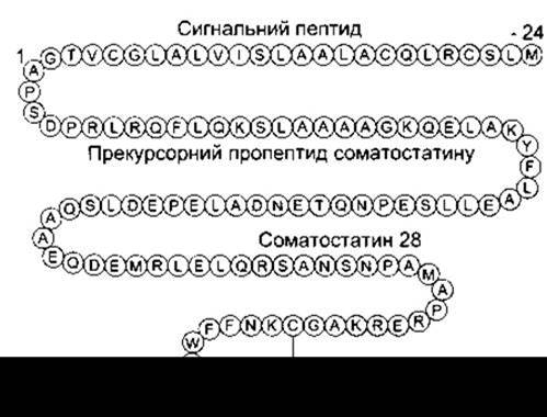
Fig. 4-30. Human preprosomatostatin. The major residues at the N-terminus where precursor Cleavage occurs to yield somatostatin-14 and somatostatin-28 are highlighted in color. Amino acid residues are designated by single-letter codes (reproduced with permission from Reisine T, Bell GI: Molecular biology of somatostatin receptors. Endocr Rev 1995; 16:427).
ATP also possesses transmitter properties, and its effects on receptors are manifested systemically. It is likely that non-structural nucleotidases are released along with ATP, thereby accelerating the clearance of ATP after it performs its function. Four purinergic receptors that bind ATP have been characterized: P2Y and P2U, which activate phospholipase via G-proteins, and P2X and P2Z, which are ligand-gated ion channels. Three P2X receptor subtypes have been identified: P2X1, P2X2, and P2X3. P2X1 and P2X2 receptors are located in the dorsal horns of the spinal cord, whereas P2X3 is found in the dorsal ROOT ganglia and trigeminal ganglion, indicating a role for ATP in sensory pathways. In addition, There is a P2T receptor, which is likely an ADP-activated ion channel. Recent studies have demonstrated that ATP mediates fast synaptic responses in the autonomic nervous system and the habenula. Purinergic receptors have also been detected in glial cells.
Cannabinoids
Two receptors with high affinity for ∆9-tetrahydrocannabinol (THC)—the psychogenic ingredient of marijuana—have been cloned. The CB1 receptor mediates a G protein-coupled decrease in intracellular cAMP levels. It is widely distributed in central neurons of pain-processing pathways, as well as in parts of the cerebellum, the hippocampus, and the cerebral cortex. The endogenous ligand for this receptor is anandamide, an arachidonic acid derivative (Fig. 4-32). This compound, much like marijuana, induces euphoria, sedation, sleepiness, drowsiness, and analgesia. CB1 receptors are also present in peripheral tissues, where their blockade leads to a reduction in the vasodilatory effect of anandamide. However, this vasodilatory effect appears to be indirect. The CB2 receptor has also been cloned, and its endogenous ligand is likely palmitoylethanolamide (PEA). The Physiological Role of this compound, however, remains undetermined.
Other Putative Transmitters
Nitric oxide (NO) is a compound released by vascular endothelial cells and acts as an endothelium-derived relaxing factor. It is also synthesized in the brain. The synthesis of NO from Arginine—a reaction catalyzed by one of three forms of NO synthase—is described in Chapter 31 (see Fig. 31-1). Nitric oxide activates guanylyl cyclase (see Chapter 1) and, unlike other transmitters, is a gas, allowing it to easily diffuse across the Cell Membrane and bind directly to guanylyl cyclase. This may serve as a signal by which postsynaptic neurons communicate with presynaptic terminals during long-term potentiation and depression ( see below). NO synthase is an NADPH-dependent enzyme. It is now known that NADPH diaphorase, the histochemical stain used for many years, is actually NO synthase. Consequently, NO synthase can be readily detected by histochemical methods in the brain and other tissues.

Fig. 4-31. Relationship between ATP and adenosine, and the metabolism of adenosine to uric acid.
Carbon monoxide (CO) is another gas that serves as a putative neurotransmitter in the brain. It is generated during heme metabolism (see Chapter 27) via one of the heme oxygenase (HO) subtypes, designated as HO2 in Fig. 4-33. Like NO, carbon monoxide activates guanylyl cyclase.
Prostaglandins are arachidonic acid derivatives (see Chapter 17) found within the nervous system. They are present in the nerve-ending fraction of brain homogenates and are released from neural tissue in vitro. The putative prostaglandin transporter features 12 membrane-spanning domains. However, prostaglandins likely exert their effects by modulating cAMP-mediated responses rather than acting as classical synaptic transmitters.
Numerous steroids are neuroactive, meaning they influence brain function although they are not neurotransmitters in the conventional sense. Circulating steroids readily cross the blood-brain barrier into the brain, where neurons harbor numerous receptors for sex steroids and glucocorticoids. In addition to their well-known DNA-binding mechanism (genomic effect), certain steroids appear to exert rapid actions by interacting directly with cell membranes (non-genomic effect). The Role of steroids in neuroendocrine regulation is discussed in Chapter 14, and their effects on GABA receptors were described above.
A growing body of evidence indicates that hormonally active steroids can be synthesized de novo within the brain. Formed from simpler steroid precursors, they are termed neurosteroids. Progesterone promotes myelination (see Chapter 2). Nevertheless, the precise Physiological Role of most neurosteroids in brain function remains to be fully elucidated.

Fig. 4-32. Anandamide.
Cotransmitters
Accumulating evidence demonstrates that individual neurons can contain and synthesize two or even three distinct transmitters. In such cases, cotransmitters frequently consist of a catecholamine or serotonin paired with a polypeptide, or a polypeptide paired with GABA or acetylcholine. Combinations of two polypeptides, or GABA paired with various catecholamines or acetylcholine, also occur. Certain brainstem neurons contain serotonin, substance P, and TRH. Many cholinergic neurons contain VIP, whereas numerous noradrenergic and adrenergic neurons contain ATP and neuropeptide Y. Neurons containing multiple transmitters are often intermingled with single-transmitter neurons. It is hypothesized that low-frequency neuronal impulses preferentially release low-molecular-weight transmitters, whereas high-frequency impulses also trigger the release of polypeptide cotransmitters. However, this hypothesis requires further validation, and the exact physiological significance of cotransmitters remains unclear. It is known, nevertheless, that VIP—co-released with acetylcholine—enhances its postsynaptic action, and neuropeptide Y potentiates the effects of norepinephrine.
Last update: 10/08/2026
Editorial and Educational Adaptation: This material has been compiled based on the primary/original source text. The project team performed an editorial review, corrected technical inaccuracies, structured sections, and adapted the content for an educational format.
What was processed:
- elimination of formatting defects (OCR errors, structural breaks, corrupted characters);
- editorial organization of content;
- standardization of terminology in accordance with academic sources;
- verification of factual statements against the original source text.
All mentions of the author, publication year, and origin of the primary text have been preserved in accordance with the source.