Medical Radiology - Lazar A.P. 2008
Radiological Examination of the Central Nervous System
Brain Tumors
Depending on the size and Location OF THE Brain tumor, clinical manifestations may include generalized cerebral symptoms (such as periodic headaches, dizziness, and syncope) as well as focal neurological deficits (such as impairments in Vision, Hearing, motor function, and sensation).
Brain tumors are detected using Diagnostic imaging Methods based on Direct and Indirect signs. Direct signs include the direct visualization of the TUMOR AS A space-occupying lesion, whereas indirect signs include perifocal edema, mass effect (displacement of adjacent brain structures and ventricular deformation), tumor calcification, destructive Changes in the cranial bones, and others (Fig. 292, 294). On CT and MRI scans, a tumor appears as a region of altered densitometric density (hyper- or hypodense) or MRI signal intensity (hyper- or hypointense) (Fig. 293). The lesion is typically irregular in shape with a heterogeneous Structure. Isodense and isointense brain tumors are identified by indirect signs and with the aid of intravenous contrast enhancement using radiopaque agents (in CT) or gadolinium (in MRI), which increases the density or signal intensity of the tumor to a greater extent than that of healthy brain tissue.
Class="center">
Fig. 292. Computed tomography scan of brain tumors (diagram):
A - intra-axial tumor,
B - intraventricular tumor, C - neurinoma of the internal acoustic meatus.
1 - hypodense peritumoral edema zone; 2 - contrast enhancement within the tumor; 3 - amputation of the ventricular cavity; 4 - Displacement of the Fourth ventricle; 5 - acoustic (vestibulocochlear) nerve schwannoma.
Due to Blood-brain barrier disruption and increased radiopharmaceutical uptake within the tumor, it appears as a "hot spot" on scintigrams, single-photon emission computed tomography (SPECT) scans, and positron emission tomography (PET) scans.
Carotid angiography reveals the following signs of a neoplasm: displacement of Blood Vessels by the tumor, dilation of tumor-feeding vessels, and their straightening.
Plain Skull radiography allows for the detection of indirect signs of brain tumors associated with elevated intracranial pressure and mass effect. Intracranial pressure rises due to tumor enlargement and brain tissue edema caused by the accumulation of free extracellular or bound intracellular fluid, resulting in the compression of the ventricles and cranial venous outflow pathways. When establishing a Diagnosis, one must also keep in mind that increased intracranial pressure can be caused by Inflammatory Diseases of the Brain and its Meninges.

Fig. 293. Meningioma on Magnetic Resonance Imaging.
A - axial T2-weighted image;
B - sagittal T1-weighted image.
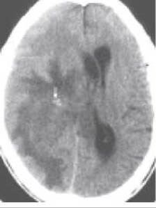
Fig. 294. Astrocytoma of the right cerebral hemisphere on MRI, T1-weighted image. The arrow indicates an area of calcification within the tumor
Radiological signs of chronic intracranial Hypertension include an enlarged skull, widening of diploic channels and emissary Veins, enhanced digital impressions, and changes in the sella turcica (enlargement, Osteoporosis, flattening of the dorsum sellae, and erosion of anatomical details). Increased intracranial pressure is more pronounced in children: the skull enlarges due to the widening of cranial sutures and thinning of the cranial vault bones, giving the skull a rounded, spherical shape.
Changes in the position, shape, and size of the ventricles and subarachnoid spaces in the Cytology/cytology/16.html">Early stages of hypertensive syndrome are detected using CT, MRI, echoencephalography, and radionuclide cisternography (Fig. 294). On CT and MRI scans, malignant tumors typically exhibit a heterogeneous structure and take up intravenously administered contrast material (Fig. 295).

Fig. 295. Glioblastoma of the left cerebral hemisphere on MRI. A - T2-weighted image;
B - T1-weighted image; C - contrast-enhanced T1-weighted image.

Fig. 296. Pituitary adenoma on CT.
1 - tumor as a hyperdense space-occupying lesion;
2 - enlarged inferior horns of the Lateral ventricles.
Pituitary tumors are predominantly adenomas developing from the anterior Pituitary Gland. Radiologically, pituitary adenomas are detected when they grow large enough to exceed the dimensions of the sella turcica. Craniograms reveal an enlarged sella (the normal sagittal dimension does not exceed 14 mm, and the frontal does not exceed 18 mm), thinning of its dorsum, and bone osteoporosis. Computed tomography scans reveal adenomas larger than 3-4 mm, which exhibit higher densitometric density (Fig. 296).
Craniopharyngiomas, which develop from remnants of the embryonic pharyngeal-pituitary pouch (Rathke's pouch), are located mostly above the diaphragma sellae near the optic pathways. Consequently, their growth rapidly leads to visual impairments. Radiological examination reveals tumor calcification in 80% of patients, expansion of the entrance to the pituitary fossa, and symptoms of intracranial hypertension.
Spinal Cord tumors are predominantly found in the thoracic region. They are best visualized using MRI (Fig. 297). During a CT scan, the densitometric density of spinal cord tumors is generally equal to the density of the Tissues they originate from; therefore, intravenous contrast enhancement is preferred for their detection using this method. On a radiograph, a tumor can be identified by indirect signs: deformation of the pedicles of the vertebral arches, indentation of the vertebral body, and widening of the intervertebral foramen.

Fig. 297. Spinal cord tumor on a sagittal slice (MRI).
A - T2-weighted image;
B - T1-weighted image.
Last update: 08/08/2026
Editorial and Educational Adaptation: This material has been compiled based on the primary/original source text. The project team performed an editorial review, corrected technical inaccuracies, structured sections, and adapted the content for an educational format.
What was processed:
- elimination of formatting defects (OCR errors, structural breaks, corrupted characters);
- editorial organization of content;
- standardization of terminology in accordance with academic sources;
- verification of factual statements against the original source text.
All mentions of the author, publication year, and origin of the primary text have been preserved in accordance with the source.