Medical Radiology - Lazar A.P. 2008
Radiation Examination of the Central Nervous System
Inflammatory Diseases of the Brain
Most commonly, intracranial infection spreads by direct contact from a focus of inflammation in the Paranasal Sinuses, ear, or in cases of open traumatic Brain injury, and less frequently via hematogenous dissemination from a distant source.
The inflammatory process may involve the brain parenchyma (cerebritis with abscess formation), the Meninges (meningitis), or present as a combined form affecting both the brain tissue and its membranes.
When the brain parenchyma is affected, a focus of brain tissue inflammation (cerebritis) first develops within the first 3 days, characterized by a central area of necrosis and surrounding edema, followed by encapsulation and abscess formation over the next 7-14 days.
X-ray imaging has low diagnostic value for Inflammatory Diseases of the brain and its meninges; it is primarily ordered to assess the sequelae of past conditions. Abscesses are clearly visualized using MRI and CT (Fig. 290). On MRI scans, cerebritis appears as a rounded necrotic center with a ring-like area of edema that is poorly demarcated from the surrounding unaffected parenchyma. Brain edema surrounding the inflammatory focus is best visualized on MRI.
On CT, an abscess appears as a rounded lesion of decreased densitometric density, which over time acquires clearer margins and is bounded by a thick capsule. A perifocal zone of increased density of inflamed brain tissue may be observed around the lesion (Fig. 291). In infants, Ultrasound examination reveals cerebritis as a rounded hyperechoic lesion, with a subsequent anechoic abscess developing with a perifocal hyperechoic zone.
Leptomeningitis involves the pia mater and arachnoid mater. Edema of the membranes is observed During the first 2-3 days, followed by The formation of an exudate that accumulates in the basal cisterns and sulci of the brain. Clinically, this presents with high fever, vomiting, headache, and photophobia. In the initial phase, CT and MRI detect meningeal edema only with intravenous contrast enhancement, which reveals hyperenhancement of the pia mater and Cerebral Cortex at the lesion site. The appearance of exudate and widening of the subarachnoid space are easily detected without contrast on CT and MRI, and via ultrasound in infants.
Class="center">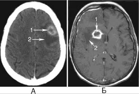
Fig. 291. Brain abscess on CT (A) and MRI (B).
1 - abscess capsule;
2 - perifocal edema.
As the inflammatory process progresses, computed and magnetic Resonance tomograms may show a lateral shift of midline structures, localized widening of the subarachnoid spaces (cystic arachnoiditis), as well as calcification after the inflammation subsides (especially of tuberculous Etiology). Infection from the meninges frequently spreads to the brain parenchyma, resulting in cerebritis.
Epidural abscess develops As a result of inflammation between the dura mater and the inner table of the cranial vault bones, appearing on CT and MRI scans as a space-occupying fluid collection. When large, an epidural abscess may cross the midline to the opposite side of the brain, which distinguishes it from a subdural abscess.
Last update: 08/08/2026
Editorial and Educational Adaptation: This material has been compiled based on the primary/original source text. The project team performed an editorial review, corrected technical inaccuracies, structured sections, and adapted the content for an educational format.
What was processed:
- elimination of formatting defects (OCR errors, structural breaks, corrupted characters);
- editorial organization of content;
- standardization of terminology in accordance with academic sources;
- verification of factual statements against the original source text.
All mentions of the author, publication year, and origin of the primary text have been preserved in accordance with the source.