Human Biochemistry, Volume 2 - Murray R. 1993
Biochemistry of Intracellular and Intercellular Communication
Adrenal Medullary Hormones
Biosynthesis of Catecholamines
Catecholamine Hormones—dopamine, norepinephrine, and epinephrine—are 3,4-dihydroxy derivatives of phenylethylamine. They are synthesized in the chromaffin Cells of The adrenal medulla. These cells earned their name because they contain granules that turn a reddish-brown color when treated with potassium dichromate. Clusters of such cells have also been found in The Heart, Liver, Kidneys, Gonads, postganglionic sympathetic adrenergic Neurons, and the Central Nervous system.
The primary product of the adrenal medulla is epinephrine. This compound accounts for approximately 80% of all medullary catecholamines. Epinephrine is not produced outside the adrenal medulla. In contrast, norepinephrine found in Organs innervated by sympathetic nerves is formed predominantly in situ (~ 80% of the total amount); the remainder is also generated mainly at nerve terminals and reaches its target sites via the bloodstream.
The conversion of Tyrosine to epinephrine involves four sequential steps: 1) ring hydroxylation, 2) decarboxylation, 3) side-chain hydroxylation, and 4) N-methylation. The catecholamine Biosynthesis pathway and its participating Enzymes are illustrated in Fig. 49.1 and 49.2.
Tyrosine Hydroxylase
Tyrosine is the direct precursor of catecholamines, and tyrosine hydroxylase is the rate-limiting enzyme for the entire biosynthesis process. This enzyme exists in both free and subcellular-bound forms. Utilizing tetrahydropteridin as a cofactor, it acts as an oxidoreductase, converting L-tyrosine to L-dihydroxyphenylalanine (L-DOPA). Various regulatory mechanisms control tyrosine hydroxylase as the rate-limiting enzyme. The most important of these is feedback inhibition by catecholamines: catecholamines compete with the enzyme for the pteridine cofactor by forming a Schiff base with it. Furthermore, tyrosine hydroxylase is competitively inhibited by several tyrosine derivatives, including a-methyltyrosine. In some cases, this compound is used to block excess catecholamine production in pheochromocytoma, though more effective treatments with milder side effects are available. Another group of compounds suppresses tyrosine hydroxylase activity by forming iron complexes, thereby depleting the available cofactor. An example of such a compound is a, a'1-dipyridyl.
Catecholamines do not cross the Blood-Brain barrier; consequently, their presence in the brain must be attributed to local synthesis. In certain central nervous system disorders, such as Parkinson's disease, impairments in dopamine synthesis occur specifically within the brain. The dopamine precursor, L-DOPA, readily crosses the blood-brain barrier, making it an effective Treatment for Parkinson's disease.
Class="center">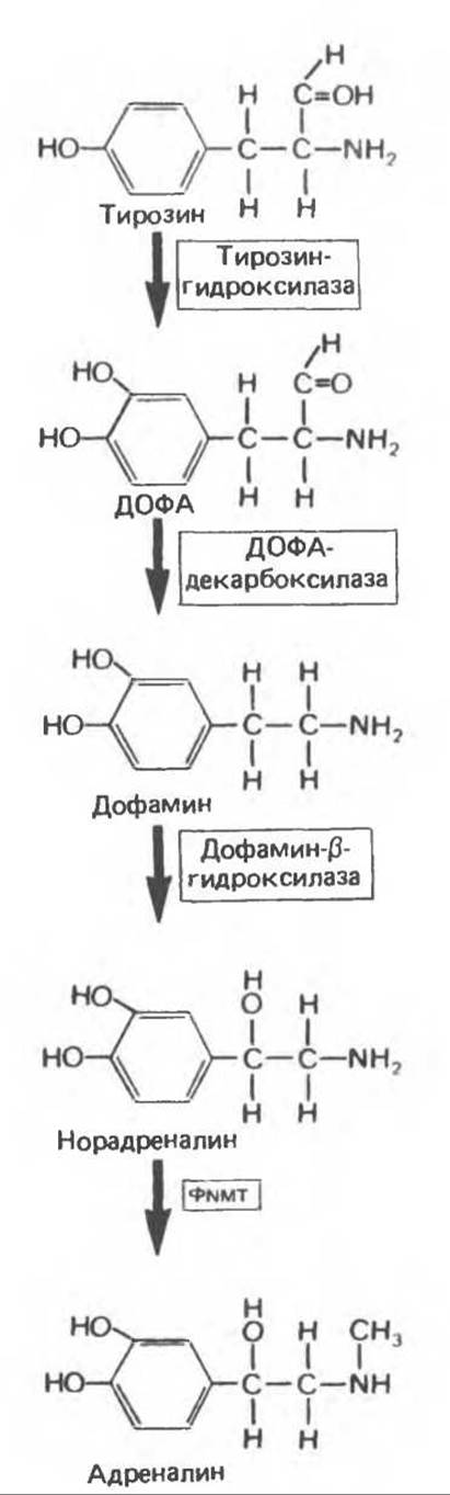
Fig. 49.1. Biosynthesis of catecholamines. PNMT — phenylethanolamine-N-methyltransferase. (Modified and reproduced, with permission, from Goldfien A. The adrenal medulla. In: Basic and Clinical Endocrinology, 2nd ed. Greenspan FS, Forsham PH [editors]. Appleton and Lange, 1986.)
DOPA Decarboxylase
Unlike tyrosine hydroxylase, which is found exclusively in Tissues capable of synthesizing catecholamines, DOPA decarboxylase is present in all tissues. This soluble enzyme requires Pyridoxal phosphate to convert L-DOPA into 3,4-dihydroxyphenylethylamine (dopamine). The reaction is competitively inhibited by compounds structurally similar to L-DOPA, such as a-methyl-DOPA. Halogenated compounds form a Schiff base with L-DOPA and likewise inhibit the decarboxylation reaction.
a-Methyl-DOPA and related compounds, such as 3-hydroxytyramine (derived from tyramine), a-methyltyrosine, and metaraminol, are successfully used to treat certain forms of Hypertension. The antihypertensive effect of these metabolites is presumably due to their ability to stimulate a-adrenergic receptors (see below) of the corticobulbar system in the central nervous system, leading to decreased peripheral sympathetic nerve activity and lowered blood pressure.
Dopamine-β-hydroxylase
Dopamine-β-hydroxylase (DBH) is a mixed-function oxidase that catalyzes the conversion of dopamine to norepinephrine. DBH utilizes ascorbate as an electron donor and fumarate as a modulator; its Active Site contains copper. DBH in adrenal medullary cells is likely localized within secretory granules. Thus, the conversion of dopamine to norepinephrine takes place inside these Organelles. DBH is released from adrenal medullary cells and nerve terminals along with norepinephrine, but (unlike the latter) it is not reuptaken by nerve terminals.
Phenylethanolamine N-Methyltransferase
The soluble enzyme phenylethanolamine N-methyltransferase (PNMT) catalyzes the N-methylation of norepinephrine to form epinephrine in epinephrine-producing Cells of the adrenal medulla. Because this enzyme is soluble, it is presumed that the conversion of norepinephrine to epinephrine occurs in the Cytoplasm. PNMT synthesis is stimulated by glucocorticoid hormones that reach the medulla via the intra-adrenal portal system. This system provides a steroid concentration in the medulla 100 times greater than that in systemic arterial blood. Such a high concentration in the adrenals appears necessary for the induction of PNMT.
Last update: 06/08/2026
Editorial and Educational Adaptation: This material has been compiled based on the primary/original source text. The project team performed an editorial review, corrected technical inaccuracies, structured sections, and adapted the content for an educational format.
What was processed:
- elimination of formatting defects (OCR errors, structural breaks, corrupted characters);
- editorial organization of content;
- standardization of terminology in accordance with academic sources;
- verification of factual statements against the original source text.
All mentions of the author, publication year, and origin of the primary text have been preserved in accordance with the source.