PHARMACEUTICAL MICROBIOLOGY - V. A. Galynkin - 2015
PART II. ANTIMICROBIAL AGENTS
CHAPTER 19. FUNDAMENTALS OF IMMUNITY
Immunity is a state of increased resistance of the Organism to living entities and substances bearing signs of genetic foreignness. These foreign substances are called Antigens. Antigens can be microorganisms, foreign Cells, Tissues, and the products of their vital activity. The organism's immune system responds to foreign substances. In addition to The Immune System, there are nonspecific defense factors whose action does not depend on the antigenic CHARACTERISTICS OF THE foreign agent. Modern immunology views the categories of specific immunity and nonspecific defense as a single functional complex whose elements are in constant interaction.
The likelihood of developing a disease As a result of a particular harmful impact depends on the organism's reactivity, i.e., its ability to neutralize the disease-causing factor (such as a microbe or its toxin), eliminate it from the body, and compensate for the resulting damage. Reactivity depends on the state of the macroorganism (sex, age, immune status, presence of other diseases), its living conditions (diet, working and living conditions), as well as the quality and intensity of the harmful factor's impact (e.g., microbial virulence, infectious dose, portal of entry).
Resistance to harmful factors is associated with the genetic, anatomical, and PHYSIOLOGICAL CHARACTERISTICS OF the macroorganism and depends on its individual state. Resistance is reduced by psychological trauma, starvation, vitamin deficiencies, radiation, and poisoning by alcohol, nicotine, or drugs.
19.1 Nonspecific Defense Factors
The organism's resistance to infections depends not only on its ability to mount an Immune Response—a highly specialized form of defense—but also on nonspecific factors directed against microorganisms regardless of their antigenic composition. Mechanical factors (Skin, mucous membranes, Lymph Nodes) prevent microbes from penetrating the body and spreading within it. Chemical factors have a detrimental effect on microbial cells. The latter include the acidic environment of The Stomach, intestinal Bile, Fatty acids in sweat and sebum secretions, Lysozyme found in saliva, tears, and mucous membrane secretions, antimicrobial substances produced by lymphocytes and Liver cells, interferon, etc.
19.1.1 Inflammation
A complex of nonspecific defense reactions is inflammation, which involves a series of changes—primarily in Blood capillaries and Connective Tissue—arising in response to a pathogenic irritant. Inflammation is accompanied by changes in tissue Structure and physiological functions, fluid leakage from Blood Vessels (exudation), and the proliferation of connective tissue. Inflammation is characterized by elevated Temperature, redness and Swelling of the affected area, and pain. Dilation of blood vessels and plasma leakage are accompanied by the precipitation of fibrin, which clogs the vessels, thereby limiting the spread of microbes through them. Phagocytic leukocytes accumulate in the inflammation site to engulf microbial cells. Leukocyte activation is promoted by special substances released from tissues, such as leukotaxine, the leukopenic factor, and histamine. The accumulation of leukocytes in the zone of inflammation leads to The formation of a protective wall that prevents microbes from spreading beyond the inflammatory focus. The elevated temperature, decreased pH (acidosis) due to the accumulation of lactic acid, and reduced partial pressure of oxygen (Hypoxia) observed in the inflammatory zone exert an unfavorable effect on microorganisms.
Inflammation is triggered by A wide variety of irritants (heat, mechanical damage, microbial infection); its symptoms are driven by specific mediators of inflammatory reactions (histamine, kinins, Leukotrienes, Prostaglandins, cytokines, etc.) that cause vasodilation, increased vascular permeability, and smooth Muscle contraction, while also activating phagocytosis.
The rise in temperature observed during inflammation or a febrile state is associated with the release of endogenous pyrogen from leukocytes, which acts on the thermoregulatory centers of the Brain. The release of endogenous pyrogen is promoted by various activators, including bacterial endotoxins, bacterial cells, and Viral Particles.
19.1.2 The Complement System
The complement system takes part in both nonspecific defense reactions and immune response reactions. It is a complex of soluble Blood Plasma Proteins and Cell surface proteins whose interactions mediate various biological effects: cell lysis, chemotaxis and opsonization during phagocytosis, and The stimulation of inflammation and hypersensitivity reactions. Most of these proteins are synthesized by hepatocytes and macrophages. They circulate in the blood in an inactive form and are activated under certain conditions via a cascade of enzymatic reactions. The classical pathway of activation begins with the binding of the C1 complement fraction to an antigen–antibody complex, after which other components are activated, resulting in the formation of a lytic complex.
The alternative pathway begins with the C3b fraction and involves a cascade of reactions mediated by properdin. This produces a protein complex that binds toglycoconjugates On the surface of microbial cells, leading to their lysis in the absence of Antibodies. Many microorganisms are able to protect themselves against the action of complement: they possess surface structures that are resistant to lysis or capable of suppressing the complement system, such as the sialic acids of streptococci, neisseriae, and treponemes.
19.1.3 The Properdin System
A potent nonspecific defense factor of the organism is the properdin system:
Blood Plasma proteins containing specific components and magnesium ions that are capable of neutralizing many Bacteria and Viruses;
lysin, released by platelets, which exhibits bactericidal properties and is thermostable;
adhesion molecules, a group of Glycoproteins that ensure Cell-to-Cell adhesion and direct the migration of leukocytes to the site of inflammation as they essentially glide along The surface of the vascular endothelial wall. In addition, they take part in phagocytosis, lymphocyte Circulation and interaction, and cytotoxic reactions;
kinins, a group of linear Polypeptides that form a Class of so-called tissue or local Hormones. Each of them is released locally by a system of diffusely distributed cells in the body; they lack specialized secretory glands and are inactivated at the site of their release. The most important among them are angiotensins, bradykinin, and substance P;
cytokines, a group of diverse proteins released by mammalian cells that act on other cells via specific receptors. The response of the target cell depends on its nature and The Nature of the cytokine: cell proliferation and differentiation, inflammation, and hemopoiesis. Cytokines produced by lymphocytes are called lymphokines; they transmit signals between different lymphocyte populations and participate in the Regulation of the immune response. Cytokines also include growth factor, colony-stimulating factor, interferons, and tumor necrosis factor;
normal antibodies, found in the blood, whose production is not linked to disease; they exert an antimicrobial effect and promote phagocytosis;
interferons (IFNs), a group of species-specific proteins and glycoproteins synthesized by body cells in response to interferonogens—viruses, bacteria, their structural elements, synthetic polyribonucleotides, and other substances that induce the derepression of genes encoding IFNs.
19.1.4 Interferons
IFNs, like other cytokines in the body, perform regulatory and control functions aimed at maintaining METABOLISM/37.html">Cellular Homeostasis. The most important among these are antiviral, antitumor, immunomodulatory, and radioprotective activities. IFNs inhibit viral Replication by stimulating the formation of a protein complex within The Cell that blocks the Translation of viral mRNA. Antitumor defense depends on the ability of IFN to activate cytotoxic lymphocytes, macrophages, and natural killer cells. In addition, they activate interferonogenesis (priming) and antibody production.
Certain factors are directed not against microbes or their toxins, but toward restoring disrupted Functions of the macroorganism. For instance, many infectious diseases involve Circulatory Disorders caused by a loss of arteriolar tone. Under The Influence of Vascular System stimulation by microbial agents, blood pressure rises, which compensates for the loss of arteriolar tone. Elevated body temperature has a detrimental effect on certain microorganisms; moreover, it activates phagocyte metabolism and their destruction of microbial cells. The Excretory Function of the Kidneys, respiratory tract, intestines, and salivary and Sweat Glands is enhanced, helping the body rid itself of various harmful factors. All these phenomena constitute a complex of defensive and adaptive reactions known as stress. Factors that induce a state of stress (stressors) can include cold, heat, radiation, psychological trauma, pathogenic microbes, and their toxins. Under the influence of stressors, the Pituitary Gland begins to secrete adrenocorticotropic hormone (ACTH), which stimulates the Adrenal Glands. Consequently, the adrenals increase their secretion of cortisone-type hormones. Cortisone reduces tissue reactivity, diminishes inflammation, and suppresses antibody production. Hormonal agents such as ACTH and cortisone are used in treating certain conditions when it is necessary to modulate the course of an inflammatory process.
19.1.5 Phagocytosis
Phagocytosis is the most evolutionarily ancient defense mechanism. It is manifested in the engulfment and Digestion of foreign particles, including bacteria and cellular debris, by phagocytes. This phenomenon was discovered by the Russian scientist Élie Metchnikoff. All phagocytic cells are colorless, which is why they are called leukocytes or, in contrast to erythrocytes, white Blood Cells; their characteristics are summarized in Table 39.
19.2 Characteristics of Phagocytic Cells
The process of phagocytosis begins with the recognition and adhesion of a foreign particle (e.g., a microbial cell) to the phagocyte membrane. During this stage, pseudopodia (false feet) extend from the phagocyte and fuse with one another, enclosing the microbe within a vacuole (phagosome), which subsequently fuses with a phagocytic lysosome. As a result, the microorganism is destroyed by the phagocyte's biocidal factors (O2, Н2О2, НСlO) and degraded by its hydrolytic Enzymes. Phagocytosis that culminates in the death and destruction of the ingested cells is termed completed (or successful) phagocytosis. Alongside this, certain infections (such as Gonorrhea, tuberculosis, leprosy, leishmaniasis, pertussis, typhoid fever, tularemia, brucellosis, and mycoses) exhibit incomplete phagocytosis, wherein microorganisms are ingested by phagocytes but survive and sometimes even multiply. This occurs because certain microbes secrete substances that block the phagocytic process. These include microbial toxins, capsular components, specific proteins, and leukocidins. Phagocytosis is activated by calcium and magnesium salts, electrolytes, complement, and antibodies. Furthermore, microorganisms may evade destruction by escaping from the phagosome before it fuses with a lysosome, thereby rendering themselves inaccessible to lytic enzymes.
In addition to destroying foreign cells, phagocytes perform the vital function of antigen-presenting cells within the immune system, facilitating the subsequent development of a specific immune response.
Natural killer (NK) cells are lymphocytes endowed with cytotoxicity against target cells (such as tumor cells, virally infected cells, and other parasites). They lack phagocytic activity, yet they eliminate their targets using cytotoxic substances. Unlike antigen-specific T killers (see below), their action is non-specific. NK cells account for 2% to 12% of human Blood Leukocytes.
19.3 Immunity
Immunity is characterized by a high degree of reaction Specificity, which is based on subtle structural and chemical differences between antigens and their corresponding antibodies (IMMUNOGLOBULINS) produced in response to The entry of an antigen into the organism [34].
Depending on its origin, immunity is classified into innate (also referred to as hereditary or species immunity) and acquired. Species immunity is the natural resistance of certain animal species to diseases that affect other species. For instance, humans do not contract rabbit snuffles or fowl cholera, while animals are immune to certain venereal diseases. Species immunity is the result of long-term evolution in the relationship between the microbe and the macroorganism, depending on their biological traits shaped through natural Selection. The degree of intensity of species immunity can be absolute or relative. For example, rat cells lack receptors for diphtheria toxin, making them absolutely resistant to diphtheria. For other animals, susceptibility to certain infectious diseases can be increased by applying unfavorable factors, such as altering body temperature, administering hormones or immunosuppressants, or exposing them to ionizing radiation. In such cases, one speaks of relative innate immunity.
Acquired immunity is not transmitted hereditarily. It develops against a specific pathogen and is strictly specific. Acquired immunity is subdivided into natural and artificial, with each of these categories further divided into active and passive. Natural active immunity develops following recovery from a disease or after subclinical infection. Natural passive immunity is the immunity of newborns, which they acquire from the mother during intrauterine development; its duration is relatively short, lasting about 6 months. Artificial active immunity arises following active immunization—that is, the administration of Vaccines and toxoids—whereas artificial passive immunity develops after the administration of immune sera or serum preparations (immunoglobulins).
19.4 Antigens
Antigens are defined as any substances that bear signs of genetic foreignness and, upon Introduction into an organism, trigger specific immunological reactions. Antigenic properties are characteristic of proteins, many Polysaccharides, Glycoconjugates (glycoproteins, lipopolysaccharides), and certain synthetic high-molecular-weight compounds.
To provoke an immune response in an organism, an antigen must possess a sufficient molecular weight (typically at least 5-10 kDa).
The fundamental concepts characterizing an antigen include antigenicity (The ability to induce an immune response), immunogenicity (the ability to induce immunity, i.e., resistance to infection), and specificity.
Specificity is determined by the specific Chemical Structure of the antigen, which distinguishes one antigen from another. The chemical grouping of the antigen molecule that dictates its specificity is called an antigenic determinant (epitope). For instance, the antigenic specificity of a protein is determined by its primary and supramolecular structures, as well as by surface-exposed groups that can act as antigenic determinants. In glycoconjugate molecules, the epitope is frequently a polysaccharide. Antigenic specificity is manifested by the fact that an antigen reacts exclusively with those antibodies and immune lymphocytes that were generated in response to its introduction.
Haptens are substances that possess specificity but fail to elicit an immune response when introduced into the body on their own. Nevertheless, they are capable of reacting with preformed antibodies. Haptens acquire The properties of complete antigens only after binding to high-molecular-weight carriers (proteins, polysaccharides, or synthetic polyelectrolytes). This binding occurs via covalent bonds or electrostatic forces. Many medicinal drugs (such as amidopyrine, quinidine, and penicillin degradation products) can act as haptens; by interacting with endogenous proteins, they can provoke an immune response, leading to drug-induced allergies.
Species specificity is the specificity through which representatives of one species differ from individuals of another species. For example, the composition of Serum proteins varies between humans and animals, a distinction that can be applied in practice, such as in forensic medicine.
Group specificity accounts for differences among individuals within the same species. Antigens that enable individuals of a single species to be distinguished from one another are termed isoantigens. Isoantigens include those present on erythrocytes that determine human Blood Groups.
The molecular COMPOSITION OF THE Major Histocompatibility Complex (MHC) is unique to each organism and defines its biological individuality, allowing it to differentiate "self" (histocompatible) from "non-self" (incompatible). Class I and Class II MHC molecules are glycoproteins that regulate the immune response and participate in T lymphocyte-mediated cytotoxicity. Class I MHC molecules are expressed on the surface of all nucleated cells, whereas Class II molecules are predominantly found on the membranes of immunocompetent cells (macrophages, B lymphocytes, and activated T lymphocytes). Class III MHC genes encode certain Components of the complement system.
Type specificity determines antigenic differences within a single species of microorganisms (serovars). For example, more than 80 serovars of pneumococci are known, differing in their polysaccharide antigens.
Heterospecificity is due to the presence of heteroantigens—antigenic determinants shared by representatives of different species. Common antigens can be found in quite phylogenetically distant species, such as humans and the plague pathogen, or the Influenza virus and other microorganisms. These are known as mimicking antigens. When the antigenic structures of a micro- and macroorganism share similarities, the Formation of the immune response is impaired. For instance, human myocardium contains chemical structures similar to streptococcal antigens; consequently, antibodies directed against streptococci can cross-react and attack Heart tissues, which is the underlying cause of complications following scarlet fever.
The antigenic structure of a microbial cell is of great scientific and practical interest, as bacterial antigens are utilized to develop vaccine preparations for inducing artificial immunity and for the serological Diagnosis of infectious diseases. Both Bacterial cells and viral particles comprise complex mixtures of substances with antigenic activity. These include high-molecular-weight proteinaceous compounds, polysaccharides, lipopolysaccharides, and others. Cellular Organelles—such as flagella, membranes, Cytoplasm, Ribosomes, and The Cell wall—also exhibit antigenic properties. Bacterial toxins likewise function as potent antigens.
Motile bacteria possess H antigens (flagellar), which are thermolabile—being destroyed at 56-80°C—and proteinaceous in nature, as well as O antigens (somatic), which are thermostable—withstanding heating up to 80-100°C—and possess a lipoprotein structure.
Capsular bacteria, such as Klebsiella, pneumococci, and others, possess polysaccharide capsular antigens.
The virulence antigen (Vi) is also a type of capsular antigen; it has been described in virulent enterobacterial species, such as Salmonella.
The protective antigen is not a permanent component of the microbial cell. It is produced within the infected host and exhibits exceptionally strong immunogenic properties.
Ribosomal antigens have served as the foundation for developing a new generation of vaccines.
Viral antigens — are proteins, glycoconjugates, and Nucleoproteins that are either virus-specific or incorporate host cell components (such as Lipids and CARBOHYDRATES). Many viruses contain a distinctive antigen known as hemagglutinin, which is detected via the hemagglutination assay. This reaction relies on the ability of viral hemagglutinin to agglutinate Human and Animal erythrocytes, thereby revealing the presence of the virus in the test sample.
The hemadsorption assay is based on the ability of erythrocytes to adsorb onto virus-infected cells. Both reactions proceed independently of antibodies and are therefore non-immunological. However, hemagglutinin can stimulate The production of antibodies, which can be detected using the hemagglutination inhibition assay.
19.5 Antibodies
Antibodies — are specialized proteins (glycoproteins) produced by the organism of vertebrates and humans in response to the introduction of antigens, possessing the ability to bind specifically to them. Antigens bound to antibodies are neutralized and eliminated from the body.
Of particular importance is the ability of antibodies to bind to a specific antigen molecule, “recognizing” even the most subtle variations in its structure—not only the substitution of a single amino acid in a protein molecule, but even the differentiation between stereoisomers of Organic compounds. The specificity of antibodies is determined by differences in their chemical structure, namely the Amino Acid Sequence of The polypeptide chains that comprise them. Alongside the subtle structural differences that define their immunological specificity, antibodies share common fundamental structural features.
Antibodies possess a globular structure and are referred to as immunoglobulins. The structural backbone of any antibody (Fig. 79) consists of a complex of four polypeptide chains: two identical heavy chains and two identical light chains. Heavy chains are designated by the letter H (heavy), and light chains by the letter L (light). The heavy and light chains are held together by Disulfide Bonds. They are folded in such a way that two identical regions, designated as Fab, are formed on the surface of the resulting structure. These regions contain the antigen-binding sites. A third region, Fc, contains structures that ensure the binding of antibodies to specific cells bearing Fc-receptor sites on their surface, such as leukocytes and mast cells. The antigen-binding sites (active centers) correspond to the epitopes of the antigen, fitting together like a lock and key. The interaction between an antigen and an antibody is driven by electrostatic, hydrophobic interactions, and Van der Waals forces. Complete antibody molecules possess at least two antigen-binding sites. Antibodies with only a single binding site are termed incomplete.
Fig. 79. Structure of an immunoglobulin monomer molecule [35, 36].

Both heavy and light chains possess two distinct structural regions based on their Amino Acid Composition: variable (V) and constant (C) regions. The variable region determines ANTIBODY SPECIFICITY AND is located within the Fab fragment, whereas the constant region resides in the Fc fragment. The Secondary structure of immunoglobulins consists of an α-Helix interspersed with complex β-structure "loops" formed by the cross-linking of amino acid residues in each chain. These folded structures are called domains and are found in both the Variable and constant regions of heavy and light chains. The antigen-binding sites (active centers) of Antibodies Are Formed by domains of the variable regions, which are localized within the hypervariable areas of the heavy and light chains.
Immunoglobulins are a heterogeneous group of proteins; this heterogeneity stems from the existence of various types of heavy and light chains. Humans have Two Types of light chains (κ and λ, kappa and lambda) and five classes of heavy chains (α, γ, μ, δ, ε). Based on the heavy chain classes, immunoglobulins are divided into five major classes (IgG, IgM, IgA, IgD, IgE), which differ in their physicochemical properties and biological activities (Table 40).
Table 40. Main Properties of immunoglobulins
Properties |
IgG |
IgA |
IgM |
IgD |
IgE |
Molecular weight, kDa |
150 |
160-400 |
900 |
180 |
190 |
Sedimentation coefficient, Svedberg units |
7 |
7 |
19 |
7 |
8 |
Serum half-life, days |
21 |
6 |
10 |
3 |
2 |
Carbohydrates, % |
3 |
7 |
12 |
13 |
11 |
Functions: |
|||||
complement activation |
++ |
+- |
++++ |
- |
- |
opsonization |
++++ |
+ |
- |
- |
- |
antiviral activity |
++ |
+++ |
+ |
? |
? |
mast cell sensitization |
- |
- |
- |
- |
+ |
IgM is a pentamer composed of 5 subunits, featuring 10 antigen-binding sites. It is phylogenetically the oldest class, the earliest to appear upon initial antigen exposure, and the primary class synthesized in newborns and infants.
IgG accounts for up to 75% of all immunoglobulins and serves as the principal class protecting the body against bacteria, viruses, and toxins. It also provides passive immunity to the fetus, as these are the only immunoglobulins capable of crossing the placental barrier.
IgA exists in two forms: serum and secretory. The majority of IgA consists of secretory immunoglobulins (sIgA), in which two or three monomers are linked by a secretory component that protects the immunoglobulin from enzymatic degradation. sIgA is present in saliva, tears, and milk, and is secreted onto the surfaces of epithelia and mucosal membranes, thereby enhancing their protective functions.
IgE specifically interacts with mast cells and basophilic leukocytes, playing a key role in The Development of allergic reactions. Its protective functions are primarily directed against helminths.
IgD functions as a membrane-bound antigen receptor on B lymphocytes.
In addition to various classes (isotypes) of immunoglobulins, allotypic and idiotypic variations also exist among them. Allotypes (markers of the constant region) are genetically determined and inherited. Idiotypes define the unique characteristics of each individual antibody. Idiotypic markers (antigenic determinants) are localized within the hypervariable regions and correspond to the antigen-binding sites of antibodies. All immunoglobulin molecules produced by a single clone of lymphocytes carry the exact same idiotype and are referred to as Monoclonal Antibodies. The Diversity of idiotypes is generated by DNA Mutations within the variable-region coding sequences, template-independent DNA Synthesis between recombining segments, and shifts in the joining sites by a few NUCLEOTIDES.
Immunity mediated by antibodies present in blood plasma and other Body Fluids is known as humoral immunity. Its mechanism relies on the ability of antibodies to neutralize pathogens and their toxins through opsonization, antitoxic action, complement activation, and other effects on the infectious agent. Opsonization occurs when antibodies bind to the surface of a microbial cell. This complex is actively engulfed by phagocytes through the interaction between the antibody's Fc fragment and the corresponding Fc receptor on the phagocyte. Antibodies can interact with cell surface receptors that bind bacteria or viruses, thereby preventing their adhesion and entry into host cells. Furthermore, antibodies exhibit catalytic activity—acting as Hydrolases and oxidoreductases—which also contributes to the neutralization of infectious agents.
19.6 The Immune System
The immune system is an integrated network of Lymphoid Organs and cells distributed throughout the body and interconnected by the Circulatory system, lymphatic drainage, and a unified immunoregulation network. The immune system possesses The unique ability to produce antibody molecules specific to any given antigen. The Organs of the immune system are subdivided into primary or central organs (Bone Marrow and thymus) and secondary or peripheral organs (Spleen, lymph nodes, and lymphoid aggregates).
Primary or Central Organs of the Immune System ensure its self-renewal. They are sites where precursor cells undergo proliferation, differentiation, and maturation, eventually transforming into immunocompetent cells that enter circulation and colonize peripheral immune organs.
Bone marrow is the site where pluripotent stem cells originate, giving rise to various hematopoietic lineages, including myeloid-monocytic and lymphocytic lineages. Bone marrow produces cytokines and, in mammals, serves as a potential site for B-lymphocyte maturation (see below).
The thymus is the primary site for the maturation and differentiation of T lymphocytes. It actively produces lymphocytes during Embryogenesis, reaches its maximum size in most vertebrates shortly after birth, and then gradually undergoes involution. In humans, peak T-lymphocyte production continues up to two years of age and then declines rapidly. However, T-lymphocyte levels remain sufficient because these are long-lived cells that can also proliferate upon encountering a specific antigen. During maturation and differentiation, T-lymphocyte precursors migrate from the bone marrow into the thymic cortex, gradually moving inward to acquire their surface markers through contact with thymic cells, locally produced mediators (specific Peptides), and cytokines.
The thymus eliminates potentially autoreactive cells. The primary function of mature T lymphocytes is to recognize foreign antigens rather than the body's own antigens (autoantigens). This elimination process works as follows: T lymphocytes carrying receptors for autoantigens (which make up 95–98% of the population) receive a signal for apoptosis, whereas T cells with receptors for foreign antigens receive a signal to proliferate. Apoptosis (programmed cell death) is the process by which a cell breaks down into distinct fragments that can be reused to build new cells.
Secondary or peripheral organs of the immune system are where immunocompetent cells encounter antigens, recognize them, and mount a specific immune response (involving cell-to-cell interactions and the synthesis of immunoglobulins).
Lymph nodes are distributed throughout the body. A single lymph node weighs about 1 g and contains approximately 2 × 10⁹ lymphocytes. Every hour, it releases a volume of lymphocytes into the lymph equivalent to three times its weight. Lymph nodes filter out microbial cells and other particulates while mounting immune responses to antigens carried in the lymph.
The spleen mounts immune responses against blood-borne antigens. It removes foreign particles, as well as Aging and damaged erythrocytes, from the bloodstream. If the spleen is removed, lymphoid organs take over its functions, though such patients experience weakened immunity.
Lymphoid tissue is associated with the mucosal linings of the gut, Pharynx, respiratory tract, and Urogenital System. Its function is to activate B lymphocytes that produce class A and E immunoglobulins.
19.7 Immunocompetent Cells
Immunocompetent cells are cells that participate in the immune response. They constantly circulate between the blood, lymph, and lymphoid organs to ensure they encounter their specific antigen, as each antigen is recognized by only a small fraction of the lymphocyte population. Lymphocytes are the sole cells capable of recognizing and responding to antigenic contact. Individual lymphocytes are specialized (committed) to respond exclusively to a specific group of structurally similar antigens. This commitment exists even before the initial contact with an antigen and is reflected in the presence of Membrane Receptors specific to particular antigenic determinants. Lymphocytes are an exceptionally heterogeneous cell population, with an estimated repertoire of at least 106 receptors featuring distinct antigen-binding sites. Furthermore, lymphocytes vary in their functions during the immune response and in other physiological characteristics.
B lymphocytes are so named because research on birds demonstrated that their differentiation occurs in a specialized lymphoid organ known as the bursa of Fabricius. In mammals, they develop in a lymphoid organ whose exact Location remains a subject of debate (the bone marrow or gut-associated lymphoid tissue).
During an immune response, B lymphocytes differentiate into immunoglobulin-producing cells. They recognize antigenic determinants using class D or M immunoglobulin receptors, alongside other receptors that detect signals during the immune response. They are capable of recognizing soluble antigens and binding protein, polysaccharide, and lipoprotein antigens.
The lifespan of the majority of the B-lymphocyte population (about 85%) does not exceed 10 days, roughly 14% live for 4–6 weeks, and about 1% persist for decades, serving as immunological memory cells.
T lymphocytes are the primary cells of immunological memory; they perform diverse functions and can be differentiated by their surface markers using serological Methods.
CD8+ T effectors produce numerous cytokines that promote their own differentiation and proliferation while also activating B lymphocytes and macrophages.
Cytotoxic T lymphocytes (CTLs, killer T cells) express the CD8+ marker and emerge through the activation and proliferation of T effectors driven by antigens. When parasites (such as Mycobacterium tuberculosis, Fungi, Protozoa, or viruses) replicate inside a host cell, their antigens form complexes with class I MHC molecules, which are recognized by CTL receptors. In response, CTLs become activated, acquiring the ability to destroy the infected cell using pre-synthesized cytotoxins: fragmentins and perforins. The former induce target cell apoptosis, while the latter are pore-forming proteins that disrupt target cell membrane permeability and pave the way for fragmentins. Contact between a CTL and its target cell is brief, after which the CTL moves on to a new target. CD8+ cells also exhibit suppressor activity, regulating the intensity of the immune response and preventing autoimmune reactions.
Regulatory CD4+ lymphocytes, or T helpers (TH), are divided into two subsets: TH1 cells (inflammatory) activate macrophages and participate in delayed-type hypersensitivity reactions, whereas TH2 cells activate B lymphocytes during the immune response.
Macrophages provide non-specific defense through phagocytosis and play a vital role in orchestrating the immune response as antigen-Processing and antigen-presenting cells. They originate from bone marrow stem cells, pass through circulating promonocyte and monocyte stages, and migrate into tissues (becoming histiocytes or tissue macrophages). They carry surface receptors (such as mannose and lipopolysaccharide receptors) required for capturing microbial cells, as well as Fc receptors for immunoglobulins and complement receptors needed to mediate antibody-dependent reactions.
Macrophages and cytotoxic lymphocytes mediate the cellular immune response (delayed-type hypersensitivity reactions, destruction of the body's own infected and tumor cells).
Dendritic cells originate in the bone marrow and populate non-lymphoid tissues. Dendritic cells found in the skin epidermis and respiratory mucosa are known as Langerhans cells. They act as antigen-presenting cells in the immune response. While similar to macrophages, only their immature forms possess phagocytic activity.
19.8 Cytokines
Cytokines are polypeptides produced by immunocompetent cells that also act as effectors on these cells, influencing their metabolism, proliferation, and secretion. Their primary functions include controlling cell proliferation, regulating inflammatory responses, modulating cellular and humoral immune responses, and exerting antiviral and cytotoxic activities.
Many cytokines are pleiotropic (Table 41). Disruptions in cytokine activity or synthesis lead to disease [34]. Certain cytokines are produced via Genetic Engineering techniques and used as therapeutic agents.
Table 41. MAIN TYPES OF Cytokines
Cytokine |
Source |
Target |
Effects |
IL-1 |
Macrophages, B cells, NK cells |
T cells, B cells, macrophages |
Lymphocyte and macrophage activation, fever, acute-phase response*, enhanced cell adhesion |
IL-2 |
T cells |
B cells, TH, NK cells, macrophages |
Lymphocyte and macrophage activation, stimulation of lymphokine secretion |
IL-3 |
T cells |
Stem cells |
Proliferation |
IL-4 |
T cells |
B cells, macrophages |
Lymphocyte proliferation, macrophage activation, regulation of antibody isotype switching: promotes IgG1 and IgE production, inhibits IgG2 and IgG3 synthesis |
IL-5 |
T cells |
B cells, stem cells |
Proliferation, differentiation, switching to IgA synthesis, eosinophilia |
IL-6 |
Macrophages, fibroblasts, tumor cells |
B cells, macrophages, myeloid precursors |
Proliferation, stimulation of Ig secretion, acute-phase response |
IL-7 |
Stromal cells |
Pre-B cells, T cells |
Proliferation |
IL-8 |
Vascular endothelium |
Neutrophils |
Inhibition of leukocyte adhesion |
IL-9 |
T cells |
T cells, mast cells |
Proliferation |
IL-10 |
T cells |
T cells |
Proliferation |
GM-CSF |
T cells, monocytes |
Stem cells, monocytes, granulocytes |
Proliferation, differentiation |
TNF-α |
Macrophages, T cells, NK cells |
B cells, macrophages, neutrophils |
Growth and differentiation, activation, enhanced adhesion, cachexia |
TNF-β (lymphotoxin) |
T cells |
Tumor cells |
Cytotoxicity |
MIF |
DTH T cells |
Macrophages |
Migration inhibition |
MAF |
DTH T cells |
Macrophages |
Activation |
MCF |
DTH T cells |
Macrophages |
Chemotaxis stimulation |
TF |
DTH T cells |
Lymphocytes |
Transfer of cell-mediated immunity |
MIF (IFL) |
DTH T cells |
Neutrophils |
Migration inhibition |
Perforin |
CTLs |
Tumor cells, graft cells |
Lysis |
IFN-α |
Leukocytes |
Somatic cells |
Inhibition of viral replication |
IFN-β |
Fibroblasts |
Somatic cells |
Inhibition of viral replication |
IFN-γ |
T cells |
Macrophages, NK cells, and other cells |
Activation, inhibition of viral replication |
Note: GM-CSF — granulocyte-macrophage colony-stimulating factor; IL — interleukin; IFN — interferon; MIF — macrophage migration inhibitory factor; MAF — macrophage-activating factor; MCF — macrophage chemotactic factor; TF — transfer factor; LIF — leukocyte migration inhibitory factor; TNF — tumor necrosis factor; TDTH — delayed-type hypersensitivity T-effectors; CTL — cytotoxic T lymphocytes; NK — natural killers; Th — T-helper cells. *Acute-phase reaction — an increase in the serum concentration of certain proteins (C-reactive protein, amyloid A, haptoglobin, ceruloplasmin, complement components) associated with acute inflammation.
19.9 Development of the Immune Response
Antigen processing is its modification (enzymatic processing, e.g., of a microbial cell) resulting in: a) making the antigenic determinant accessible to lymphocytes; b) conversion of large protein molecules into peptides, thereby smoothing out conformational differences and requiring less diversity of T-cell receptors; c) deep proteolysis enabling the recognition of internal antigenic structures of the microbial cell, which are less prone to mimicry of "self" than external ones. Macrophages, B lymphocytes, dendritic cells, liver Kupffer cells, etc., perform the function of ANTIGEN PROCESSING AND presentation to the T lymphocyte. The processed antigen, in a complex with an MHC class II molecule, is expressed on the surface of the antigen-presenting cell and recognized by a T-helper cell.
Interaction of immunocompetent cells (Fig. 80). Recognition of the antigenic complex by a T-helper cell leads to its activation, as a result of which the lymphocyte synthesizes IL-1; under the action of IL-1, the synthesis of IL-2 occurs, which stimulates proliferation.
Fig. 80 Interaction of immunocompetent cells. Antigen: a — B-cell epitope, b — T-cell epitope; 1 — antigen-presenting cell (macrophage); 2 — non-activated T-helper cell; TCR — T-cell receptor; 3 — activated T-helper cell: CR — cytokine receptor; 4 — B lymphocyte.
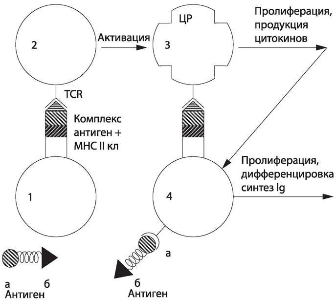
The B lymphocyte recognizes its "self" antigen, processes it, and presents a fragment of it on its surface in a complex with an MHC class II molecule. This complex is recognized by the activated T-helper cell, which in response secretes A number of interleukins (IL-2, IL-4, IL-5, gIFN), under the action of which the B cell multiplies, forming a clone of plasma cells. Plasma cells synthesize immunoglobulins; their secretion is stimulated by IL-6 released by activated T-helper cells.
19.10 Allergy
Usually, when speaking of immunity, one means defense reactions beneficial to the organism. However, immune reactions can also result in pathological alterations within the body. This altered reactivity arising under the influence of antigens is termed an allergy, and the substances causing it are called allergens. Allergens are subdivided into household allergens (down pillow dust, pet dander and fur), plant allergens (pollen), occupational allergens (cotton and wool dust, Dyes, varnishes, etc.), food allergens (eggs, wild strawberries, citrus fruits, chocolate, etc.), and drug allergens (acetylsalicylic acid, sulfonamides, Antibiotics, etc.). Allergic reactions are classified into 5 main types.
19.10.1 Type 1 Allergic Reactions
Type 1 reactions (anaphylactic) can be triggered by plant pollen and organic components of dust. Allergens activate a specific population of T-helper cells, which in turn activate B lymphocytes that produce IgE. These antibodies are capable of binding tightly to Fc receptors on target cells (mast cells, basophils).
An allergen entering the body repeatedly interacts with IgE fixed on cells, triggering a chain reaction in the target cell, which begins to release mediators (histamine, kinins, heparin, chemotaxis factors) that affect smooth muscle cells, blood vessels, and Endocrine glands. As a result, the clinical picture of anaphylactic diseases develops, the symptoms of which depend on the localization of the sensitized cells: rhinitis, Conjunctivitis, Bronchial Asthma, and anaphylactic Shock.
19.10.2 Type II Allergic Reactions
Type II allergic reactions are referred to as cytotoxic; they are associated with the production of IgG against antigenic components of the body's cell membranes. Such components can be autoantigens of the body's cells or antigens secondarily fixed on cell membranes, such as drug allergens. The IgG complex with these antigens is capable of binding complement and activating it via the classical pathway. As a result, the cell dies (complement-dependent cytolysis). This is The Mechanism of allergic reactions to penicillin, sulfonamides, and those occurring during blood transfusion, transplant rejection, and autoimmune diseases. At the same time, reactions of this type play a protective role, ensuring the elimination of damaged, tumor, and parasite-infected cells.
19.10.3 Type III Allergic Reactions
Type III reactions are those caused by the formation of immune complexes (ICs). The formation of ICs is a continuously occurring physiological reaction, and pathological reactions to ICs may be associated with impaired mechanisms of their clearance by Cells of the phagocytic system. ICs are capable of activating plasma components (complement and Blood Coagulation systems, chemotaxis) and certain cells (granulocytes, platelets, etc.). Activated cells release biogenic amines, enzymes, kinins, and other mediators that drive the pathological process. Depending on the type of antigen and its localization, various clinical manifestations of the disease are observed.
Endogenous antigens cause autoimmune diseases: systemic lupus erythematosus, rheumatoid Arthritis, Pemphigus, etc. Responses to exogenous antigens manifest as serum sickness, the Arthus phenomenon, and a number of infectious diseases. Serum sickness develops upon the administration of sera and other drugs; its clinical manifestations include arthritis, endocarditis, Glomerulonephritis, etc. The Arthus phenomenon is a local reaction that develops at the site of antigen entry (insect bite, drug administration) in the skin and adjacent tissues. The Pathogenesis of INFECTIOUS DISEASES OF various etiologies is associated with IC formation: viral (hepatitis B, measles), bacterial (streptococcal, meningococcal, mycoplasmal, etc.), protozoal (malaria, trypanosomiasis), and helminthiases. In addition, ICs are involved in the pathogenesis of tumor diseases and transplant rejection.
The Three types of allergic reactions discussed are antibody-mediated and develop within a few minutes after antigen administration; therefore, they are termed immediate-type reactions. They differ significantly from Type IV cell-mediated reactions, or delayed-type hypersensitivity (DTH) reactions, which manifest no earlier than 6–8 hours, usually 24–48 hours after antigen administration. The basis of these reactions is cellular rather than humoral immunity. T lymphocytes bearing receptors specific for the given antigen take part in the reaction. Recognition of the antigen complex with MHC class II molecules initiates lymphocyte proliferation, the release of lymphokines, and the execution of a cytotoxic effect. Lymphokines (Phospholipids, peptides), which are mediators of cellular immunity, activate macrophages or act directly on target cells. Activated macrophages exhibit enhanced phagocytic and microbicidal activity. Activated lymphocytes (T killers) come into close contact with the target cell due to the simultaneous binding of the antigen and MHC molecules by their respective receptors. The Enzymes of the T lymphocyte are activated, disrupting the permeability of the target cell membrane, which subsequently undergoes lysis. The contact between the T lymphocyte and the target cell lasts about 1 hour, lymphokines appear within 1–12 hours, and the first necrosis appears after 24–48 hours.
T-cell cytotoxicity manifests in anti-tumor, antiviral, and transplantation immunity. Delayed-type hypersensitivity develops in tuberculosis, leprosy, brucellosis, pneumococcal and streptococcal infections, diphtheria, mycoses, and helminthiases. The development of DTH is possible upon contact with haptens — chemical substances, including drugs, that form complex antigens with skin proteins.
19.10.4 Type IV Allergic Reactions
Type IV allergic reactions perform not only pathogenetic but also protective functions by enhancing cell-mediated immunity. Active immunization against tuberculosis is built upon this principle, wherein children in the first hours of life are administered an attenuated culture of the tubercle bacillus (BCG vaccine), which increases the body's reactivity and prevents the development of the disease.
Sensitized T lymphocytes persist in the body for years and react with the antigen upon its re-entry. Skin diagnostic tests for infectious diseases (tuberculosis, mycoses) are based on this principle.
Under natural conditions, combined forms of cellular and humoral allergic reactions are frequently observed.
19.10.5 Type V Allergic Reactions
Type V reactions are caused by the formation of antibodies against receptors or mediators of certain physiological responses, such as Hormone Receptors, resulting in a disruption of the body's hormonal regulation.
Treatment and Prevention of allergies involve identifying allergens and avoiding contact with them, administering immunosuppressive drugs (immunosuppressants), using nonspecific agents (such as novocaine, diphenhydramine, or calcium chloride) in cases of anaphylaxis, or employing desensitization methods. One desensitization approach is the fractional administration of the antigen. Small, fractionated doses of the antigen bind circulating antibodies in the blood, thereby preventing an allergic reaction. Immune complexes can be removed via plasmapheresis. In industrial settings, safety measures must be strictly observed to prevent exposure to allergens (microbial cells, their byproducts, and chemicals). These measures include equipment hermetization, industrial process automation, and The Use of personal protective equipment.
19.11 Tolerance and Autoimmunity
Tolerance (unresponsiveness) ensures the absence of an Immune Response to the body's own antigens; that is, the immune system remains tolerant to the vast majority of the body's tissue antigens (autoantigens). In some instances, unresponsiveness to a foreign antigen is also observed. A variety of mechanisms maintain the state of tolerance, including T-suppressor cells, genetic restriction of the immune response, deletion of T- and B-lymphocyte clones expressing the corresponding receptor idiotype, and limitations on antigen-presenting cells and lymphocytes.
Tolerance to autoantigens is natural. Artificial tolerance to foreign antigens can be achieved through specific immunization protocols: either the fractional administration of escalating doses of the antigen or a single high-dose administration.
Autoimmunity
When tolerance to self-antigens breaks down, autoimmune diseases develop, such as systemic lupus erythematosus and rheumatoid arthritis. Several Mechanisms for the abrogation of tolerance are known.
1. Damage to cell membranes, such as occurs during viral infections.
2. Introduction into the organism of an antigen whose epitopes closely resemble those of an autoantigen (cross-reactive or mimicking antigens).
3. Binding of foreign antigens to host cells (see type III allergic reactions).
4. Acute tissue trauma can lead to the release of antigens normally sequestered from the immune system; for example, eye injury can provoke an immune response against its own antigens, which are then recognized as foreign.
5. Activation of immunocompetent cells by mitogens.
6. Dysregulation of the immune system (suppressor cell deficiency, aberrant expression of class II MHC molecules).
Last update: 13/08/2026
Editorial and Educational Adaptation: This material has been compiled based on the primary/original source text. The project team performed an editorial review, corrected technical inaccuracies, structured sections, and adapted the content for an educational format.
What was processed:
- elimination of formatting defects (OCR errors, structural breaks, corrupted characters);
- editorial organization of content;
- standardization of terminology in accordance with academic sources;
- verification of factual statements against the original source text.
All mentions of the author, publication year, and origin of the primary text have been preserved in accordance with the source.