Fundamentals of Biochemistry - Filippovich, Y. B. 1999
Lipids and Their Metabolism
Lipid Metabolism
Lipid (triglyceride) METABOLISM. Fat Hydrolysis. The first phase of lipid (triglyceride) metabolism is hydrolysis, which yields glycerol and Higher Fatty acids. The triglyceride hydrolysis reaction is accelerated by glycerol ester hydrolase, commonly known as lipase.
The hydrolysis of triglycerides proceeds stepwise: first, the two outer ester bonds (α-bonds) are cleaved. The equation for this reaction is given above (see p. 129). This is how triglycerides are hydrolyzed, for instance, in the digestive tract of humans and animals through the catalytic action of pancreatic lipase (M = 48,000, monomeric). β-Monoglycerides are absorbed by the intestinal wall and either undergo resynthesis back into triglycerides directly within the intestinal mucosa or are further degraded by non-specific esterases capable of accelerating the hydrolysis of secondary alcohol esters. An example is the hydrolysis of β-monoglyceride in the presence of Liver ali-esterase:
Class="center">
In the plant kingdom, lipases are widespread in seeds and vegetative Organs. Their Specificity toward α- and β-glycerides has not been definitively established, whereas a lipase isolated from Yeast Cells attacks both α- and β-bonds equally well (M = 55,000, monomer, containing 7% CARBOHYDRATES). Multiple Forms of lipases have been identified among microscopic Fungi. A distinction is made between simple lipases, which catalytically accelerate the release of higher fatty acids from free triglycerides, and lipoprotein lipases, which facilitate the hydrolysis of protein-bound Lipids.
The Tertiary Structure of yeast lipase has been elucidated. Its polypeptide chain (consisting of 430 amino acid residues) is folded into a globule (7×7×5 nm) with an active center located at its core, containing a Histidine residue. Hypotheses have also been put forward regarding The structure of the active center of pancreatic lipase: histidine, Serine, dicarboxylic amino acid, and isoleucine radicals play a leading role in it. As with other Hydrolases, the histidine residue serves as a proton donor/transfer agent, while the serine residue acts as an acceptor for the acyl group released during the Cleavage of the ester bond in the triglyceride molecule. The isoleucine residue interacts with the hydrocarbon radical of the higher fatty acid residue, helping to anchor the triglyceride molecule within the enzyme's Active Site (Fig. 122). It has been established that lipase activity is regulated via phosphorylation-dephosphorylation:

Breakdown of glycerol and higher fatty acids. Lipid Metabolism is characterized by the extensive reuse of their breakdown products for resynthesis. Therefore, a significant portion of the β-monoglycerides, glycerol, and free higher fatty acids liberated during triglyceride hydrolysis is utilized for the resynthesis of new triglycerides, albeit with a somewhat different Composition and Structure characteristic of a given Organism (when dietary fats are used) or tissue (during lipid remodeling within the organism).
Since newly formed fats inevitably differ from the degraded triglycerides in their structure and The ratio of higher fatty acid residues (in accordance with species or tissue specificity), a fraction of the higher Fatty Acids and a certain proportion of glycerol undergo further degradation.

Fig. 122. Mechanism of triglyceride hydrolysis (explanations in the text)
Regardless of whether glycerol is channeled into lipid resynthesis or subjected to further breakdown, it is first phosphorylated. ATP serves as the phosphate group donor in this reaction. The process is catalyzed by the corresponding phosphotransferase:

Glycerophosphate is primarily directed toward the synthesis of new triglyceride molecules, though a portion of it is oxidized to form dihydroxyacetone phosphate:
Dihydroxyacetone phosphate is isomerized into 3-phosphoglyceraldehyde, which then enters the metabolic pathways discussed previously (see Chapter VIII).
Of greatest interest in the metabolism of triglyceride hydrolysis products is The Fate of higher fatty acids. The earliest hypotheses concerning The Mechanism of their degradation were put forward at the beginning of the last century (F. Knoop, 1904). These concepts were subsequently refined and expanded through the work of the laboratories of F. Lynen, D. Green, S. Ochoa, H. Lardy, and A. Lehninger. Current data on this subject can be summarized as follows. It is generally accepted that higher Fatty acids are degraded predominantly via β-oxidation. Unsaturated higher fatty acids (oleic, linoleic, linolenic, etc.) are first reduced to saturated acids. The Oxidation of Saturated higher fatty acids proceeds stepwise through the sequential cleavage of two-carbon fragments from their molecules. All reactions of this multi-stage oxidation are accelerated by specific Enzymes; starting from the third phase (see below), they are organized into a metabolon with an M = 260,000 Da.
The first phase in the Breakdown of Higher fatty acids involves their activation via The formation of a high-energy bond compound with coenzyme A (CoA). This high-energy bond presumably facilitates a smoother course of oxidation reactions for the resulting compound, known as acyl-coenzyme A (acyl-CoA). The interaction between higher fatty acids and CoA is catalyzed by specific ligases—Three types of acyl-CoA synthetases specific for short-, medium-, and long-chain hydrocarbon radical acids, respectively. They are localized in the membranes of the Endoplasmic reticulum and the outer mitochondrial membrane. Apparently, all acyl-CoA synthetases are multimeric; for instance, the enzyme from liver microsomes has an M = 168,000 and consists of 6 identical subunits with an M = 28,000.
The equation for the activation reaction of higher fatty acids prior to their oxidation is as follows:


The pyrophosphate generated in this reaction is rapidly hydrolyzed to H3PO4 with the participation of pyrophosphatase, which drives the equilibrium of the entire process strongly to the right.
The second phase of higher fatty acid degradation involves The oxidation of acyl-CoA mediated by acyl-CoA dehydrogenase, which contains flavin adenine dinucleotide (FAD, see p. 120) as a coenzyme:

There are at least three acyl-CoA dehydrogenases, showing a preference for short, medium, and long acyl radicals, respectively.
The third phase of higher Fatty acid oxidation involves The addition of a Water molecule across the double bond of dehydroacyl-CoA. This reaction is accelerated by corresponding hydrolases. Since the addition of water (Hydration) occurs across a double bond (conventionally denoted by the suffix '-en-'), these enzymes are referred to in modern nomenclature as enoyl-CoA hydratases. One of them is specific for cis-isomers of dehydroacyl-CoA, while the other is specific for trans-isomers:

The fourth phase of higher fatty acid degradation involves another oxidation step, driven by the removal of two hydrogen atoms from the ß-carbon atom (hence the entire mechanism examined here is termed ß-Oxidation). As In the second phase of the process, the removal of hydrogen atoms is carried out by an oxidoreductase, but with NAD+ acting as a coenzyme. The enzyme is specific solely for the L-form of ß-hydroxyacyl-CoA:

Finally, the last, fifth phase of degradation reduces to The transfer of the newly formed acyl group from the ß-ketoacyl-CoA molecule to a CoA molecule. This process is accelerated by the corresponding acyltransferase, which is preferably called thiolase, since the reaction itself essentially represents the cleavage of a C—C bond with the addition of the HS-group elements at the cleavage site (thiolysis):

As a result of the reactions described above, the higher fatty acid molecule (stearic acid in our example) is shortened by two carbon atoms, yielding palmitic and acetic acids in the form of their CoA derivatives (palmitoyl- and acetyl-CoA). This process repeats multiple times (Fig. 123). The final product of ß-oxidation of higher fatty acids with an even number of carbon atoms is acetyl-CoA, whereas for those with an odd number, it is propionyl-CoA.
If acetyl-CoA were to accumulate in the organism, the reserves of HSKoA would soon be depleted, and the oxidation of higher fatty acids would halt. However, this does not happen because CoA is rapidly released from acetyl-CoA. This is achieved through a series of processes: acetyl-CoA enters the tricarboxylic and Dicarboxylic Acid Cycle (see Fig. 117) or the closely related Glyoxylate cycle (see below), or, finally, acetyl-CoA is utilized for the synthesis of polycyclic alcohols (sterols) and compounds containing isoprenoid groups, and so forth.
ß-Oxidation of higher fatty acids takes place in the Cell/35.html">Mitochondria. Naturally, since the Enzymes of the respiratory cycle—which transfer hydrogen atoms and electrons to oxygen coupled with Oxidative Phosphorylation—are also localized there, the ß-oxidation of higher fatty acids can serve as an energy source for ATP synthesis.
In some cases, higher fatty acids are the sole substances whose oxidation serves as an energy source for oxidative phosphorylation (such as Protein Biosynthesis in the silk-secreting gland of the silkworm, or insect flight).
The unstructured part of The Cell content also contains enzyme systems capable of oxidizing higher fatty acids. The oxidation occurring here proceeds at the a-carbon atom and is termed a-oxidation. It involves H2O2 and an enzyme, peroxidase:

The higher fatty acid aldehyde is oxidized by means of a dehydrogenase into a higher fatty acid, and the process repeats:

This is how the chains of higher fatty acids containing from 15 to 18 carbon atoms are shortened. This additional pathway of a-oxidation of higher fatty acids is characteristic exclusively of plants. There is also an enzyme system that ensures ω-oxidation, i.e., oxidation at the CH3 group of the higher fatty acid radical. It has been studied in the liver microsomal fraction and in microorganisms. First, under the action of a monooxygenase (see Chapter X), an ω-hydroxy acid is formed, followed by a dicarboxylic higher fatty acid. The latter is shortened from either end via ß-oxidation reactions.
Acetyl-CoA metabolism. As noted above, acetyl-CoA is rapidly consumed, releasing free HSKoA. Consequently, in the ß-oxidation reactions of higher fatty acids, HSKoA and its acyl derivatives, acting as Coenzymes, perform a catalytic function.
One of the processes that results in the regeneration of HSKoA is the formation of acetoacetic acid. This pathway of acetyl-CoA conversion is widespread in liver mitochondria. First, two molecules of acetyl-CoA condense to form β-ketobutyryl-CoA with the release of one molecule of free HSKoA:

Next, HSKoA is released from β-ketobutyryl-CoA. Several reactions are known to lead to this result. Among them, the reaction involving yet another molecule of acetyl-CoA predominates:

The ß-hydroxy-ß-methylglutaryl-CoA formed as a result of the Condensation reaction is a crucially important compound, as it can be used to synthesize mevalonic acid, a key intermediate in the synthesis of sterols and Isoprenoids (see p. 402). However, in this specific case (i.e., in liver mitochondria), ß-hydroxy-β-methylglutaryl-CoA breaks down into acetoacetic acid and acetyl-CoA:

As a result of the reactions listed above, one molecule of acetoacetic acid is synthesized from two molecules of acetyl-CoA, and two molecules of HSKoA are released.
Another common pathway of acetyl-CoA metabolism is its interaction with the enol form of oxaloacetic acid to form citryl-CoA, i.e., entry into the tricarboxylic and dicarboxylic acid cycle. Upon hydrolysis of citryl-CoA, HSKoA is released, and citric acid is further metabolized According to the scheme considered earlier (see Fig. 117). This pathway of acetyl-CoA metabolism is characteristic of the mitochondria of the vast majority of Tissues—Kidneys, Muscles, etc., with the exception of the liver.
Many other processes are known that lead to the release of HSKoA from acetyl-CoA. Acetyl-CoA is a universal donor of acetyl groups for Acetylation reactions. There are more than ten specific acetyltransferases that accelerate the transfer reactions of acetyl residues (the synthesis of acetylcholine, N-acetylglucosamine, etc.). In all cases, free HSKoA is liberated.
The release of HSKoA from acetyl-CoA may be accompanied by the accumulation of oxaloacetic acid. This occurs when acetyl-CoA is metabolized via the so-called glyoxylate cycle. To a large extent, the chemical processes occurring during The glyoxylate cycle coincide with those of the dicarboxylic and Tricarboxylic Acid Cycle (see Fig. 117). Everything proceeds identically up to the formation of isocitric acid. However, in the glyoxylate cycle, isocitric acid is cleaved into succinic and glyoxylic acids:

Succinic acid is converted into oxaloacetic acid in the same way as in the dicarboxylic and tricarboxylic acid cycle. Glyoxylic acid, in turn, condenses with a new molecule of acetyl-CoA, ultimately forming free HSKoA and malic acid:

The latter undergoes dehydrogenation to give rise to a molecule of oxaloacetic acid. Thus, via the glyoxylate cycle, acetyl-CoA is converted into oxaloacetic acid and free HSKoA. This process is of paramount importance for supplying the body with synthesized carbohydrates derived from The breakdown of higher fatty acids.
Propionyl-CoA metabolism. Propionyl-CoA, the end product of the β-oxidation of higher fatty acids with an odd number of carbon atoms, is converted into succinyl-CoA through two consecutive reactions:

Subsequently, succinyl-CoA is utilized via the tricarboxylic and dicarboxylic acid cycle.
Synthesis of higher fatty acids. For a long time, it was believed that the synthesis of higher fatty acids proceeded via the Reversal of the β-oxidation pathway. However, this view was abandoned after it was discovered that the process requires not only acetyl-CoA but also CO2 (which form malonyl-CoA via an ATP-dependent reaction), and that the process itself is accelerated by fatty acid synthase localized in the soluble cell fraction. In the 1960s, the works of F. Lynen and co-workers played a monumental role in deciphering the mechanism of higher FATTY ACID BIOSYNTHESIS.
Current concepts regarding the Biosynthesis of Higher fatty acids in the organism are illustrated in Scheme 10.
The initial stage of higher fatty acid biosynthesis, leading to the formation of malonyl-CoA, is catalyzed by a multifunctional enzyme ($M = 225,000$ Da) containing a biotin carboxylase domain, a biotin carboxyl carrier domain, and a transcarboxylase domain. The first domain facilitates the carboxylation of biotin (Fig. 124), which is attached via a Lysine residue to the second, biotin carboxyl carrier domain. Possessing a high degree of mobility, the carboxylated biotin transfers CO2 to the active center of the third domain—transcarboxylase—which removes CO2 from it and directly transfers it to acetyl-CoA, producing malonyl-CoA:

Scheme 10. Mechanism of higher fatty acid biosynthesis. Repeated cycles of two-carbon unit elongation lead to the synthesis of acids containing 16 or more carbon atoms.

Fig. 124. Mechanism of malonyl-CoA biosynthesis (explanations in the text)
In its monomeric state, acetyl-CoA carboxylase is inactive and only acquires The ability to carboxylate CH3CO~SKoA after the monomers assemble into a filamentous oligomer with a Molecular Weight of several hundred million and a length of about 500 nm. The oligomerization process is allosterically regulated by the binding of citric acid.
Furthermore, The activity of acetyl-CoA carboxylase is regulated by its phosphorylation (inactivation) and dephosphorylation (activation). Thus, The rate of the tricarboxylic and dicarboxylic acid cycle and the level of protein kinase and protein phosphatase reactions determine the extent of higher fatty acid biosynthesis, the subsequent stages of which are carried out by a second multifunctional enzyme: fatty acid synthase.
In highly organized forms (mammals, birds, insects), this complex is characterized by $M = 400,000–560,000$, whereas in lower organisms (mycobacteria, lower fungi, flagellates), it is $1.4 imes 10^6–2.3 imes 10^6$. It concentrates all the catalytic activities required to drive the multi-step biosynthesis of higher fatty acids, as well as an acyl carrier domain whose function is to transport the acyl group from one subdomain to another in strict accordance with the chemical mechanism of the process. Figures 125 and 126 provide An Overview of fatty acid synthase function.
In the first case (Fig. 125, chicken liver synthase), each polypeptide chain, approximately 2,300 amino acid residues long, forms 3 domains and 8 subdomains, each associated with a specific function. However, one of the subdomains—specifically the one possessing β-ketoacyl synthase activity—Functions only as a pair with another identical polypeptide chain positioned relative to the first in a "HEAD-to-tail" orientation. It transfers the acetyl (first synthesis cycle) or acyl (subsequent cycles) group from its Cysteine residue (Fig. 125) to the malonyl residue attached to the HS group of the pantetheine "arm" of the acyl-transferring subdomain on the adjacent subunit. The resulting β-ketoacyl residue is moved by the same pantetheine arm across the remaining three subdomains of domain II (which reduces the β-ketoacyl to An acyl group). Next, the acetyl (acyl) transferase domain of elongation domain I transfers this acyl group to the cysteine HS residue of the 3-ketoacyl synthase subdomain, and a new chain elongation cycle begins, this time on the adjacent subunit of fatty acid synthase. Once the acyl radical reaches a length of 16 carbon atoms, it is cleaved by thioesterase in the form of acyl-CoA.

Fig. 125. Structure and MECHANISM OF ACTION of chicken liver fatty acid synthase:
numbers indicate molecular weights (in kDa) of subdomains. Other explanations are in the text
In the second case (yeast synthase), THE PRINCIPLE OF coordinated and interdependent operation of the fatty acid synthase subunits remains valid (Fig. 126, explanations in the caption).

Fig. 126. Structure of yeast fatty acid synthase
The enzyme represents an α6β6 complex with $M = 2,469,000$ Da. Each subunit possesses its own set of catalytic activities. ACP is the acyl carrier domain featuring a pantetheine "arm" [designated as SH (P)]. SH—(C) is the cysteine residue of β-ketoacyl synthase from which the acetyl (first cycle) and acyl (subsequent cycles) groups are transferred to the malonyl residue attached to the HS group of the pantetheine "arm". Top: folded conformation; bottom: unfolded conformation of the enzyme. The mechanism of action is completely analogous to that of chicken liver fatty acid synthase
Triglyceride synthesis. Triglycerides can be obtained in vitro from glycerol and higher fatty acids through the catalytic action of lipase. Consequently, it was once hypothesized that in vivo lipase might exhibit not only hydrolytic but also synthetic activity, thereby generating triglycerides via the reversal of hydrolysis. In recent years, a fundamentally different scheme of triglyceride biosynthesis has been proven, in which the starting Materials are acyl-CoA and phosphoglycerol, and the enzymes are Acyltransferases. Given the general evolutionary trend toward a clear demarcation and distinct Separation of Catabolic and anabolic pathways for Major Classes of Organic compounds in biological systems (cf. the Synthesis and Breakdown of Proteins, Nucleic Acids, and Polysaccharides), it is unquestionable that fat synthesis via the reversal of hydrolysis is scarcely represented in nature, and that the primary pathway for triglyceride formation relies on transacylation reactions.
As mentioned above, the starting materials for the synthesis of triglycerides via transacylation reactions are α-phosphoglycerol and various acyl-CoAs. The former is produced either by the phosphorylation of glycerol or by the reduction of phosphodihydroxyacetone. Direct phosphorylation of glycerol is characteristic of animal kidneys and microorganisms, whereas the reduction of phosphodihydroxyacetone occurs in muscles, the intestinal mucosa, etc. The latter are synthesized either by the activation of higher fatty acids or by *de novo* formation from acetyl-CoA (see above). Initially, Phosphatidic acid is synthesized through transacylation reactions:

With the participation of phosphatase, phosphatidic acid is hydrolyzed to yield a diglyceride and phosphoric acid:

Diglyceride reacts with acyl-CoA once again to form a triglyceride. This reaction is catalyzed by transacylase:

Enzymes that accelerate the synthesis of triglycerides according to the equations above are found in the liver, intestinal mucosa, adipose tissue, and other organs. An interesting feature of all these enzymes is their lipoprotein nature (with the exception of glycerol kinase). They catalyze the synthesis of triglycerides on the membranes of the cell's endoplasmic reticulum. As they are formed, triglycerides migrate and are taken up by lipid droplets within the cell. From tissues that actively synthesize triglycerides (such as the liver), they pass into tissues where active synthesis is absent (such as the Blood). In the animal body, there are typically several fat depots with slowly exchanging triglycerides.
The mechanism of triglyceride biosynthesis via phosphatidic acids as intermediates is not the only pathway. In the intestinal mucosa, the synthesis of triglycerides proceeds from ß-monoglycerides through the action of a highly active monoglyceride transacylase:

It goes without saying that the diglyceride is subsequently converted into a triglyceride with the catalytic participation of diglyceride transacylase (see the previous equation). The monoglyceride pathway of biosynthesis is energetically twice as efficient as the phosphatidic acid route. In addition, dihydroxyacetone phosphate acyltransferase has recently been discovered, which may provide yet another pathway for The biosynthesis of acylglycerols.
Metabolism of sterides. Upon entering the degradative pathway, sterides are immediately hydrolyzed into a Fatty acid and a sterol. Since sterides are chemically esters of higher fatty acids and polycyclic alcohols (sterols), the hydrolysis reaction is accelerated by Cholesterol esterase, which also acts on the esters of other sterols (see p. 381).
Cholesterol esterase has been isolated from the Pancreas of humans and A number of animals; it exists as a monomer with M = 65,000–69,000, which has a tendency to oligomerize (M = 300,000–800,000).
As for the higher fatty acids released from sterides during hydrolysis, they can subsequently either be used for the resynthesis of lipids, including sterides, or broken down into acetyl-CoA and further into СО2 and Н2О. Therefore, let us examine the further metabolism of sterols, the second component formed during the hydrolysis of sterides.
Catabolism of sterols. That fraction of sterols which is not utilized for the resynthesis of sterides undergoes modification. The simplest modification involves the reduction of sterols at their double bonds. For instance, cholesterol in humans and higher animals is converted into dihydrocholesterol (cholestanol), which is excreted from the body as a conformer (coprostanol — see p. 380):

A more complex modification of sterols occurs via oxidation. First, OH groups appear at positions 7 and 12 of the cyclopentanoperhydrophenanthrene ring, after which the side chain is oxidized to form a СООН group (position 24). As a result, cholic acids are formed. It has been estimated that up to 80% of cholesterol is converted in the liver into various cholic acids. More extensive oxidation of sterols can yield Steroid Hormones (see p. 444). Thus, a portion of sterols is converted through oxidation into various compounds that perform vital functions in the organism.
Synthesis of sterols and sterides. The mechanism of sterol biosynthesis remained a mystery for a long time, although it had long been known that sterols are synthesized unhindered in most biological forms (with the exception of insects, for example). Only the application of the radiotracer method made it possible to decipher this rather complex process, the MAIN STAGES OF which appear to be identical across A wide variety of organisms.
Sterol synthesis proceeds from acetyl-CoA as the starting material. The Initial Stages of biosynthesis coincide with the reactions described above in the Discussion of acetyl-CoA metabolism. Recall that as a result of two consecutive reactions, one molecule of ß-hydroxy-ß-methylglutaryl-CoA is formed from three molecules of acetyl-CoA. This compound is enzymatically reduced to mevalonic acid; the reduction proceeds via a high-energy bond and is accompanied by the release of free HSKoA:

Hydroxymethylglutaryl-CoA reductase from rat liver microsomes has an M of 32,000, whereas the molecular mass is three times higher (97,092 Da, 887 amino acid residues) for the enzyme from Chinese hamster Ovary cells. Both are active only in the dephosphorylated state, whereas a protein kinase reaction completely inactivates them. This enzyme is considered key in sterol biosynthesis because it successfully competes for the substrate with enzymes of other metabolic pathways.
Mevalonic acid is phosphorylated twice at the 8-hydroxyl group. ATP serves as the donor of the phosphoric acid residues in these reactions. The process is accelerated by specific phosphotransferases:

Pyrophosphomevalonic acid undergoes decarboxylation. Simultaneously, a dehydration reaction takes place, yielding isopentenyl pyrophosphate:

Isopentenyl pyrophosphate is converted into dimethylallyl pyrophosphate with the participation of the enzyme isopentenyl pyrophosphate isomerase (see p. 135).
The synthesis of sterols proceeds from the two aforementioned compounds: isopentenyl pyrophosphate and dimethylallyl pyrophosphate. First, these compounds combine to form geranyl pyrophosphate:

This reaction is accelerated by the enzyme dimethylallyl transferase, which facilitates the transfer of the dimethylallyl radical to the opening double bond of the isopentenyl pyrophosphate molecule. Concurrently, pyrophosphate is released, accepting a hydrogen atom from a neighboring methylene group. The same enzyme catalyzes the transfer of the geranyl radical from geranyl pyrophosphate to the next isopentenyl pyrophosphate molecule:

These transalkylation reactions are driven by the concomitant hydrolysis of pyrophosphate, mediated by pyrophosphatase:

Two molecules of farnesyl pyrophosphate condense at the sites of their pyrophosphate groups, which are cleaved off in the process. NADPH serves as the source of hydrogen atoms for the Formation of the pyrophosphate molecules. The equation for this reaction can be represented by the following scheme:

The reaction yields an unsaturated hydrocarbon, squalene, composed of 6 isoprenoid units. The process is accelerated by squalene synthase, The Study of which has been hindered by its tight association with The endoplasmic reticulum and its phospholipid component. However, it has been established that upon extraction of the enzyme from the microsomal fraction without detergents (using ultrasonic disintegration, etc.), its M = 450,000, whereas with detergent extraction, it is 54,500. Squalene synthase possesses two farnesyl pyrophosphate binding sites, presumably located on different subunits.
The squalene molecule easily adopts a spatial configuration closely resembling that of sterols and is readily oxidized at the terminal double bond to form squalene-2,3-oxide through the action of squalene epoxidase, a member of the monooxygenase subclass (see p. 419). Protonation of the epoxide group induces shifts in electron density within squalene's double bond system, leading to the closure (indicated by arrows) of the six-membered and five-membered rings characteristic of sterols. The scheme of this conversion of squalene oxide into a sterol is presented below:

A distinctive feature of this process is that during the closure of ring C of the sterol, the migration of the CH3 group from position 8 to position 13 and the elimination of a proton from the 9th carbon atom of the ring inevitably occur.
Such is the course of the process in the endoplasmic reticulum of liver cells. In plants and other organisms, the cyclization of squalene-2,3-oxide proceeds via different cyclizing enzyme systems, yielding end products other than lanosterol.
Through the modification of lanosterol and other primary cyclization products, a diverse array of individual sterols characteristic of the animal and plant kingdoms is formed. This transformation is multi-stepwise; for instance, the removal of just the two methyl groups at the 4th carbon atom of ring A (via oxidation followed by decarboxylation) involves 12 distinct steps.
The biosynthesis of sterides proceeds via the transfer of a higher fatty acid residue from an acyl-CoA molecule to the hydrogen position of the sterol's OH group, catalyzed by cholesterol acyltransferase:

Phosphatidylcholine can also serve as a source of acyl groups during steride biosynthesis. For example, human Lymph and Blood Plasma cholesterides are synthesized in this manner with the participation of phosphatidylcholine-sterol acyltransferase.
In Conclusion, it should be emphasized that dimethylallyl pyrophosphate and isopentenyl pyrophosphate serve as universal precursors for the biosynthesis of various other polyisoprenoids, such as carotenoids, rubber, etc.
Phosphatide metabolism. Pathways of phosphatide degradation. Current views on the pathways of phosphatide degradation in the organism are based primarily on thorough investigations of the transformations that phosphatides undergo *in vitro* upon Treatment with various enzymes. Therefore, when discussing phosphatide degradation pathways, one refers to potential rather than actual pathways of their destruction. These pathways have not yet been sufficiently explored directly in biological objects. However, it is known that the half-lives of phosphatidylglycerol and diphosphatidylglycerol in Bacteria are 1 and 2 hours, respectively, while the half-lives of phosphoinositides and sphingomyelins in rat Brain are 12.5 and 40 days, respectively.
Phosphatides break down hydrolytically into their constituent structural units: higher fatty acids, phosphoric acid, nitrogenous bases, and glycerol. The hydrolysis reactions leading to the cleavage of ester bonds within the phosphatide molecule are accelerated by enzymes known as phospholipases, which belong to the esterase subclass (hydrolase class). Depending on which of the four ester bonds in the phosphatide molecule is hydrolyzed, the phospholipase is classified as phospholipase A, B, C, or D (Scheme 11).
As seen from the scheme, phospholipases A1 and A2 accelerate the cleavage of the α- and β-acyl radicals in the phosphatide molecule; they are characteristic of animals and are localized respectively in the endoplasmic reticulum and mitochondria, yielding β-acyl-lysolecithin and α-acyl-lysolecithin upon hydrolysis. Phospholipases A can also be secreted and are present, for example, in snake venoms. Animals also possess phospholipase B, which acts on both bonds. The phosphatide degradation pathway initiated by phospholipase C is typical of microorganisms, whereas that initiated by phospholipase D is typical of plants.

Scheme 11. Pathways of phospholipid degradation
Phospholipases A2 from snake venoms and other sources have been studied in the most detail, including their Primary and secondary (Fig. 127, A) structures. With molecular weights ranging from 11,000 to 15,000 and at least 4 disulfide bridges, they feature an active center containing histidine and aspartic acid residues that operates via a mechanism typical of hydrolytic reaction catalysis (see p. 332). It has also been established that these phospholipases function as dimers, where one subunit performs the catalytic act and the other removes the cleaved higher fatty acid residue (Fig. 127, B).

Fig. 127. Hypothetical scheme of phosphatide hydrolysis by phospholipase A2 from Central Asian cobra venom:
A — Introduction/11.html">Secondary structure of phospholipase A2; C — scheme of hydrolysis: at the 1st reaction step (k3), an enzyme-substrate complex is formed, in which the acyl group at the a-carbon atom of the glycerol residue is located in the substrate center, while the phosphoric acid residue with the attached nitrogenous base is located in the catalytic center of phospholipase A2. At the 2nd step (k2), a second enzyme molecule joins the enzyme-substrate complex, binding in its substrate center the acyl radical at the ß-carbon atom of the glycerol residue of the phosphatide molecule. At the 3rd reaction step (k3), hydrolysis of the ester bond at the ß-carbon atom and removal of the higher fatty acid acyl radical from the reaction zone take place. At the 4th step (k4), the phospholipase A2 dimer dissociates: the a-lysophosphatide remains bound to one of the protomers, and the higher fatty acid to the other. At the 5th step (k5), the final reaction products are released
It should be particularly emphasized that the action of phospholipases on the membranes of subcellular particles undoubtedly leads to significant shifts in the functional activity of the latter. Recently, an increasingly important role has been assigned to these phospholipases in metabolism and its regulation.
Further metabolism of phosphatide breakdown products—higher fatty acids and glycerol—was discussed earlier. Therefore, we shall consider here only the subsequent transformations of Choline.
One of the most important reactions involving choline, at least in animal Nerve Tissue, is its acetylation. The source of the acetyl group in this process is acetyl-CoA, and the reaction itself is catalyzed by a specific enzyme—choline acetyltransferase:

Acetylcholine is physiologically active because it participates in the transmission of nerve impulses. This is presumably why phosphatides, particularly choline phosphatides, are an essential component of nerve tissue.
Another reaction of major metabolic significance is the oxidation of choline to betaine, which in turn serves as an excellent donor of CH3 groups in Transmethylation reactions (see p. 170):

Betaine, entering into a transmethylation reaction with homocysteine, forms Methionine:

As noted above, methionine in the form of S-adenosylmethionine serves as a universal source of methyl groups in transmethylation reactions (see p. 270).
It is quite possible that dimethylglycine loses its remaining two methyl groups at the nitrogen atom and is converted into Glycine.
Mechanism of phosphatide biosynthesis. As in many previously noted cases, the biosynthesis of phosphatides proceeds via a completely different pathway than the reversal of their hydrolysis reactions. The initial stages of phosphatide biosynthesis coincide with those of triglyceride synthesis. Everything proceeds identically up to the formation of phosphatidic acid, and from it—diglyceride. However, further on in the biosynthesis of phosphatides, a phosphocholine residue is attached to the free OH group of the diglyceride, being transferred from cytidine diphosphate-choline (CDP-choline) (see the upper reaction equation on p. 409).
This pathway of phosphatide biosynthesis was discovered by E. Kennedy and S. Weiss (1956). The reaction is accelerated by a specific enzyme—CDP-choline-1,2-diacylglycerol cholinephosphotransferase, which is localized in the Cytosol (M = 200,000) and oligomerizes in the presence of diacylglycerols with a sevenfold increase in activity, thus being autoregulated. Similarly, the transfer of a phosphoethanolamine residue from CDP-ethanolamine to diglyceride takes place with the participation of a specific enzyme as well. Consequently, this biosynthesis pathway is fully established for lecithins (choline phosphatides) and cephalins (colamine phosphatides).
The question arises as to how such a complex compound as CDP-choline is synthesized in the organism. The mechanism of its biosynthesis is as follows (see the lower reaction equation on p. 409).
CMP, interacting with ATP, is converted back into CTP and, combining with phosphocholine, again forms CDP-choline. Consequently, CDP-choline performs a catalytic function in this process by transferring phosphocholine residues to diglyceride. Comparing this process with the synthesis of oligo- and polysaccharides, UDP-glucose played a similar function there with respect to the glucose residue. This provides grounds for asserting that in biosynthetic reactions, nucleoside diphosphate radical-type compounds play an outstanding role as Donors of various organic residues. This role of nucleoside diphosphate derivatives is presumably related to their ability to convert the stable energy of the high-energy bond between phosphoric acid residues into the mobile excitation energy of electrons in interacting molecules, which ensures the reaction proceeds.
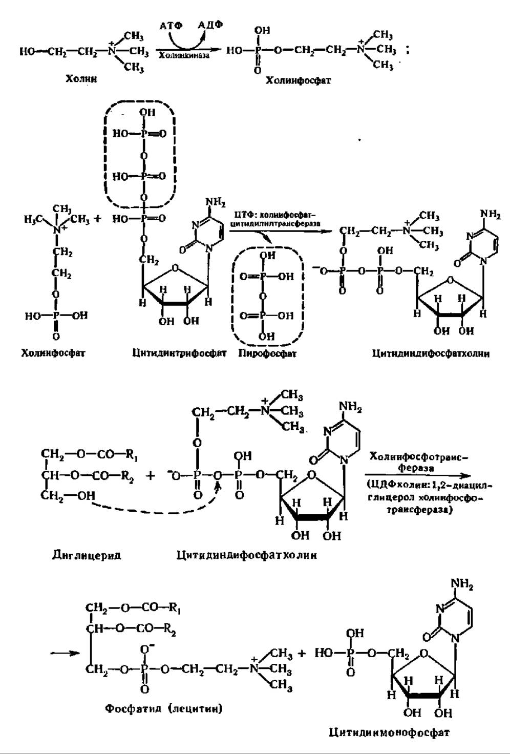
Enzymes and Intermediates of the reaction cycle described above have been found in most animal tissues, bird livers, carrots, etc. They are particularly prominent in the brain. However, in yeast, for example, the synthesis of phosphatidylcholine occurs mainly via the methylation of phosphatidylethanolamine; this is the second pathway of lecithin biosynthesis, discovered by J. Bremer and G. Greenberg (1960). Phosphatidylglycerol, diphosphatidylglycerol, phosphatidylinositol, and phosphatidylserine are synthesized via cytidine diphosphate diacylglycerols, which are formed from CTP and diglycerides under the action of CDP-diacylglycerol synthase (M = 114,000, dimer). Interacting with glycerol phosphate, Inositol, and serine, CDP-diacylglycerols form the aforementioned phosphatides with the participation of corresponding enzymes.
As for the metabolism of certain Other types of lipids (Sphingolipids, Glycolipids, etc.), it proceeds in accordance with the principles noted when considering the metabolism of triglycerides and phosphatides. The breakdown of sphingolipids, glycolipids, etc., is carried out with the participation of hydrolases, and the subsequent metabolism of their hydrolysis products proceeds via standard degradation pathways for compounds of the respective classes: carbohydrates, higher fatty acids, etc., discussed earlier. The biosynthesis of sphingolipids and glycolipids proceeds with the extensive participation of various acyl- and Glycosyltransferases.
Intermembrane lipid transfer. In recent years, a new direction in the study of lipid metabolism has emerged. It concerns the rather vigorous process of intermembrane lipid transfer, especially Phospholipids, from mitochondria to the endoplasmic reticulum and vice versa, from The cell membrane fraction to Liposomes, from liposomes of one composition to liposomes of another composition, from the inner lipid layer of the membrane to the outer one and vice versa, etc. The Significance of this dynamically occurring renewal and Modification of the membrane Lipid Composition is enormous, as it regulates the METABOLIC ACTIVITY OF the cellular membrane apparatus and subcellular structures.
Importantly, intermembrane lipid transfer is carried out by specific proteins that are ubiquitous. For example, a protein that transfers phosphatidylcholine from one membrane to another has been isolated from the cytosol of bovine liver cells. The molecular mass of this protein, which binds and transfers a single phosphatidylcholine molecule, is 22,000, pI = 5.8, and it contains 190 amino acid residues, 38% of which are polar.
Last update: 06/08/2026
Editorial and Educational Adaptation: This material has been compiled based on the primary/original source text. The project team performed an editorial review, corrected technical inaccuracies, structured sections, and adapted the content for an educational format.
What was processed:
- elimination of formatting defects (OCR errors, structural breaks, corrupted characters);
- editorial organization of content;
- standardization of terminology in accordance with academic sources;
- verification of factual statements against the original source text.
All mentions of the author, publication year, and origin of the primary text have been preserved in accordance with the source.