Molecular Biology of the Cell - Volume 2 - Alberts B., Bray D., Lewis J., Raff M., Roberts K., Watson J. 1993
Cytoskeleton
Cytoskeleton Organization
Up to this point, we have treated microtubules, Actin filaments, and Intermediate filaments as if they were independent Components of the Cytoskeleton. In reality, of course, these various cytoskeletal elements must be linked into an integrated whole, and their Functions coordinated so that The Cell can execute various types of movement and alter its shape. For example, when a fibroblast in culture rounds up in preparation for division, its entire cytoskeleton undergoes a global reorganization: stress fibers and Cytoplasmic microtubules disappear, a mitotic spindle forms, followed by a contractile ring, with all of these events unfolding as a tightly controlled sequence.
In this section, we examine the interactions among the major cytoskeletal filament systems in relation to three of its primary functions. First, we explore how the cytoskeleton organizes the Contents of the Cytoplasm, including components traditionally viewed as freely dissolved. Next, we turn to how the coordinated action of the cytoskeleton drives the directed locomotion of animal Cells across a solid substrate. Finally, we discuss how the cytoskeleton generates the myriad morphological changes that occur during embryonic development. This Overview will highlight just how fragmentary our current understanding of the molecular mechanisms underlying these fundamental processes remains.
11.6.1. The cytoplasm contains a complex three-dimensional network of protein filaments [58]
We already know that the cortical cytoplasm of many animal cells contains networks of cross-linked actin filaments. Similar networks, formed by interacting actin filaments, microtubules, and intermediate filaments, permeate the entire cytoplasm. They are most clearly visualized following extraction of cells with a nonionic detergent, which removes Phospholipids and soluble Proteins. If cells prepared in this manner are rapidly frozen and deep-etched, metal Replication of the resulting specimen reveals a striking view of the cytoskeleton (Fig. 11-78). Different types of protein filaments can be distinguished by their thickness and, in some cases, by the arrangement of their protein subunits. Neighboring filaments are frequently seen to be interconnected by finer threads, the proteins of which have also been identified in several cases using Antibodies. These have turned out to be various types of microtubule-associated proteins and long, flexible side-arms projecting from the subunits of certain intermediate filament types (see Fig. 11-77A). However, in most instances, the proteins forming these cross-links remain unknown.
In cells not treated with detergent, the cytoplasmic Structure is even more complex. The space between cytoskeletal filaments is filled with a granular "ground substance" thought to represent a highly concentrated mixture of the "soluble" proteins present in the living cell. Various membrane-bounded Organelles are also embedded in this dense matrix and connected to cytoskeletal filaments by fine protein threads. Both granular material and organelles become increasingly abundant closer to the central region of the cell, where microtubules and intermediate filaments are concentrated, and where—as can be observed using video-enhanced light Microscopy—the bulk of cytoplasmic transport takes place. In more peripheral regions, the network of actin filaments is considerably denser, effectively excluding most membrane-bounded organelles and perhaps a fraction of the granular material as well (Fig. 11-79). This dense network is anchored to The Plasma Membrane and corresponds to the actin-rich cell cortex described earlier (Section 11.2).
Class="center">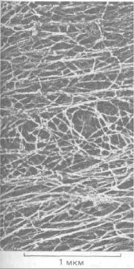
Fig. 11-78. Cytoplasm of a fibroblast extracted with a nonionic detergent (deep-etch electron micrograph). Most of the straight fibers running from left to right in loose bundles are actin filaments, whereas the entangled fibers in the middle are predominantly intermediate filaments. (Courtesy of John Heuser and Marc Kirschner.)
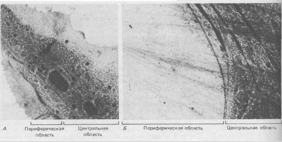
Fig. 11-79. Transmission electron micrograph of a rapidly frozen (" cryo-fixed") fibroblast, revealing an organelle-free zone at the cell periphery and an organelle-rich region toward the center (A). The boundary between these two regions is more clearly resolved at higher magnification (B). (R. S. Bridgman, B. Kachar, T. S. Reese, J. Cell Biol. 102: 1510-1521, 1986. By permission of the Rockefeller Univ. Press.)
11.6.2. How ordered is the Organization OF THE cytoplasm? [59]
When membrane-bounded organelles in the cytoplasm undergo rapid repositioning, they move along protein "tracks" to which they are linked by specialized bridges. As discussed previously, movement along microtubules is mediated by kinesin and dynein-like proteins (Section 11.4.9), whereas movement along actin filaments is driven by Myosin-like proteins (Section 11.2.4). Ribosome clusters in the Cytosol are also frequently associated with filaments; upon extraction of cells with nonionic detergents, a significant portion of the protein-synthesis machinery remains bound to the cytoskeleton. Even soluble Enzymes, including certain glycolytic enzymes, appear to be tethered to specific sites on myofibrils in Muscle cells and stress fibers in fibroblasts, where they can be visualized using fluorescent antibodies.
The degree of structural organization within the cytosol remains a subject of debate. Our knowledge of cytosolic processes is derived primarily from biochemical studies, which invariably begin with cell homogenization—a prerequisite for assaying enzyme activity and isolating enzymes in purified form. The successes of this approach have led many biochemists to view the cytosol as a simple solution of enzymes. Other investigators, however, are inclined to believe that virtually all cytosolic enzymes are organized into functional groups corresponding to specific biochemical pathways and anchored to the cytoskeleton to ensure more rapid and efficient metabolite flux along each pathway. Because such attachments are likely labile and easily disrupted, their convincing demonstration may require novel methodologies, such as the microinjection of fluorescently labeled proteins into living cells (Section 4.2.3).
11.6.3. Stretching of the actin cortex can trigger the cell polarization required for directed cell migration [60]
The cytoskeleton not only provides a structural framework for the attachment and transport of cytoplasmic components, but also makes cell migration possible. Despite recent advances in elucidating cytoskeletal Structure and function, the mechanism by which animal cells crawl across a substrate surface remains poorly understood. Efficient locomotion requires the cell to be polarized: its entire plasma membrane must remain relatively quiescent, except at the leading edge, where the cell periodically extends lamellipodia and filopodia as it advances.
When an unpolarized migrating cell initially attaches to The surface of a culture dish, filopodia and lamellipodia extend in all directions, pulling the cell in opposing directions and leaving it essentially stationary. However, if two oppositely directed lamellipodia happen to attach sufficiently firmly to the substrate, the resulting tension placed on the cell cortex between them inhibits the further formation of filopodia and lamellipodia within the stretched region—presumably because the actin filaments in this area lie parallel to the plasma membrane (Fig. 11-80) rather than perpendicular to it, which is required for the protrusion of lamellipodia and filopodia (Section 11.2.11). Thus, the absence of cytoplasmic protrusions in stretched regions can be explained on purely mechanical grounds.
Subsequent "competition" between lamellipodia resembles a tug-of-war: those pulling with less force detach from the substrate and are incorporated into the passive, stretched cortex. Eventually, only a single active region of the plasma membrane remains; this becomes the leading edge of the now-polarized cell, which initiates movement in a single direction (Fig. 11-81).
When a cell rests on a flat, uniform surface, the choice of direction (i.e., which lamellipodia "wins") is essentially random, depending on stochastic variations in cytoskeletal organization. On a non-uniform surface, however—such as a culture substrate with a gradient of an adhesion-promoting molecule—the Selection of lamellipodia and filopodia is guided by environmental cues; these protrusions act as tactile sensors that determine where the cell should move.
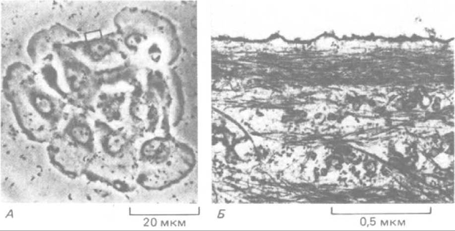
Fig. 11-80. Organization of actin filaments in a region of stretched cell cortex. The pulling forces exerted by opposing lamellipodia in a group of eight epithelial cells shown in the light micrograph (A) create a quiescent zone in one of the cells where protrusion formation is suppressed (indicated by the rectangle). Examination of this region by Electron microscopy (B) reveals bundles of actin filaments running parallel to the plasma membrane—that is, perpendicularly to the orientation required for The formation of lamellipodia and filopodia. (John Kolega, J. Cell Biol. 102: 1400-1411, 1986. By permission of the Rockefeller Univ. Press.)
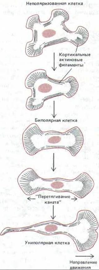
Fig. 11-81. Emergence of polarity in a migrating cell. Although a cultured cell is depicted here, similar processes likely play a fundamental role in the morphogenesis of many Tissues. Upon adhering to the culture dish surface, the cell extends lamellipodia in all directions. As these pull against one another, regions arise where the cortex is stretched and further lamellipodium formation is inhibited (see Fig. 11-80). This "tug-of-war" continues until one lamellipodium gains the upper hand—presumably by chance, for instance because the substrate beneath it happens to be more "sticky," or owing to a shallow chemotactic gradient that favors forward movement on one side of the cell (Section 11.2.13). The cell thereby becomes unipolar and migrates in the direction of the "successful" lamellipodium. Through a analogous mechanism, cells in developing tissues move toward those cells or Extracellular matrix elements to which they adhere most strongly, often choosing among substrates that differ only slightly in adhesiveness (Section 14.3.9).
11-34
11.6.4. The relative roles of Actin filaments and microtubules in cell migration depend on cell type [61]
During cell migration, microtubules and the actin cytoskeleton generally function in concert, making their individual contributions difficult to dissect. The degree to which microtubules participate in directed cell locomotion varies among different cell types. If fibroblast microtubules are depolymerized with colchicine, the cell loses its directional persistence and begins to extend lamellipodia in random directions. By contrast, the directed migration of neutrophils is virtually unaffected by colchicine. Furthermore, if a neutrophil is briefly warmed to 42 °C, a significant portion of its cortical cytoplasm detaches from the rest of the cell and initiates autonomous movement. Although these motile cell fragments lack microtubules and a nucleus, they remain capable of crawling across a culture dish for a day or longer before undergoing degeneration (Fig. 11-82). Initially, these fragments exhibit chemotactic responses comparable to those of intact neutrophils; for example, they migrate up a concentration gradient of N-formylated Peptides (Section 11.2.13). However, migration ceases if the actin filaments within the neutrophils (or their derived fragments) are depolymerized with cytochalasin. Evidently, these cells require actin filaments, but not microtubules, for directed migration.
By contrast, the movement of neuronal growth cones appears to depend on both microtubules and actin filaments. Axons and dendrites (collectively termed neurites) contain exceptionally ordered bundles of microtubules. Each neurite is "outgrown" from the neuronal cell body driven by tensile forces generated by the growth cone at its tip. When cultured Nerve Cells are treated with colchicine, neurite outgrowth halts, indicating that microtubules are required for growth cone advancement. The growth cone is functionally equivalent to the leading edge of a neutrophil or fibroblast, and the formation of lamellipodia and filopodia upon it likewise depends on actin filaments and is abolished by cytochalasin Treatment. Nevertheless, nerve cells growing on a highly adhesive surface (such as a culture dish coated with the polycation poly-L-Lysine) will extend neurites even in the presence of cytochalasin, although the direction of their growth becomes randomized. Apparently, lamellipodia and filopodia serve to guide the growth cone and enable it to navigate low-adhesiveness surfaces. The organization of microtubules and actin filaments in a growth cone and a fibroblast leading edge is compared in Fig. 11-83.
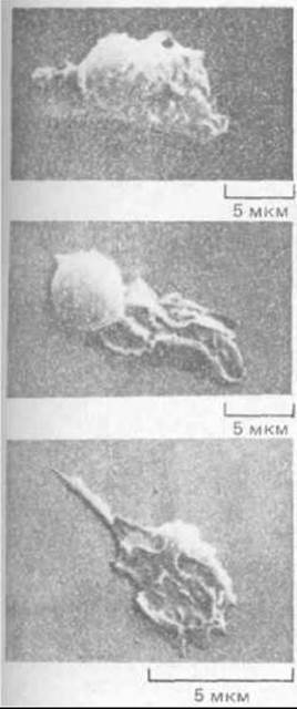
Fig. 11-82. A neutrophil from which a large fragment capable of continuous movement and chemotaxis was obtained by brief heat treatment (42°C), even though it lacked a nucleus and Mitochondria. Scanning electron micrograph. (S. R. Malawista and A. DeBoisfleury Chevance, J. Cell Biol. 95: 960–973, 1982; courtesy of Rockefeller Univ. Press.)
11.6.5. Tension in the Actin Cortex May Help Drive Animal Cell Locomotion [62]
What is the Molecular Mechanism of forward cell locomotion? There is still no definitive answer to this crucial question. One hypothesis, which assigns the primary role to the actin cortex, is illustrated in Fig. 11-84. There is clear evidence that the cortex in animal cells is under tension. This cortical tension, which has been measured in very large cells such as sea urchin eggs, tends to give suspended cells a spherical shape (i.e., one that minimizes their surface area). In addition, cells appear capable of "relaxing" the cortex in specific regions of their surface, such as the leading edge, although The Mechanism of this relaxation remains unknown. As a result, lamellipodia form periodically at the leading edge—presumably due to the activation of specialized capping proteins in the plasma membrane, which allows actin subunits to add to the plus ends of actin filaments in that region (see Section 11.2.15). Lamellipodia that fail to attach firmly to the substrate are pulled backward by cortical tension, leading to "ruffling" (see Fig. 11-42). However, if a lamellipodium attaches firmly, its actin filaments become linked to the substrate via transmembrane junctional proteins (see Fig. 11-43); the very same cortical tension that pulled the free lamellipodium backward will now propel the entire cell forward toward the new attachment site (see Fig. 11-84).
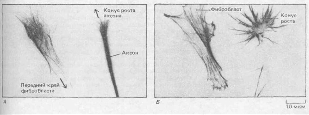
Fig. 11-83. Organization of microtubules and actin filaments in Two Types of motile cells. Immunofluorescence staining with fluorescein-labeled antibodies against tubulin (A) reveals microtubules as a fine network throughout the fibroblast cytoplasm and as a dense bundle running along the axon axis. Staining the same cells with rhodamine-phalloidin (B) reveals actin filaments as bundles that extend toward the leading edge in fibroblasts and concentrate in the microspikes of the growth cone. (Courtesy of Peter Hollenbeck.)
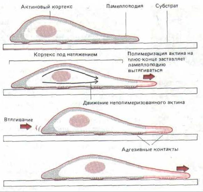
Fig. 11-84. A model showing how an actin-rich cortex could propel a cell forward. Actin polymerization leads to the extension of a lamellipodium at the leading edge; upon attaching to the substrate, the lamellipodium stretches the actin cortex, and the resulting cortical tension pulls the cell body forward while partially relieving this tension. This cycle can repeat over and over, advancing the cell step by step.
By analogy with muscle—the best-studied actin-based motility system—one might expect the contractile forces in the cortex to be generated by the interaction of Actin and myosin filaments. However, experiments with the cellular slime mold Dictyostelium discoideum argue against this possibility (Section 14.3.1). Researchers successfully generated mutants of this slime mold in which the normal Gene for conventional myosin was replaced by an artificially modified gene. A long protein-coding region was deleted from this gene (see Section 4.6.14), leaving these mutants devoid of myosin filaments. Unsurprisingly, mutant cells were unable to form a contractile ring and thus became giant multinucleated cells that divided only rarely, simply by pinching in two. Nevertheless, these cells retained The ability to migrate and even to exhibit a chemotactic response to cAMP (Section 14.3.2), although both processes were markedly impaired. Evidently, coordinated cell movement, like cortical tension, does not depend entirely on bipolar myosin filaments; tension may be generated by an elastic network of actin filaments (acting somewhat like a rubber sheet) or by other contractile forces powered, for example, by actin filament depolymerization or mini-myosin.
11.6.6. The Endocytic Cycle May Facilitate the Movement of the Leading Edge in Migrating Cells [63]
As discussed in Chapter 6, all animal cells continuously "swallow" small patches of their plasma membrane and return them to the cell surface in a process known as the endocytic cycle (Section 6.5). Evidence suggests that in polarized cells crawling along a substrate, membrane patches are internalized from the entire cell surface and returned predominantly to the leading edge. This Asymmetry in the endocytic cycle of a migrating cell presumably assists in advancing the leading edge (Section 6.5.13). The return of internalized membrane patches to the leading edge of a polarized cell likely depends on oriented microtubules and actin filaments, both of which can use accessory proteins to direct The Active Transport of membrane vesicles toward their plus ends (Sections 11.1.10 and 11.4.9). Thus, a migrating cell possesses at least two types of directed "engines" driving its locomotion: (1) an actin-filament-based mechanism in the cell cortex that protrudes lamellipodia and generates cortical tension; and (2) an internal cellular mechanism requiring oriented microtubules, actin filaments, or both, which provides active transport of membrane vesicles to the cell's leading edge (Fig. 11-85).
11.6.7. Microtubules May Act as General Cytoskeletal Organizers
In addition to participating in cell motility, microtubules play a pivotal role in determining cell shape. All Eukaryotic cells exhibit a characteristic geometry manifested both in their overall external structure and in the arrangement of their organelles. While all other cytoskeletal components merely reflect this geometry, microtubules often appear to play a unique role in establishing it. It is well known, for example, that microtubules typically align along the long axis of the cell and that in many cases their presence is required to maintain the elongated shape of The Cell as a whole.
As previously mentioned, microtubules dictate the positioning of the Golgi apparatus and Endoplasmic reticulum in each cell (Section 11.4.10) and influence the distribution of intermediate filaments, which collapse into a perinuclear cap upon colchicine Treatment of the cell (Section 11.5.3); furthermore, the distribution of actin filaments may also depend on them. The contractile ring, which is built of actin filaments and pinches the cell in two at the completion of Cell Division, always forms in a plane perpendicular to the mitotic spindle axis. If the forming spindle is mechanically displaced, the site of subsequent contractile ring formation shifts accordingly, indicating that THE POSITION OF this ring is determined by the spindle (Section 13.5.13).
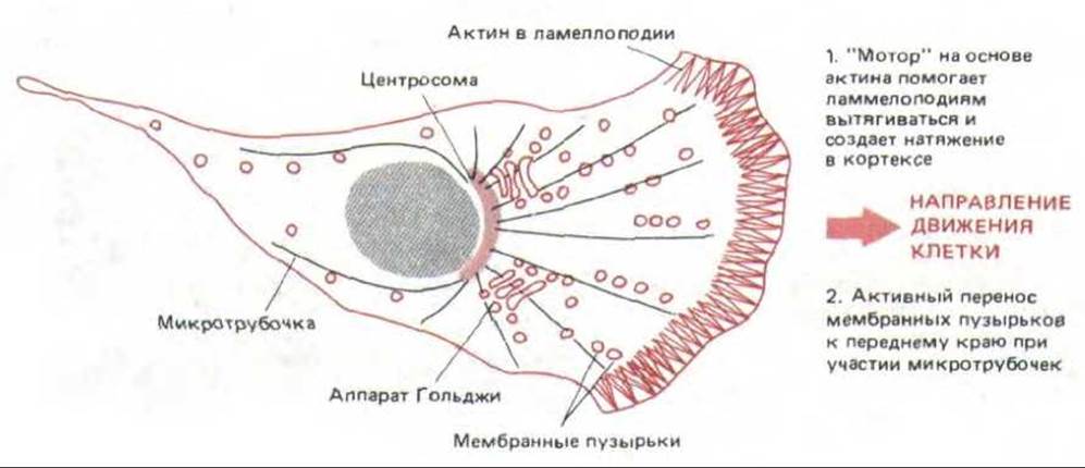
Fig. 11-85. Two directed "engines" in a migrating cell: one driven by cortical actin filaments, and the other based on The transport of membrane vesicles toward the leading edge along microtubules (and possibly actin filaments). These mechanisms presumably work together to drive directed cell movement.
The mechanisms by which microtubules influence the positioning of cortical actin filaments and intermediate filaments remain unknown, although they presumably involve cross-linking proteins that bridge different types of protein filaments.
11.6.8. The Structural organization of One Cell's Cytoskeleton Can Be Transmitted to Neighboring Cells [64]
The cytoskeleton of a given cell can influence the cytoskeleton of its neighbors. This mode of Intercellular Communication is thought to play a vital role in determining Tissue and organ Morphology. One of the simplest types of cytoskeletal interaction can be observed when the leading edges of two migrating cells Touch one another. In most cell types, this triggers an immediate "paralysis" of the leading edge in both cells—a phenomenon known as contact inhibition of locomotion. As a result, two colliding fibroblasts in vitro cease extending microspikes and lamellipodia at the contact zone and begin producing them everywhere except at this site, so that the cells gradually "walk away" from each other, reversing their direction of movement (Fig. 11-86). This response is apparently linked to rapid Changes in the cortical actin cytoskeleton at the contact zone, although the molecular mechanisms underlying these changes remain elusive.
Contact inhibition of locomotion should not be confused with contact inhibition of cell division; the latter is observed in cultured cells that continue dividing until they cover the entire surface of the culture dish. As we will see in Chapter 13, the arrest of growth and proliferation under these conditions depends not only on cell-cell contact but also on the shape cells are forced to adopt when crowded, as well as on an increasing nutrient deficit (Section 13.3.5).
Contact inhibition of locomotion plays a crucial role in wound healing. Epithelial cell sheets at the wound edges extend lamellipodia and begin migrating rapidly to "crawl" over the damaged surface; this movement ceases as soon as cells from opposite edges make contact, closing the wound gap. Now that the continuous cell sheet has been restored, Intercellular junctions form between new neighbors, serving as attachment points for protein filaments that link the cytoskeletons of all cells within the sheet (Section 14.1.2). Contact inhibition of locomotion may also promote the selective fasciculation of axons in the developing Nervous system: growth cones of central Neurons stall and even retract upon encountering Peripheral Nervous System axons, whereas they continue to grow avidly along the axons of other central neurons.
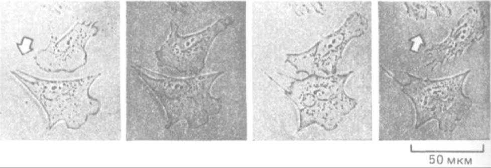
Fig. 11-86. Contact inhibition of fibroblast locomotion. When two fibroblasts crawling across the surface of a culture dish collide, their lamellipodia are paralyzed at the point of contact. After 10–15 minutes, the cells typically begin moving away from each other in opposite directions. (Courtesy of Graham Dunn.)
Another mechanism by which one cell's cytoskeleton can influence that of its neighbors involves interactions between the cytoskeleton and the extracellular matrix secreted by the cell. As has been established (see Chapter 14), a cell with an oriented cytoskeleton frequently deposits a similarly oriented extracellular matrix, which in turn influences the cytoskeletal orientation of other cells contacting that matrix (see Fig. 14-84). Thus, through intercellular interactions mediated by cell junctions and the extracellular matrix, the cytoskeletal organization of an individual cell is often determined not autonomously by the cell itself, but by the organization of the tissue as a whole.
11.6.9. Coordinated Cytoskeletal Contractions Drive the Folding of Epithelial Sheets in the Embryo [65]
The interaction between the cytoskeletons of neighboring cells underlies the bending of cell sheets, one of the fundamental morphogenetic processes in animals. How is this interaction realized? It is likely mediated by mechanical stimuli transmitted from Cell to Cell. Imagine, for example, a layer of epithelial cells in which a transient tension across the entire layer leads to the contraction of apical bundles of actin filaments located in zonula adherens (sealing desmosomes) (Section 14.1.3). If such a bundle contracts in a single cell, tension will arise in its neighboring cells, which may trigger the contraction of analogous structures in those cells as well, thereby propagating a wave of contraction throughout the entire cell sheet. Computer models based on this simple mechanism produce A wide variety of final shapes, depending on the initial geometry of the sheet. For instance, a layer of epithelial cells surrounding a spherical cavity will invaginate to form a bilayered structure, which is remarkably reminiscent of Gastrulation in sea urchin embryos (Fig. 11-87).
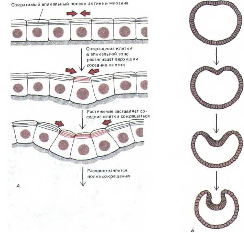
Fig. 11-87. Computer model of gastrulation based on a wave of cytoskeletal contractions propagating from cell to cell. Each cell of the epithelial sheet possesses a zonula adherens (an intercellular junction with a contractile bundle of actin filaments that is postulated to contract in response to stretch). Because the zonula adherens is located at the apical end of the cell, contraction changes the cell's shape from cylindrical to conical. In a sheet composed of contiguous cells, the contraction of one cell will stretch its neighbors, forcing them to contract in response (A). If this principle operates in a spherical or cylindrical layer of cells, it will lead to invagination (B). The exact geometric shape of this invagination will depend on the mechanical Properties of the model cell system; the system shown on the right (B) is designed to simulate gastrulation in the sea urchin embryo. (G. Odell et al., Dev. Biol. 85: 446-462, 1981.)
Although these models are entirely speculative, there is no doubt that the cytoskeleton plays a fundamental role in tissue morphogenesis. The complex shape and organization of cells in vertebrate tissues and Organs depend on cytoskeletal proteins in their diverse structural combinations. Often, the element that appears to direct morphogenetic movements is microtubules. In other cases, actin filaments play the leading role, such as during The Development of Hair cells in the vertebrate ear.
11.6.10. The development of hair cells in the cochlea of the Inner ear depends on precise control of actin polymerization [66]
Hair cells are specialized epithelial cells located in the cochlea and Vestibule of the inner ear. These cells are extremely sensitive to the slightest movement, whether it is vibration caused by sound waves or fluid displacement in the semicircular canals during HEAD position changes. These movements are detected by bundles of stereocilia arranged in a hexagonal pattern On the surface of each hair cell; within each such bundle, the stereocilia are arranged in " graded height" rows, much like the pipes of an organ (Fig. 11-88). The subtle vibrations of the stereocilia caused by sounds are converted by the hair cell into electrical signals, which are then transmitted to the Brain (Section 19.6.2).
The refinement of the structural organization of stereocilia is expressed not only in their strictly ordered arrangement on the surface of the hair cell, but also in the systematic variation of their size, number, and orientation from one end of the cochlea to the other. In chickens, each hair cell at the proximal end of the cochlea bears more than 250 stereocilia with an average length of 1.7 µm, whereas at the opposite end, each cell has only 50 stereocilia with an average length of 5.2 µm. These and other structural gradients reflect the functional organization of the hair cells, which are "tuned" to low frequencies at one end of the cochlea and to high frequencies at the other. The dimensions of the stereocilia bundle on any given cell are remarkably precise and reproducible, serving as a reliable marker for locating the cell's precise position between the two ends of the cochlea.

Fig. 11-88. Stereocilia are arranged on the surface of hair cells like organ pipes. Scanning electron micrograph. (Courtesy of Lewis Tilney.)
Stereocilia are very large, specialized microvilli that bear no relation to true cilia. They form as protrusions of the plasma membrane and contain a bundle of actin filaments (Fig. 11-89). Thus, the Formation of the cochlea clearly demonstrates the ability of cells to control the number, Location, and length of actin filaments in a highly specific manner throughout the tissue space.
Using a Scanning Electron microscope, one can observe that in the chick embryo, the growth of stereocilia occurs in three distinct stages (Fig. 11-90). First, short stereocilia appear more or less simultaneously on the surface of each hair cell. Then they begin to elongate; those that will become the longest in the mature bundle elongate first, followed by the future "second-tallest," and so on. Once they begin to grow, the stereocilia of a given row continue to elongate at a constant, slow rate (about 0.5 µm per day), apparently by The addition of actin monomers at the distal end (plus end, see Section 11.2.10) of each actin filament. After three to four days of such uniform growth, the surface of each hair cell resembles a freshly mown lawn of small stereocilia.
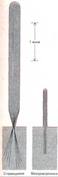
Fig. 11-89. Comparison of the sizes of a typical stereocilium and a typical microvillus; The structure of both is supported by an internal bundle of actin filaments.
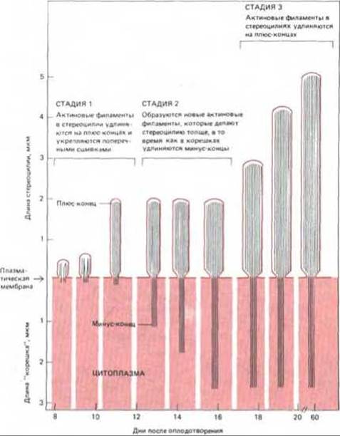
Fig. 11-90. Three stages of growth of an individual stereocilium in the chick cochlea. All stereocilia undergo the same three developmental stages, but the onset and duration of growth vary both among individual stereocilia of a single hair cell and from cell to cell. This creates a highly precise stepped gradient of stereocilia lengths. Chicks hatch on the 21st day. (From L.C. Tilney, M.S. Tilney, Hearing Research 22: 55-77, 1986.)
During the subsequent six days, the addition of actin monomers apparently occurs exclusively at the Base of the stereocilium, so that the actin filaments grow from their "roots" anchored in the cortex of the hair cell. At this stage, growth evidently takes place at the minus ends of the actin filaments; thus, when necessary, the cell can also utilize this unusual mode of growth. Concurrently, stereocilia in specific Regions of the cochlea thicken through the incorporation of new actin filaments into their core bundles.
Finally, during the Third Stage of growth (from embryonic day 17 to hatching), the addition of actin monomers to the plus ends of the filaments within the bundles resumes, and the stereocilia begin to elongate once again. Hair cells at the distal end of the cochlea now grow more rapidly, ceasing growth only when the gradient of stereocilia length characteristic of the adult chicken is established.
The mechanism that enables cells to regulate actin filament assembly with such precision remains unknown. What strictly determines the initial number of stereocilia on the surface of each hair cell? How does the cell control the final length of each actin filament bundle while ensuring the strict gradation of stereocilia lengths? These are just two of the many intriguing questions facing researchers of the cytoskeleton.
Actin filaments, microtubules, intermediate filaments, and their associated proteins are capable of self-assembly into a complex network of protein filaments that structure the cytoplasm. The cytoskeleton plays a leading role in determining cell shape and polarity, as well as cell motility. When an animal cell moves, a bundle of actin filaments periodically "pushes out" lamellipodia and microspikes at one side of the cell (the leading edge) and stretches the cell cortex, polarizing the cell and helping it to advance. This polarity is maintained by microtubules or actin filaments, which direct the flow of plasma membrane material toward the leading edge of the cell.
The cytoskeleton of a single cell can influence the cytoskeleton of its neighbors both through Intercellular Junctions and indirectly via the extracellular matrix. In this way, changes in cell shape are coordinated during the development of tissues and organs.
General
Bershadsky A.D., Vasiliev J.M. Cytoskeleton. New York: Plenum Press, 1988.
Lackie J.M. Cell Movement and Cell Behaviour. London: Allen and Unwin, 1985.
Schliwa M. The Cytoskeleton. New York, Springer-Verlag, 1986.
Shcterline P. MECHANISMS OF CELL Motility. London, Academic Press, 1983.
Cited
1. Amos L. A. Structure of muscle filaments studied by electron microscopy. Annu. Rev. Biophys. Biophys. Chem., 14, 291-313, 1985.
Huxley A. F. Reflections on Muscle. Princeton, NJ, Princeton University Press, 1980.
Squire J. M. Muscle: Design, Diversity and Disease. Menlo Park CA, Benjamin-Cummings, 1986.
2. Cooke R. The Mechanism of muscle contraction. CRC Crit. Rev. Biochem., 21, 53-118, 1986.
Huxley H. E. The mechanism of muscular contraction. Science, 164, 1356-1366, 1969.
3. Cohen C., Parry D. A. D. a-Helical coiled coils a widespread motif in proteins. Trends Biochem. Sci., 11, 245-248, 1986.
Davis J. S. Assembly processes in vertebrate skeletal thick filament formation. Annu. Rev. Biophys. Biophys. Chem., 17, 217 239, 1988.
4. Bessman S. P., Carpenter C. L. The creatine-creatine phosphate energy shuttle. Annu. Rev. Biochem., 54, 831-862, 1985.
5. Warrick H. M., Spudich J. A. Myosin structure and function in cell motility. Annu. Rev. Cell Biol., 3, 379-421, 1987.
6. Pollard T. D., Cooper J. A. Actin and actin-binding proteins. A critical evaluation of mechanisms and functions. Annu. Rev. Biochem., 55, 987-1035, 1986.
7. Irving M. Muscle mechanics and probes of the crossbridge cycle. In: Fibrous Protein Structure (J. M. Squire, P. J. Vibert, eds.). San Diego, CA, Academic Press, 1987.
Pollard T. D. The myosin crossbridge problem. Cell, 48, 909, 910, 1987.
8. Katz B. Nerve, Muscle and Synapse. New York, McGraw-Hill, 1966. [Russian Translation: Katts B. Nerv, myshtsa i sinaps. Moscow, Mir, 1969].
Lai F. A., Erickson H. P., Rousseau E., Liu O.-Y., Meissner G. Purification and reconstruction of the calcium release channel from Skeletal Muscle. Nature, 331, 315-319, 1988.
Saito A., Inui M., Radermacher M., Frank J., Fleischer S. ULTRASTRUCTURE OF THE calcium release channel of sarcoplasmic reticulum. J. Cell Biol., 107, 211 219, 1988.
9. Murray J. M., Weber A. The cooperative action of Muscle Proteins. Sci. Am., 230(2), 59-71, 1974.
Phillips G. N., Fillers J. P., Cohen C. Tropomyosin crystal structure and muscle regulation. J. Mol. Biol., 192, 111-131, 1986.
Zot A. S., Potter J. D. Structural aspects of troponin-tropomyosin regulation of skeletal Muscle contraction. Annu. Rev. Biophys. Biophys. Chem., 16, 535-559, 1987.
10. Wang K. Sarcomere-associated cytoskeletal lattices in striated muscle. Review and hypothesis. In: Cell and Muscle Motility (J. W. Shay, ed.). Vol. 6, pp. 315-369. New York, Plenum Press, 1985.
11. Fawcett D. W. A Textbook of Histology, 11th ed. Philadelphia, Saunders, 1986.
12. Korn E. D., Hammer J. A. Myosins of nonmuscle cells. Annu. Rev. Biophys. Biophys. Chem., 17, 23-45, 1988.
Sellers J. R., Adelstein R. S. Regulation of contractile activity. In: The Enzymes (P. Boyer, E. G. Krebs, eds.), Vol. 18, pp. 381-418. San Diego, CA, Academic Press, 1987.
13. Citi S., Kendrick-Jones J. Regulation of non-muscle myosin structure and function. Bioessays, 7, 155- 159, 1987.
14. ByersH.R., Fujiwara K. Stress fibers in cells in situ: immunofluorescence visualization with anti-actin, anti-myosin and anti-alpha-actinin. J. Cell Biol., 93, 804-811, 1982.
Langanger B. et al. The Molecular organization of myosin in stress fibers of cultured cells. J. Cell Biol., 102, 200-209, 1986.
Schroeder Т.Е. Actin in dividing cells: contractile ring filaments bind heavy meromyosin. Proc. Natl. Acad. Sci. USA, 70, 1688-1692, 1973.
15. Buckingham M.E. Actin and myosin multigene families: their expression during the formation of skeletal muscle. Essays Biochem., 20, 77-109, 1985.
Emerson C. P., Bernstein S. I. Molecular Genetics of myosin. Annu. Rev. Biochem., 56, 695-726, 1987.
Otey C.A., Kalnoski M.H., Bulinski J.C. Identification and quantification of actin isoforms in vertebrate Cells and Tissues. J. Cell. Biochem., 34, 113-124, 1987.
16. Breitbart R.E., Andreadis A., Nadal-Ginard B. Alternative Splicing: a ubiquitous mechanism for the generation of multiple protein isoforms from single genes. Annu. Rev. Biochem., 56, 467-495, 1987.
17. Bray D., Heath J., Moss D. The membrane-associated "cortex" of animal cells: its structure and mechanical properties. J. Cell Sci. Suppl. 4, 71-88, 1986.
Korn Е. D. Actin polymerization and its regulation by proteins from nonmuscle cells. Physiol. Rev., 62, 672-737, 1982.
Pollard Т.О., Cooper J. A. Actin and actin-binding proteins. A critical evaluation of mechanisms and functions. Annu. Rev. Biochem., 55, 9871035, 1986.
Tilney L. G. Interactions between actin filaments and membranes give spatial organization to cells. In: Modern Cell Biology, Vol. 2. Spatial Organization of Eukaryotic Cells (J. R. Mclntosh, В. Н. Satir esdl.) pp. 163-199. New York: Liss, 1983.
18. Sato M., Schwartz W.H., Pollard Т.О. Dependence of the mechanical properties of actin/alpha-actinin gels on deformation rate. Nature, 325, 828-830, 1987.
Stossel T. P. et el. Non-muscle actin binding proteins. Annu. Rev. Cell Biol., 1, 353-402, 1985.
19. Matsudaira P., Janmev P. Pieces in the actin-severing protein puzzle. Cell, 54, 139-140, 1988.
Yin H. L. Gelsolin: calcium and polyphosphoinositide-regulated actin modulating protein. Bioessays, 7, 176 179, 1987.
20. Korn E. D., Hammer J. A. Myosins of nonmuscle cells. Annu. Rev. Biophys. Biophys. Chem., 17, 23-45, 1988.
Warrick H.M., Spudlich J.A. Myosin structure and function in cell motility. Annu. Rev. Cell Biol., 3, 379-421, 1987.
21. Adams R. J., Pollard T.D. Propulsion of organelles isolated from Acanthamoeha along actin filaments by myosin-I. Nature, 322, 754 756, 1986.
Sheetz M. P., Spudich J. A. Movement of myosin-coated fluorescent beads on actin cables in vitro. Nature, 303, 31-35, 1983.
22. Bennett V. The membrane Skeleton of human erythrocytes and its implications for more complex cells. Annu. Rev. Biochem., 54, 273-304, 1985.
23. Conzelman K.A., Mooseker M.S. The 110-kD protein-calmodulin complex of the intestinal microvillus in an actin-activated MgATPase. J. Cell Biol., 105, 313-324, 1987.
Mooseker M. S. Organization, chemistry, and assembly of the cytoskeletal apparatus of the intestinal brush border. Annu. Rev. Cell. Biol., 1, 209-241, 1985.
24. Burridae K. et al. Focal adhesions: transmembrane junctions between the extracellular matrix and cytoskleton. Annu. Rev. Cell Biol., 4, 487-525, 1988.
Horwitz A., Duyaan K., Buck C., Beckerle M.C., Bur ridge K. Interaction of plasma membrane Fibronectin receptor with talin-a transmembrane linkage. Nature, 320, 531-533, 1986.
25. Bonder E. M., Fishkind D.J., Mooseker M.S. Direct measurement of critical concentrations and assembly rate constants at the two ends of an actin filament. Cell, 34, 491-501, 1983.
Korn E.D., Carlier M.-F., Pantaloni D. Actin polymerization and ATP Hydrolysis. Science, 238, 638-644, 1987.
26. Abercrombie M. The crawling movement of metazoan cells. Proc. R. Soc. Lond. (Biol.), 207, 129-147, 1980.
Small J. V., Rinnerthaler G., Hinssen H. Organization of actin meshworks in cultured cells: the leading edge. Cold Spring Harbor Symp. Quant. Biol., 46, 599-611, 1982.
Wany Y. Exhange of actin subunits at the leading adge of living fibroblasts: possible role of treadmiling. J. Cell Biol., 101, 597 602, 1985.
27. Tilney L. G., Inoue S. Acrosomal reaction of Thyone sperm. II. The kinetics and possible mechanism of acrosomal process elongation. J. Cell Biol., 93, 820-827, 1982.
28. Carson M., Weber A., Zigmond S. H. An actin-nucleating activity in poly-morphonuclear leukocytes is modulated by chcrnotactic peptides, J. Cell Biol., 103, 2707-2714, 1986.
Devreotes P., Zigmond S. Chemotaxis in eukaryotic cells. Annu. Rev. Cell Biol., 4, 649-686, 1988.
Tilney L. G., Bonder E. M., DeRosier D.J. Actin filaments elongate from their membrane-associated ends. J. Cell Biol., 90, 485-494, 1981.
29. Cooper J.A. Effects of Cytochalasin and phalloidin on actin. J. Cell Biol., 105, 1473-1478, 1987.
30. Dustin P. Microtubules, 2nd ed., pp. 127-164. New York, Springer-Verlag, 1984.
Gibbons I.R. Cilia and flagella of eukaryotes. J. Cell Biol., 91, 107s-124s, 1981.
Roberts K., Hyams J.S., eds. Microtubules. New York. Academic Press, 1979. Satir P. How cilia move. Sci. Am., 231(4), 44-63, 1974.
31. Amos L. A., Baker T.S. The Three dimensional structure of tubulin protofilaments. Nature, 279, 607-612, 1979.
Mandelkow E.-M., Schultheiss R., Rapp R., Muller M., Mandelkow E. On the surface lattice of microtubules: helix starts, protofilament number, seam and handedness. J. Cell Biol., 102, 1067-1073, 1986.
Raff E.C. Genetics of microtubule systems. J. Cell Biol., 99, 1-10, 1984.
Sulivan К. F. Structure and utilization of tubulin isotypes. Annu. Rev. Cell Biol., 4, 687-716, 1988.
32. Linek R. W., Amos W. B. Localization of tektin filaments in microtubules of sea urchin sperm flagella by immunoelectron microscopy, J. Cell Biol., 100, 126-135, 1985.
33. Goodenough U. W., Heuser J. E. Substructure of inner dynein arms, radial spokes, and the central pair/projection complex of cilia and flagella.
J. Cell Biol., 100, 2008 2018, 1985.
34. Summers K. E., Gibbons 1. R. ATP-induced sliding of tubules in Trypsin-treated flagella of sea urchin sperm. Proc. Natl. Acad. Sci. UDA, 68, 3092-3096, 1971. Warner F. D., Satir P. The structural basis of ciliary bend formation. J. Cell Biol., 63, 35-63, 1974.
35. Johnson K. A. Pathway of the microtubule-dynein ATPase and structure of dynein: a comparison with Actomyosin. Annu. Rev. Biophys. Biophys. Chem., 14, 161-188, 1985.
36. Brokaw C.J. Future directions for studies of mechanisms for generating flagellar bending waves. J. Cell Sci., Suppl. 4, 103 113, 1986.
Brokaw C. J., Luck D. J. L., Huang B. Analysis of the movement of Chlamydomonas flagella: the function of the radial-spoke system is revealed by comparison of wild-type and mutant flagella. J. Cell Biol., 92, 722-732, 1982.
37. Afzelius B. A. The immotile-cilia syndrome: a microtubule-associated defect. CRC Crit. Rev. Biochem., 19, 63-87, 1985.
Huang B. Chlamydomonas reinhardtii: a model system for genetic analysis of flagellar structure and motility. Int. Rev. Cytol., 99, 181-215, 1986.
Luck D. J. L. Genetic and biochemical dissection of the eukaryotic flagellum. J. Cell Biol., 98, 789-794, 1984.
38. Lefebvre P. A., Rosenbaum J. L. Regulation of the synthesis and assembly of ciliary and flagellar proteins during regeneration. Annu. Rev. Cell Biol., 2, 517-546, 1986.
Wheatley D. N. The Centriole: A Central Enigma of Cell Biology. New York, Elsevier, 1982.
39. Karsenti E., Maro B. Centrosomes and the spatial distribution of microtubules in animal cells. Trends Biochem. Sci., 11, 460-463, 1986. Ramanis Z., Luck D. J. L. Loci affecting flagellar assembly and function map to an unusual linkage group in Chlamydomonas reinhardtii. Proc. Natl. Acad. Sci. USA, 83, 423-436, 1986.
Vorobjev I. A., Chentsov Y. S. Centrioles in the Cell Cycle. 1. Epithelial cells. J. Cell Biol., 93, 938-949, 1982.
40. Dustin P. Microtubules, 2nd ed. Berlin, Springer-Verlag, 1984.
41. De Brabander M. et al. Microtubule dynamics during the cell cycle: the effects of taxol and nocodazole on the microtubule system of PtK2 cells at different Stages of the mitotic cycle. Int. Rev. Cytol., 101, 215-274, 1986.
Inoue S. Cell division and the mitotic spindle. J. Cell Biol., 91, 131s-147s, 1981.
Salmon E. D., McKeel M., Hays T. Rapid rate of tubulin dissociation from microtubules in the mitotic spindle in vivo measured by blocking polymerization with colchicine. J. Cell Biol., 99, 1066-1075, 1984.
42. Farrell K. W., Jordan M. A., Miller H. P., Wilson L. Phase dynamics at microtubule ends: the coexistence of microtubule length changes and treadmilling. J. Cell Biol., 104, 1035-1046, 1987.
McIntosh J. R., Euteneuer U. Tubulin hooks as probes for microtubule polarity: an analysis of the method and evaluation of data on microtubule polarity in the mitotic spindle. J. Cell Biol., 98, 525-533, 1984.
43. Carlier M.-F. Role of nucleotide hydrolysis in the polymerization of actin and tubulin. Cell Biophys., 12, 105-117, 1988.
Horio H., Hotani H. Visualization of the dynamic instability of individual microtubules by Dark field Microscopy. Nature, 321, 605-607, 1986. Mitchison T., Kirschner M. Dynamic instability of microtubule growth. Nature, 312, 237-242, 1984.
44. Karsenti E., Maro B. Centrosomes and the spatial distribution of microtubules in animal cells. Trends Biochem. Sci., 11, 460-463, 1986. Mitchison T., Kirschner M. Microtubule assembly nucleated by isolated centrosomes. Nature, 312, 232-237, 1984.
45. Kirschner M., Mitchison T. Beyond self-assembly: from microtubules to morphogenesis. Cell, 45, 329-342, 1986.
Sammak P. J., Borisy G. G. Direct observation of microtubule dynamics in living cells. Nature, 332, 724-726, 1988.
46. Barra H. S., Arce C. A., Argarana C. E. Posttranslational tyrosination and detyrosination of tubulin. Molec. Neurobiol., 2, 133-153, 1988. Gundersen G. G., Khawja S., Bulinski J. C. Postpolymerization detyrosination of [alpha]-tubulin: a mechanism for subcellular differentiation of microtubules. J. Cell Biol., 105, 251-264, 1987.
Maruta H., Greer K., Rosenbaum J. L. The Acetylation of a-tubulin and its relationships to the assembly and disassembly of microtubules. J. Cell Biol., 103, 571-579, 1986.
Schulze E., Asai D. J., Bulinski J. C., Kirschner M. Post-translational modification and microtubule stability. J. Cell Biol., 105, 2167-2177, 1987.
47. Olmsted J. B. Microtubule-associated proteins. Annu. Rev. Cell Biol., 2, 421-457, 1986.
Vallee R. B., Bloom G. S., Theurkauf W. E. Microtubule-associated proteins: subunits of the cytomatrix. J. Cell Biol., 99, 38s-44s, 1984.
48. Allen R. D. The microtubule as an intracellular engine. Sci. Am., 256(2), 42-49, 1987.
Allen R. D. et al. Gliding movement of and bidirectional transport along single native microtubules from squid axoplasm: evidence for an active role of microtubules in cytoplasmic transport. J. Cell Biol., 100, 1736-1752, 1985.
49. Vale R. Intracellular Transport using microtubule-based motors. Annu. Rev. Cell Biol., 3, 347-378, 1987.
Vale R.D., Reese T.S., Sheetz M.P. Identification of a novel force-generating protein, kinesin, involved in microtubule-based motility. Cell, 42, 39 50, 1985.
Vallee R. В., Wall J. S., Paschal B. M., Shpetner H. S. Microtubule-associated protein 1C from brain is a two-headed cytosolic dynein. Nature, 332, 561 563, 1988.
50. Allan V.J., Kreis Т.Е. A microtubule-binding protein associated with membranes of the Golgi apparatus. J. Cell Biol., 103, 2229-2239, 1986. Dabora S. L., Sheet- M. P. The microtubule-dependent formation of a tubulovesicular network with CHARACTERISTICS OF THE ER from cultured cell extracts. Cell, 54, 27-35, 1988.
Lee C., Chen L. B. Dynamic behavior of endoplasmic reticulum in living cells. Cell, 54, 37-46, 1988.
Lucocq J. M., Warren G. Fragmentation and partitioning of the Golgi apparatus during mitosis in HeLa cells. EMBO J., 6, 3239-3246, 1987.
51. Geiger B. Intermediate filaments: looking for a function. Nature, 329, 392-393, 1987. Steinert P. M., Roop D. R. MOLECULAR AND CELLULAR biology of intermediate filaments. Annu. Rev. Biochem., 57, 593-626, 1988.
Traub P. Intermediate Filaments: A Review. New York, Springer-Verlag, 1985. Wang E., Fischman D., Liem B.K.H., Sun T.-T., eds. Intermediate Filaments. Ann. N.Y. Acad. Sci., 455, 1985.
52. Osborn M., Weber K. Tumor Diagnosis by intermediate filament typing: a novel tool for surgical pathology. Lab. Invest., 48, 372-394, 1983.
53. Ip W., Hartzer M. K., Pang S. Y.-Y., Robson R. M. Assembly of vimentin in vitro and its implications concerning the structure of intermediate filaments. J. Mol. Biol., 183, 365-375, 1985.
Quintan R. A. ct al. Characterization of dimer subunits of intermediate filament proteins. J. Mol. Biol., 192, 337-349, 1986.
54. Geuens G., De Brabander M., Nuydens R., De Mey J. The interaction between microtubules and intermediate filaments in cultured cells treated with taxol and nocodazole. Cell Biol. Int. Rep., 7, 35-47, 1983.
Goldman R. et al. Intermediate filaments: possible functions as cytoskeletal connecting links between The Nucleus and the cell surface. Ann. N. Y. Acad. Sci., 45, 1-17, 1985.
55. Geisler N., Weber K. Phosphorylation of desmin in vitro inhibits formation of intermediate filaments: identification of three kinase A sites in the aminoterminal head domain. EMBO J., 7, 15 20, 1988.
Inagaki M., Nishi Y., Nishizawa K., Matsuyama M., Sato C. Site-specific phosphorylation induces disassembly of vimentin filaments in vitro. Nature, 328, 649 652, 1987.
56. Aebi U., Cohn J., Buhle L., Gerace L. The nuclear lamina is a meshwork of intermediate-type filaments. Nature, 323, 560-564, 1986.
McKeon F.D., Kirschner M. W., Caput D. Homologies in both Primary and secondary structure between nuclear envelope and intermediate filament proteins. Nature, 319, 463-468, 1986.
57. Abercrombie M. The crawling movement of metazoan cells. Proc. R. Soc. Lond. (Biol.), 207, 129-147, 1980.
Bridgman P. Structure of cytoplasm as revealed by modern electron Microscopy Techniques. Trends Neurosci., 10, 321-325, 1987.
Singer S. J., Kupfer A. The directed migration of eukaryotic cells. Annu. Rev. Cell Biol., 2, 337-365, 1986.
Trinkaus J. P. Cells into Organs: The Forces that Shape the Embryo. 2nd ed. Englewood Clifis, NJ, Prentice-Hall, 1984.
58. Bridgman P. C., Reese T. S. The structure of cytoplasm in directly frozen cultured cells. 1. Filamentous meshworks and the cytoplasmic ground substance. J. Cell Biol., 99, 1655-1668, 1984.
Heuser J., Kirschner M. W. Filament organization revealed in platinum replicas of freeze-dried cytoskeletons. J. Cell Biol., 86, 212-234, 1980.
59. Fulton A.B. How crowded is the cytoplasm? Cell, 30, 345-347, 1982.
Luby-Phelps K., Taylor D. L., Lanni F. Probing the STRUCTURE OF THE cytoplasm. J. Cell Biol., 102, 2015-2022, 1986.
60. Kolega J. Effects of mechanical tension on protrusive activity and microfilament and intermediate filament organization in an epidermal epithelium moving in culture. J. Cell Biol., 102, 1400 1411, 1986.
Trinkam J. P. Cells into Organs: The Forces That Shape the Embryo, 2nd ed., pp. 157 244. Englewood Cliffs, NJ. Prentice-Hall, 1984.
61. Bray D., Hollcnbeck P.J. Growth cone motility and guidance. Annu. Rev. Cell Biol., 4, 43-62, 1988.
Euteneuer U., Schliwa M. Persistent, directional motility of cells and cytoplasmic fragments in the absence of microtubules. Nature, 310, 5861, 1984.
Malawista S. E., De Boisfleury Chevance A. The cytokinetoplast: purified, stabile, and functional motile machinery from human Blood polymorphonuclear leukocytes. J. Cell Biol., 95, 960-973, 1982.
Marsh L., Letourneau P. C. Growth of neurites without filopodial or lamellipodial activity in the presence of cytochalasin B. J. Cell Biol., 99, 2041-2047, 1084.
Vasiliev J. M. et at. Effect of colcemid on the locomotion of fibroblasts. J. Embryol. Exp. Morphol., 24, 625-640, 1970.
62. Bray D., White J.G. Cortical flow in animal cells. Science, 239, 883-888, 1988.
De Lozannc A., Spudich J.A. Disruption of the Dictyostelium myosin heavy chain gene by Homologous Recombination. Science, 236, 10861091, 1987.
Knecht D. A., Loomis W. F. Antisense RNA inactivation of myosin heavy chain Gene Expression IN Dictyostelium discoideum. Science, 236, 1081 1086, 1987.
63. Bergmann J.E., Kupfer A., Singer S.J. Membrane insertion at the leading edge of motile fibroblasts. Proc. Natl. Acad. Sci., USA, 80, 1367-1371, 1983.
Bretscher M.S. How animal cells move. Sci. Am., 257(6), 72 90, 1987.
64. Lackie J.M. Cell Movement and Cell Behaviour, pp. 253-275. London, Alien and Unwin, 1986.
Trinkaus J. P. Cells into Organs: The Forces That Shape the Embryo, 2nd ed., pp. 157 244. Englewood Cliffs, NJ. Prentice-Hall, 1984.
65. Ode/I G. M., Oster G., Alberch P., Burnside B. The mechanical basis of morphogenesis. 1. Epithelial folding and invagination. Dev. Biol., 85, 446-462, 1981.
66. Tilncy L. G., Tilney M. S., Cotanche D. A. Aclin filaments, stereocilia, and hair Cells of the cochlea. V. How the staircase pattern of stereociliary lengths in generated. J. Cell Biol., 106, 355-365, 1988.
Tilney L. G., De Rosier D. J. Actin filaments, stereocilia, and hair cells of the bird cochlea. IV. How the actin filaments become organized in developing stereocilia and in the cuticular plate. Dev. Biol., 116, 119-129, 1986.
Last update: 12/08/2026
Editorial and Educational Adaptation: This material has been compiled based on the primary/original source text. The project team performed an editorial review, corrected technical inaccuracies, structured sections, and adapted the content for an educational format.
What was processed:
- elimination of formatting defects (OCR errors, structural breaks, corrupted characters);
- editorial organization of content;
- standardization of terminology in accordance with academic sources;
- verification of factual statements against the original source text.
All mentions of the author, publication year, and origin of the primary text have been preserved in accordance with the source.