BIOCHEMISTRY - L. Stryer - 1984
VOLUME 2
PART II GENERATION AND STORAGE OF METABOLIC ENERGY
CHAPTER 14. OXIDATIVE PHOSPHORYLATION
14.15. Three-Dimensional Structure of Cytochrome c
Cytochrome c is the only protein electron carrier that can be detached from The inner mitochondrial membrane by mild treatments. The solubility of this peripheral membrane protein in Water facilitates its purification and crystallization. Indeed, much more is known about The Structure of cytochrome c than about that of any other electron-transfer protein.
Cytochrome c consists of a single 104-residue polypeptide chain covalently attached to a heme group. The three-dimensional STRUCTURE OF THE ferro- and ferri- forms of cytochrome c was solved by Richard Dickerson at near-atomic resolution (Fig. 14.14). The protein is nearly spherical with a diameter of 34 A. The heme group is surrounded by numerous tightly packed hydrophobic side chains. The iron atom is coordinated to the sulfur atom of a Methionine residue and the nitrogen atom of a Histidine residue (Fig. 14.15). The hydrophobic Nature of the heme environment accounts for the higher positive Redox Potential of cytochrome c (corresponding to a higher electron affinity) compared with the same heme complex in an aqueous medium. Removing an electron from the heme within cytochrome c is energetically less favorable than removing it from heme in water because the dielectric constant near the iron atom is lower in cytochrome c.
Class="center">Fig. 14.14. Three-dimensional structure of reduced tuna cytochrome c. The heme group (red), methionine-80 (blue), histidine-18 (blue), and α-carbon atoms are shown.
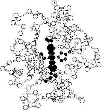
Fig. 14.15. The iron atom of the heme group in cytochrome c is bonded to methionine and histidine side chains.

The overall structure of the molecule can be described as a single-residue-thick shell tightly enveloping the heme. Hydrophobic side chains form the interior of the shell. Next comes the main chain, followed by charged side chains located On the surface. There is a very short α-Helix, and β-pleated sheets are absent. Essentially, the polypeptide chain is wrapped around the heme. Residues 1 through 47 lie on the histidine-18 side of the heme group (designated as the right side), and residues 48 through 91 are on the methionine-80 side (designated as the left side). Residues 92 through 104 loop back across the heme to its right side.
14.16. Interaction of Cytochrome c with Its Reductase and Oxidase
As noted earlier, cytochrome c transfers electrons from the QH2-cytochrome c reductase complex (the second energy-conserving site) to the cytochrome c oxidase complex (the third energy-conserving site). How does cytochrome c interact with its reductase and subsequently with its oxidase? An important approach to addressing this question is studying the distribution of charged residues on the protein surface. In cytochrome c molecules from all species studied to date, there are clusters of Lysine side chains surrounding the heme crevice on one surface of the protein (the front of Fig. 14.14). The surface charge distribution of cytochrome c presumably plays a role in the recognition and binding of the reductase and oxidase. For instance, the interaction between cytochrome c and cytochrome oxidase is disrupted by the Modification of lysine-13. Furthermore, polylysine competes with cytochrome c for binding to both Enzymes.
How does cytochrome c accept electrons from the reductase that it then donates to the oxidase? A priori, two mechanisms are possible. Electron transfer between the heme groups of different Proteins could be mediated by aromatic side chains. Another possibility is direct Electron transfer from one heme to another. It is important to note that the electron carried by the heme is not necessarily localized on its iron atom; rather, it is partially delocalized over the entire conjugated ԉ-electron System of the heme. Consequently, an electron can be transferred from one heme to another if their edges are sufficiently close (less than 8 A apart) and their planes are approximately parallel. The direct electron-transfer mechanism appears more likely due to the very large Free energy required to form an aromatic side chain free-radical anion. Moreover, one of the heme edges in cytochrome c (the front edge in Fig. 14.14) is accessible for direct electron-transfer reactions.
14.17. The Conformation of Cytochrome c Has Remained Largely Constant for Billions of Years
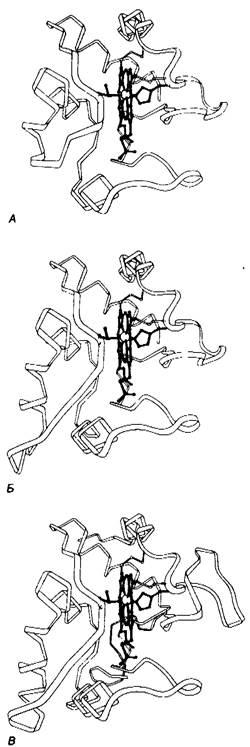
Cytochrome c is present in all organisms possessing mitochondrial respiratory chains: plants, animals, and eukaryotic microorganisms. This electron carrier arose more than 1.5 billion years ago, prior to the divergence of the PLANT AND ANIMAL kingdoms. Its function has remained unchanged throughout this period, as evidenced by the fact that cytochrome c from any eukaryotic species reacts in vitro with cytochrome oxidase from any currently studied species. For example, cytochrome c from wheat germ reacts with cytochrome oxidase from human tissue. A second criterion for functional conservation is the close similarity of redox potentials for all cytochrome c molecules (about +0.25 V). A third criterion is the near-identical absorption spectra of cytochrome c molecules from different species. Indeed, the Cytochromes of certain prokaryotes—such as cytochrome c2 from photosynthetic Bacteria and cytochrome c550 from denitrifying bacteria—are remarkably similar to cytochrome c from tuna Heart Cell/35.html">Mitochondria (Fig. 14.16).
Fig. 14.16. Conservation of the three-second structure of cytochrome c during evolution, illustrated by the conformational similarity of cytochrome c from tuna heart mitochondria (A), cytochrome c2 from the photosynthetic bacterium Rhodospirillum rubrum (B), and cytochrome c550 from the denitrifying bacterium Paracoccus denitrificans (C).
Emil Smith, Emanuel Margoliash, and coworkers determined the Amino acid sequences of Cytochromes c from more than eight widely divergent eukaryotic species. They made a striking discovery: 26 of the 104 residues have remained invariant over more than 1.5 billion years of evolution. Now that the three-dimensional structure of the molecule is known, the reasons for this constancy in most of these residues become apparent. As might be expected, the heme ligands methionine-80 and histidine-18 are invariant, as are the two cysteines covalently linked to the heme. An 11-residue sequence (from position 70 to 80) is nearly identical in all cytochrome c molecules. Many hydrophobic residues contacting the heme are invariant. Throughout evolution, cytochrome c has conserved most of its Glycine residues. As mentioned previously (Section 9.6), glycine residues are crucial due to their small size. The compact folding of the peptide chain requires the presence of glycine at specific positions. Several invariant lysine and Arginine residues are localized in positively charged clusters on the molecular surface. One such cluster interacts with cytochrome c reductase, and another with cytochrome c oxidase.
Last update: 06/08/2026
Editorial and Educational Adaptation: This material has been compiled based on the primary/original source text. The project team performed an editorial review, corrected technical inaccuracies, structured sections, and adapted the content for an educational format.
What was processed:
- elimination of formatting defects (OCR errors, structural breaks, corrupted characters);
- editorial organization of content;
- standardization of terminology in accordance with academic sources;
- verification of factual statements against the original source text.
All mentions of the author, publication year, and origin of the primary text have been preserved in accordance with the source.