BIOCHEMISTRY: A TEXTBOOK FOR HIGHER EDUCATION - E. S. Severin - 2004
SECTION 15. BIOCHEMISTRY OF THE EXTRACELLULAR MATRIX
In Multicellular Organisms, most Cells are surrounded by an extra- or intercellular matrix. The Extracellular matrix is a complex network of interconnected macromolecules. These macromolecules (Proteins and Heteropolysaccharides) are typically secreted by the cells themselves, and within the extracellular matrix, they assemble into an ordered network. The extracellular matrix surrounding cells influences their attachment, development, proliferation, Organization, and METABOLISM.
The extracellular matrix, together with the various types of cells residing within it (fibroblasts, chondro- and osteoblasts, mast cells, and macrophages), is often referred to as Connective Tissue.
The extracellular matrix performs A wide variety of functions in the body:
✵ provides a structural framework for Organs and Tissues;
✵ acts as a universal "biological" glue;
✵ participates in The regulation of Water and Salt Metabolism;
✵ forms highly specialized structures (bones, Teeth, Cartilage, tendons, basement membranes).
The Main Components of the extracellular matrix include structural proteins such as Collagen and Elastin, glycosaminoglycans, Proteoglycans, as well as non-collagenous structural proteins (fibronectin, Laminin, tenascin, osteonectin, etc.).
I. Collagen
Collagen is the primary structural protein of the extracellular matrix. It accounts for 25% to 33% of the total protein content in the body, which is approximately 6% of body weight. The term "collagen" encompasses a family of closely related Fibrillar Proteins that serve as the main protein framework of the Skin, bones, tendons, cartilage, Blood Vessels, and teeth. Different tissues are dominated by Different types of collagen, which in turn is determined by the specific role collagen plays in a given organ or tissue. For example, in lamellar Bone tissue—which makes up most of the flat and tubular BONES OF THE Skeleton—collagen fibers have a strictly oriented direction: longitudinal in the central part of the lamellae, and transverse or angled in the peripheral regions. This ensures that even if the lamellae delaminate, the fibrils of one lamella can continue into adjacent ones, thereby creating a unified fibrous Bone Structure. Transversely oriented collagen fibers can weave into the intermediate layers between bone lamellae, providing the tissue with mechanical strength. In tendons, collagen forms dense, parallel fibers that enable these structures to withstand heavy mechanical loads. In the cartilage matrix, collagen forms a fibrillar network that imparts strength to the cartilage, while in the Cytology/practical/76.html">Cornea of the eye, collagen participates in forming the hexagonal lattices of Descemet's membranes, ensuring corneal transparency and the refraction of light rays. In the dermis, collagen fibrils are oriented to form a network that is especially well-developed in areas of the skin subjected to high pressure (such as the soles, elbows, and palms), whereas in healing wounds, they aggregate in a highly chaotic manner. The Amino Acid Composition and conformation of collagen are described in subsection "Fibrillar Proteins" of Section 1.
Here, we will examine the synthesis and maturation of collagen, the structures it forms and their functions, as well as DISEASES ASSOCIATED WITH the disruption of these processes.
Collagen Polymorphism
Collagen is a prominently polymorphic protein. Currently, 19 types of collagen are known, which differ in the Introduction/19.html">Primary Structure of their peptide chains, their functions, and their localization in the body. There are far more than 19 Variants of the α-chains that form the triple helix (around 30). Each type of collagen is designated by a specific formula in which the collagen type is written in Roman numerals in parentheses, while Arabic numerals are used to designate the α-chains. For example, type II and type III collagens are formed by identical α-chains, with their respective formulas being [α1(II)]3 and [α1 (III)]3; type I and type IV collagens are heterotrimers typically formed by two different types of α-chains, with their respective formulas being [α1(I)]2α2(I) and [α1(IV)]2α2(IV). The subscript outside the parentheses indicates the number of identical α-chains. The distribution of collagens across organs and tissues is presented in Table 15-1.
Class="center">Table 15-1. Distribution of collagen in tissues and organs
Types |
Genes |
Tissues and organs |
I |
СОL1А1, СОL1А2 |
Skin, tendons, bones, cornea, Placenta, Arteries, Liver, dentin |
II |
СОL2А1 |
Cartilage, intervertebral discs, vitreous body, cornea |
III |
СОL3А1 |
Arteries, Uterus, fetal skin, stroma of parenchymatous organs |
IV |
СОL4А1-СОL4А6 |
Basement membranes |
V |
СОL5А1-СОL5А3 |
Minor component of tissues containing type I and II collagens (skin, cornea, bones, cartilage, intervertebral discs, placenta) |
VI |
СОL6А1-СОL6А3 |
Cartilage, blood vessels, ligaments, skin, uterus, Lungs, Kidneys |
VII |
СOL7А1 |
Amnion, skin, Esophagus, cornea, chorion |
VIII |
СОL8A1-СОL8А2 |
Cornea, blood vessels, endothelial culture medium |
IX |
СOL9А1-СOL9А3 |
Tissues containing type II collagen (cartilage, intervertebral discs, vitreous body) |
X |
СОL10А1 |
Cartilage (hypertrophic) |
XI |
СОL1А1-СОL11А2 |
Tissues containing type II collagen (cartilage, intervertebral discs, vitreous body) |
XII |
СОL12А1 |
Tissues containing type I collagen (skin, bones, tendons, etc.) |
XIII |
СОL13А1 |
Many tissues |
XIV |
СОL14А1 |
Tissues containing type I collagen (skin, bones, tendons, etc.) |
XV |
СОL15А1 |
Many tissues |
XVI |
СОL16А1 |
Many tissues |
XVII |
СОL17А1 |
Skin hemidesmosomes |
XVIII |
СОL18А1 |
Many tissues, e.g., liver, kidneys |
XIX |
СОL9А1 |
Rhabdomyosarcoma cells |
Collagen genes are named according to their corresponding collagen types and are written with Arabic numerals—for example, COL 1 for the type I collagen Gene, COL 2 for the type II collagen gene, COL 7 for the type VII collagen gene, and so on. This symbol is appended with the letter A (denoting the α-chain) and an Arabic numeral (denoting the specific type of α-chain). For example, COL1A1 and COL1A2 encode the α1 and α2 chains of type I collagen, respectively.
A. Stages of Collagen Synthesis and Maturation
Collagen synthesis and maturation is a complex, multi-step process that begins inside The Cell and concludes in the extracellular matrix. It involves a series of post-translational modifications (Fig. 15-1):
✵ hydroxylation of Proline and Lysine to form hydroxyproline (Hyp) and hydroxylysine (Hyl);
✵ glycosylation of hydroxylysine;
✵ partial proteolysis—Cleavage of the "signal" peptide as well as the N- and C-terminal propeptides;
✵ triple helix formation.
Fig. 15-1. Synthesis and maturation of collagen.

Synthesis of Collagen Polypeptide Chains
Collagen polypeptide chains are synthesized on polyribosomes associated with the membranes of The Endoplasmic reticulum (ER) as precursors longer than mature chains, known as prepro-α-chains. These precursors feature a hydrophobic "signal" peptide at the N-terminus comprising approximately 100 Amino Acids.
The primary function of the signal peptide is to direct the Synthesis of the peptide chains into the ER lumen. Once this function is fulfilled, the signal peptide is immediately cleaved off. The synthesized procollagen molecule contains additional regions—the N- and C-terminal propeptides—consisting of about 100 and 250 amino acids, respectively. The propeptides include Cysteine residues that form intra- and interchain (only in C-Peptides) S–S bonds. Rather than forming a triple helix, the terminal propeptides fold into globular domains. The absence of N- and C-terminal peptides in the procollagen structure disrupts the proper Formation of the triple helix.
Post-Translational Modifications of Collagen
Hydroxylation of Proline and Lysine. The Role of Vitamin C
The hydroxylation of proline and lysine begins during the Translation of collagen mRNA on Ribosomes and continues along the growing polypeptide chain until its release from the ribosomes. Once the triple helix is formed, further hydroxylation of proline and lysine residues ceases.
Hydroxylation reactions are catalyzed by microsomal membrane-bound oxygenases. Proline and lysine residues at the Y-position of the (Gly-X-Y)n peptide are acted upon by prolyl-4-hydroxylase and lysyl-5-hydroxylase, respectively. Prolyl-3-hydroxylase targets specific proline residues in X-positions. Essential Cofactors for this reaction include α-ketoglutarate, O2, and vitamin C (ascorbic acid). The oxygen atom attached to C-4 of proline originates from an O2 molecule, while the second oxygen atom from O2 is incorporated into succinate, which is formed during the decarboxylation of α-ketoglutarate, with CO2 being generated from the carboxyl group of α-ketoglutarate (see Scheme A).
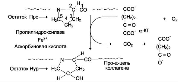
Proline and lysine hydroxylases contain an Fe2+ iron atom in their Active Site. A reducing agent is required to maintain the iron atom in its ferrous state. This role is performed by the hydroxylase coenzyme, ascorbic acid, which is readily oxidized to dehydroascorbic acid. The reverse reduction is driven enzymatically by reduced Glutathione (see Scheme B).

Proline hydroxylation is essential for stabilizing the collagen triple helix, as the OH groups of hydroxyproline (Hyp) participate in hydrogen bonding. Meanwhile, lysine hydroxylation is crucial for the subsequent formation of covalent bonds between collagen molecules during collagen fibril assembly. Scurvy, a disease caused by Vitamin C Deficiency, impairs the hydroxylation of proline and lysine residues. Consequently, weaker and less stable collagen fibers are produced, leading to fragile and brittle blood vessels characteristic of the condition. Clinically, scurvy manifests as multiple petechial hemorrhages under the skin and mucous membranes, bleeding Gums, tooth loss, and anemia.
Glycosylation of Hydroxylysine
Following the completion of hydroxylation, carbohydrate groups are introduced into the procollagen molecule through the action of specific Glycosyltransferases. These CARBOHYDRATES are most commonly galactose or the disaccharide galactosylglucose (Fig. 15-2).
Fig. 15-2. Carbohydrate components of collagen.

They form a covalent O-glycosidic bond with the 5-OH group of hydroxylysine. Glycosylation of hydroxylysine takes place in collagen that has not yet undergone helix formation and is completed after the triple helix assembles. The number of carbohydrate units in a collagen molecule varies depending on the tissue type. For example, tendon collagen (type I) contains 6 such units, whereas lens capsule collagen (type IV) contains 110. The precise function of these carbohydrate groups remains unclear; however, it is known that in a hereditary disorder caused by lysyl hydroxylase deficiency (Ehlers-Danlos syndrome type VI), the levels of hydroxylysine and carbohydrates in the resulting collagen are reduced, which is likely responsible for the compromised mechanical Properties of the skin and ligaments in affected individuals.
Formation of Procollagen and Its Secretion into the Intercellular Space
After hydroxylation and glycosylation, each pro-α-chain forms Hydrogen Bonds with two other pro-α-chains, yielding the procollagen triple helix. These events occur within the ER lumen, initiating after interchain disulfide bridges are formed in the region of the C-terminal propeptides. From the ER, procollagen molecules move to the Golgi apparatus, where they are packaged into secretory vesicles and secreted into the extracellular space.
Formation of Tropocollagen and Associated Disorders
In the extracellular matrix, the terminal propeptides of types I, II, and III collagens are cleaved by specific procollagen peptidases, producing tropocollagen molecules, which serve as the structural units of collagen fibrils. When The activity of these Enzymes is reduced (Ehlers-Danlos syndrome type VII), the procollagen terminal propeptides are not cleaved, impairing tropocollagen formation and subsequently disrupting the assembly of normal collagen fibrils. Under a Microscope, collagen fibers appear as disorganized bundles. Clinically, this presents as short stature, spinal curvature, recurrent joint dislocations, and skin hyperextensibility.
For certain types of collagen (IV, VIII, X), the terminal propeptides are not cleaved. This is because these collagens do not form fibrils, but instead assemble into network-like structures, in which the N- and C-terminal peptides play a critical role.
B. Structural and functional Features of Different Collagen Types
The 19 collagen types are subdivided into several classes based on the structures they are capable of forming. These structures are summarized in Table 15-2.
Table 15-2. Classification of collagens According to the types of structures they form
Structure |
Type |
Fibrils |
I, II, III, V, XI |
Fibril-associated |
IX, XII, XIV, XVI, XIX |
Networks |
IV, VIII, X |
Microfibrils |
VI |
Anchoring fibrils |
VII |
Transmembrane domains |
XIII, XVII |
Other |
XV, XVIII |
Fibril-forming types (I, II, III, V, and XI)
Types I, II, and III collagens account for 95% of all collagen in The Human Body, forming extremely strong fibrils. The high Abundance of these specific collagen types is because they serve as the primary Structural components of organs and tissues subject to constant or periodic
mechanical stress (bones, tendons, cartilage, intervertebral discs, blood vessels), and also participate in forming the stroma of parenchymal organs. Consequently, types I, II, and III collagens are often referred to as interstitial collagens. Minor types V and XI collagens also belong to the fibril-forming class.
Structure of collagen fibrils and their formation
The Structural organization of collagen fibrils is based on staggered parallel arrays of tropocollagen molecules shifted by 1/4 relative to each other (Fig. 15-3).
Fig. 15-3. Diagram of the staggered arrangement of collagen molecules within a collagen fibril.

The diagram clearly shows that collagen molecules are not linked end-to-end; instead, There is a gap of 35 — 40 nm between them. It is hypothesized that in bone tissue, these gaps act as mineralization centers where calcium phosphate crystals are deposited. Under Electron Microscopy, fixed and stained collagen fibrils appear striated with a 67 nm periodicity, comprising one dark and one light band. This architecture is believed to maximize the resistance of the entire aggregate to tensile stress.
Collagen fibrils are formed spontaneously via self-assembly. However, these fibrils are not yet mature, as they lack sufficient tensile strength (it is known that a mature collagen fiber 1 mm thick can withstand a load of up to 10 kg).
The newly formed collagen fibrils are strengthened by intra- and interchain covalent cross-links (which occur exclusively in collagen and elastin). These cross-links are formed as follows:
✵ the extracellular copper-containing enzyme lysyl oxidase catalyzes the Oxidative Deamination of ε-amino groups in certain lysine and hydroxylysine residues, yielding reactive aldehydes (allysine and hydroxyallysine). Vitamins PP and B6 are required for these reactions (Fig. 15-4).
Fig. 15-4. Formation of cross-links in collagen. A — formation of an aldol cross-link from two lysine side chains; B — formation of Schiff bases from lysine and allysine side chains.

✵ the resulting reactive aldehydes participate in forming covalent bonds with one another, as well as with other lysine or hydroxylysine residues of adjacent tropocollagen molecules, producing transverse "Lys-Lys cross-links" that stabilize the collagen fibrils (Fig. 15-5).
Fig. 15-5. Intra- and intermolecular cross-links in collagen.

The number of cross-links in collagen fibrils depends on tissue function and age. For instance, cross-links are particularly abundant between collagen molecules in the Achilles tendon, as high mechanical strength is crucial for this structure. With advancing age, the number of cross-links in collagen fibrils increases, leading to a slower rate of collagen turnover in middle-aged and elderly individuals.
A decrease in lysyl oxidase activity, as well as a deficiency in copper, Vitamin PP, or vitamin B6, impairs cross-link formation and consequently reduces the strength and elasticity of collagen fibers. Structures such as skin, tendons, and blood vessels become brittle and easily ruptured.
These issues are discussed in further detail below in the subsection on elastin.
Fibril-associated collagens
This class comprises collagens that perform a vital function: they regulate the size of fibrils formed by interstitial collagens (primarily types I and II) and participate in organizing the extracellular matrix in bones, skin, cartilage, and tendons. This group includes types IX, XII, XIV, and XVI collagens. While collagens of this class do not form fibrils on their own, they are directly associated with the fibrils formed by interstitial collagens. The function of these collagens can be illustrated by type IX collagen, which in cartilage binds to type II collagen fibrils in an antiparallel orientation with a periodicity of ~67 nm (Fig. 15-6).
Fig. 15-6. Structure of type II collagen fibrils and associated type IX collagen.

Type IX collagen consists of three collagenous (fibrillar) domains (Col1 —> Col3) and four non-collagenous (globular) domains (NC1 —> NC4) numbered from the C-terminus (Fig. 15-7).
Fig. 15-7. Model of type IX collagen structure. COL1–COL3 are collagenous domains; NC3–NC4 are non-collagenous structures.

Type IX collagen is linked to type II collagen fibrils via transverse Lys-Lys cross-links within the COL1 and COL2 domains, as well as the NC1, NC2, and NC3 domains.
The NC4 domain is not bound to type II collagen fibrils; its key feature is the presence of numerous positively charged groups, which allow it to bind negatively charged glycosaminoglycans such as hyaluronic acid and chondroitin sulfate. These properties enable type IX collagen to participate in the ORGANIZATION OF THE extracellular matrix in cartilage.
Network-Forming Collagens
This class includes types IV, VIII, and X collagens. The structural and functional features of these Proteins can be examined using types IV and VIII collagens, which are currently the most thoroughly studied.
Type IV collagen is a key structural component of basement membranes, which represent a specialized form of the extracellular matrix. It is secreted by various cell types, including epithelial, endothelial, Muscle, nerve, and fat cells. A distinctive feature of type IV collagen is that its repeating helical regions with the (Gly-X-Y)n sequence are frequently interrupted by short non-helical segments. This likely enhances The flexibility of type IV collagen and facilitates The formation of mesh-like structures (Fig. 15-8).
Fig. 15-8. Organization of type IV collagen. A. Triple helix of a collagen monomer: 7S is the N-terminus; NC1 is the C-terminus. B. Polymerization of type IV collagen: 1 — monomer; 2 — dimers formed by joining monomers at their NC1 domains; 3 — tetramers formed by joining monomers at their 7S segments in parallel and antiparallel orientations; 4 — formation of a mesh-like structure from oligomeric forms of type IV collagen.

Molecules of this collagen cannot associate laterally to form fibrils because their N- and C-terminal propeptides are not cleaved off. However, these very fragments participate in the formation of oligomeric collagen structures due to the presence of potential binding sites (cysteine and lysine residues). Disulfide bridges and transverse lysine cross-links stabilize the resulting oligomers. Additionally, lateral interactions between the helical regions of different molecules can occur, leading to the formation of supercoils. In the basement membrane, these components form a mesh-like structure with hexagonal units measuring 170 nm.
Types VIII and X collagens belong to the so-called short-chain collagens. Each molecule consists of a short collagenous domain, which is approximately half the length of interstitial collagens, and non-collagenous fragments at the N- and C-termini.
Type VIII collagen is the major component of Descemet's membrane in the corneal endothelium. Molecules of this collagen assemble in an antiparallel orientation to form tetramers, which then form hexagonal lattices that ensure corneal transparency (Fig. 15-9).
Fig. 15-9. Proposed mechanism of hexagonal lattice formation by type VIII collagen molecules. 1 — monomer; 2 — dimer; 3 — tetramer; 4 — hexagonal lattices.

Aside from the cornea, type VIII collagen is present in many other tissues, with another primary localization being blood vessels, where it is predominantly found in the subendothelial matrix. Whether this collagen also forms hexagonal lattices in blood vessels remains unknown. It is possible that in the vasculature, type VIII collagen forms network-like structures similar to those formed by type IV collagen in basement membranes.
Microfibril-Forming Collagens
This class includes type VI collagen, which is a short-chain protein. It forms microfibrils that are located between the larger fibrils of interstitial collagens. This collagen is widely distributed in the cartilage matrix, but is most abundant in the intervertebral discs, where it accounts for ~20% of the total collagen in The Nucleus pulposus. Two molecules of this collagen associate in an antiparallel manner to form a dimer. Dimers then assemble into tetramers, which are secreted from the cell and extracellularly linked end-to-end to form microfibrils (Fig. 15-10).
Fig. 15-10. Organization of type VI collagen. 1 — monomer; 2 — dimer; 3 — fully assembled tetramer; 4 — partially assembled tetramer; 5 — end-to-end connected microfibrils.

The functions of type VI collagen are not yet fully understood, although it is known that its microfibrils can bind to numerous Components of the extracellular matrix, including interstitial collagen fibrils, hyaluronic acid, and proteoglycans. The molecule of this collagen contains multiple Arg-Gly-Asp (RGD) sequences, suggesting its potential involvement in Cell Adhesion through binding to membrane adhesion molecules, such as the α1β1 and β1 Integrins.
Anchoring Fibril-Forming Collagens
This class includes types VII and XVII collagens, also referred to as epithelial-associated collagens, as they are typically found at the junctions between the epithelium and subepithelial layers.
Type VII collagen is the primary structural component of anchoring fibrils. Each molecule of this protein contains two non-collagenous domains (NC1 at the C-terminus, NC2 at the N-terminus) and a single collagenous domain situated between them. Monomers associate to form dimers, with the molecules joining via their NC2 domains in an antiparallel orientation relative to one another. Subsequently, the NC2 domains are cleaved, and the dimers join side-by-side to form fibrils (Fig. 15-11).
Fig. 15-11. Organization of type VII collagen. 1 — type VII collagen monomer, with NC1 and NC2 representing the non-collagenous domains at the C- and N-termini; 2 — type VII collagen dimer, with molecules assembled in an antiparallel orientation with overlap at the N-terminus; 3 — type VII collagen dimers following the removal of NC2 domains; 4 — fibril formed by side-by-side association of type VII collagen dimers.

These fibrils play a crucial role in anchoring the epidermis to the dermis, as one of their ends attaches to the lamina densa—upon which the skin epithelium rests—while the other end penetrates deeper subepidermal skin layers and binds to specialized structures known as anchoring discs.
Type XVII collagen is a transmembrane protein typically localized within epidermal hemidesmosomes. It is believed to interact with other hemidesmosomal molecules, thereby participating in the attachment of the epidermis to the dermis.
C. Collagen Catabolism
Like any protein, collagen functions in the body for a defined period. It is classified as a slow-turnover protein, with a T1/2 ranging from weeks to months. The degradation of collagen fibers is mediated by reactive oxygen species and/or enzymatic (hydrolytic) cleavage.
Collagenases and Their Functional Characteristics
Native collagen is not hydrolyzed by standard peptidases. The primary enzyme involved in its catabolism is collagenase, which cleaves peptide bonds at specific sites within the helical regions of collagen. Two Types of collagenases are known.
Tissue collagenase is found in various human organs and tissues. Under normal conditions, it is synthesized by connective tissue cells, primarily fibroblasts and macrophages. Tissue collagenase is a metalloenzyme containing a Zn2+ ion at its active center. Currently, four isoforms of this enzyme are known. Collagenase activity depends on The ratio of its activators to inhibitors within the extracellular matrix. Among the activators, plasmin, kallikrein, and cathepsin B play a particularly important role (see Section 14). Tissue collagenase exhibits high Specificity, cleaving the collagen triple helix at a precise Location—approximately 1/4 of the distance from the C-terminus, between Glycine and leucine (or isoleucine) residues (see the scheme below).

The resulting collagen fragments are water-soluble; at body Temperature, they undergo spontaneous Denaturation and become accessible to other Proteolytic Enzymes. Impaired collagen catabolism leads to fibrosis of organs and tissues (primarily The Liver and lungs). Conversely, accelerated collagen breakdown occurs in autoimmune diseases (such as rheumatoid Arthritis and systemic lupus erythematosus) As a result of excessive collagenase synthesis during the Immune Response.
Bacterial collagenase is synthesized by certain microorganisms. For example, Clostridium histolyticum (the CAUSATIVE AGENT OF gas gangrene) secretes a collagenase that cleaves the collagen peptide chain at more than 200 sites. This enzyme hydrolyzes the specific -X-Gly-Pro-Y- bond between the X and Gly residues.
This mechanism destroys connective tissue barriers in the human body, facilitating the penetration (or invasion) of the microorganism and promoting the onset and progression of gas gangrene. The pathogen itself does not contain collagen and is therefore resistant to collagenase action.
Medical Applications of Collagenases
Collagenase is used in medical practice for the Surgical Treatment of burn injuries and in ophthalmology for treating purulent eye diseases.
Determination of Hydroxyproline in Human Physiological Fluids as an Indicator of Collagen Degradation Rate
As a result of collagen breakdown, free hydroxyproline appears in the blood and urine. The majority of this amino acid is catabolized by the enzyme hydroxyproline oxidase, while a fraction is excreted in the urine. Consequently, hydroxyproline serves as a marker amino acid used to assess The rate of collagen degradation.
In certain diseases associated with connective tissue pathology, hydroxyproline excretion increases due to accelerated collagen turnover. This is observed in Paget's Disease, hyperparathyroidism, collagenoses, and various infectious diseases. In cases of impaired hydroxyproline catabolism—typically caused by a deficiency in hydroxyproline oxidase—urinary hydroxyproline excretion may exceed 1 g/day.
Characteristics of Collagen Turnover
In young individuals, collagen turnover is highly active. With Aging (particularly in the elderly), it decreases significantly because the accumulation of cross-links in older adults restricts the accessibility of collagen to collagenase. Thus, while urinary hydroxyproline levels in young individuals aged 10–20 years can reach up to 200 mg/day, this excretion drops to 15–20 mg/day in advanced age.
In certain situations, collagen synthesis increases markedly. For instance, fibroblasts migrate into a healing wound and actively synthesize the major extracellular matrix components in the affected area. The result of these processes is the formation of a connective tissue scar rich in randomly arranged collagen fibrils. A similar replacement of dying cells by connective tissue occurs in the liver during cirrhosis, in arterial walls during atherosclerosis, and in Muscles during muscular dystrophy.
D. Regulation of Collagen Turnover
Collagen synthesis is regulated through multiple mechanisms. First, collagen itself and its cleaved N-propeptides inhibit collagen translation via a negative feedback loop. Ascorbic acid stimulates the synthesis of collagen and proteoglycans, as well as fibroblast proliferation.
Hormones play a particularly important role in regulating collagen synthesis. Glucocorticoids inhibit collagen synthesis, firstly by decreasing procollagen mRNA levels, and secondly by suppressing the activity of prolyl and lysyl hydroxylase enzymes. Inadequate hydroxylation of proline and lysine residues increases the susceptibility of collagen to cleavage by collagenase and non-specific proteases. Macroscopically, the suppressive effect of glucocorticoids on collagen synthesis manifests as a reduction in dermal thickness and skin atrophy at sites of prolonged parenteral administration of these hormones.
Collagen synthesis is also influenced by Sex Hormones, whose receptors are found not only in reproductive organ stroma but also in fibroblasts of other organs and tissues. Uterine collagen turnover is under strict hormonal control. Furthermore, skin collagen synthesis depends on estrogen levels, which is supported by the fact that postmenopausal women exhibit a reduced collagen content in the dermis.
E. Diseases Associated with Impaired Collagen Synthesis and Maturation
There is a group of disorders linked to abnormalities in collagen structure or synthesis. The primary cause is Mutations in collagen genes, which are widely distributed across various Chromosomes. These genes are exceptionally large and contain numerous short exons separated by large introns.
Since about 50% of all collagen proteins are found in Skeletal Tissues, roughly 40% in the skin, and 10% in the stroma of Internal Organs,
the Clinical presentation of disorders caused by defects in collagen synthesis and maturation tends to be highly polymorphic. Many of these conditions feature not only musculoskeletal pathology or skin changes, but also prominent visceral manifestations (affecting the intestines, kidneys, lungs, Heart, and blood vessels).
To date, numerous hereditary disorders have been described that stem from defects in various types of collagen (see Table 15-3 below).
Table 15-3. Disorders associated with impaired synthesis and maturation of collagen
Type of collagen |
Gene |
Tissue localization of collagen |
Disorders |
Cause |
Clinical manifestations |
I |
СОLА1 СОL1А2 |
Bones, skin, ligaments, tendons, sclera, cornea, stroma of internal organs |
Osteogenesis imperfecta |
Mutations in the genes (over 160), most commonly deletions and substitutions. The most detrimental is the substitution of glycine with another amino acid, which introduces a kink or bend in the procollagen molecule and prevents the formation of a normal triple helix |
Increased bone fragility, dental anomalies, triangular face, joint hypermobility, blue sclerae |
II |
СОL2А1 |
Cartilage, intervertebral discs, vitreous body |
Kniest Dysplasia Stickler and Wagner syndromes |
A gene deletion leading to the synthesis of shortened collagen chains Formation of a premature termination codon, resulting in the synthesis of half-length collagen molecules in the vitreous body |
Limb shortening and deformities, joint stiffness, kyphoscoliosis, high myopia Progressive myopia, frequently retinal detachment; joint pathology resembling chronic osteoarthritis |
III |
СОL3А1 |
Skin, blood vessels, stroma of parenchymal organs, uterus |
Ehlers-Danlos-Rusakov syndrome, type IV |
Mutations in the gene (over 20) involving deletions, insertions, and substitutions. This results in the synthesis of a collagen molecule with a disrupted primary structure and reduced stability. The fibrils formed by such collagen molecules are thinner than normal and less organized |
Spontaneous rupture of major vessels, intestinal perforations, rupture of the gravid uterus, Spontaneous pneumothorax |
IV |
СОL4А3- СОL4А6 |
Basement membranes (kidneys and lungs) |
Alport syndrome Goodpasture syndrome |
Mutations in genes that impair the formation of basement membranes Production of Antibodies against type IV collagen molecules |
Predominant renal involvement manifested by Hematuria and proteinuria; certain forms concurrently develop diffuse esophageal leiomyomatosis (a benign smooth muscle tumor of the esophagus). Glomerulonephritis, pulmonary hemosiderosis |
VII |
СOL7А1 |
Skin |
Epidermolysis bullosa |
Mutations in the gene leading to a reduced total number of anchoring fibrils in the skin, as well as the synthesis of defective fibrils |
The epidermis is weakly attached to the dermis, detaches easily, and forms blisters (bullae) that are prone to trauma, leading to subsequent erosion |
Last update: 06/08/2026
Editorial and Educational Adaptation: This material has been compiled based on the primary/original source text. The project team performed an editorial review, corrected technical inaccuracies, structured sections, and adapted the content for an educational format.
What was processed:
- elimination of formatting defects (OCR errors, structural breaks, corrupted characters);
- editorial organization of content;
- standardization of terminology in accordance with academic sources;
- verification of factual statements against the original source text.
All mentions of the author, publication year, and origin of the primary text have been preserved in accordance with the source.