BIOCHEMISTRY - V. V. Emelyanov - 2016
SECTION 3. CARBOHYDRATE METABOLISM
3.1. Biological Role of CARBOHYDRATES
Carbohydrates, along with Proteins and Lipids, are essential chemical compounds that make up living organisms. In humans and animals, carbohydrates perform several vital Functions:
- energetic (serving as the primary cellular fuel). They account for more than 50% of the daily calorie requirement. Glucose and Glycogen play The Central Role in METABOLISM/26.html">Energy Metabolism;
- structural (acting as an obligatory component of most intracellular structures). These include the pentoses of NUCLEOTIDES and Nucleic Acids, as well as the carbohydrates of Glycoproteins and Glycolipids. In the form of glycosaminoglycans, carbohydrates are constituents of the Extracellular matrix;
- protective (IMMUNOGLOBULINS involved in immune defense contain a carbohydrate component). Glucuronides participate in the detoxification of endogenous toxins and xenobiotics;
- biosynthetic precursors, as compounds of other classes—specifically lipids and Certain Amino Acids—can be synthesized from carbohydrates within the Organism.
Thus, aside from their primary energetic role, carbohydrates perform A wide variety of functions, each of which is vital to the organism.
3.2. Digestion AND ABSORPTION of Carbohydrates
Carbohydrate Metabolism in The Human Body begins with The breakdown of dietary Polysaccharides and Disaccharides into Monosaccharides within the digestive tract, followed by the absorption of these monosaccharides from the intestine into the Blood.
During DIGESTION IN THE gastrointestinal tract, the Enzymatic Hydrolysis of glycosidic bonds takes place, yielding monosaccharides, primarily glucose. Starch hydrolysis starts in the Oral Cavity through the action of salivary amylase, which partially cleaves internal α-1,4-glycosidic bonds to form smaller molecules called dextrins. Hydrolysis then continues in the upper intestine under The Influence of pancreatic amylase, which also cleaves α-1,4-glycosidic bonds, resulting in The formation of maltose and isomaltose disaccharide residues. The hydrolysis of all disaccharides occurs On the surface of small intestinal Cells and is catalyzed by specific Enzymes: sucrase, lactase, maltase, and isomaltase.
The absorption of monosaccharides from the intestine into the bloodstream occurs via Facilitated Diffusion. When the intestinal glucose concentration is low, its transport can be driven by the sodium ion concentration gradient established by Na+, K+-ATPase.
Glucose entering from the intestinal lumen is carried by the HEPATIC PORTAL VEIN to the Liver, where a portion is retained, while the remainder is transported via the general Circulation to other Organs and Tissues.
The transport of glucose from the blood into cells is regulated by Insulin, a pancreatic hormone. The Action of Insulin triggers the translocation of transporter proteins from the cellular Cytosol to Cell/30.html">The Plasma Membrane, allowing glucose to enter The Cell down its concentration gradient.
The Brain and liver are exceptions. The rate of glucose uptake in these organs is independent of insulin and is determined solely by blood glucose concentration. These tissues are referred to as insulin-independent.
3.3. Glucose Metabolism
Glucose plays a central role in metabolism because it serves as the primary energy source.
The intracellular conversion of glucose begins with the formation of glucose-6-phosphate via The transfer of the terminal phosphate group of ATP to the C-6 position of glucose (Fig. 12). Because glucose-6-phosphate formation is driven by the Cleavage of a high-energy bond in the ATP molecule, the reaction is effectively irreversible. It is catalyzed by the enzyme hexokinase, which is present in almost all animal, plant, and bacterial cells.
Class="center">Fig. 12. Glucose phosphorylation reaction
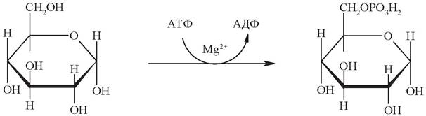
The hexokinase reaction is of major biological significance. Unlike free glucose, glucose-6-phosphate cannot cross The cell membrane, effectively "trapping" glucose inside the cell. This creates the conditions necessary for the further metabolism of the internalized glucose.
Moreover, the phosphorylation of glucose disrupts the symmetrical and stable cyclic conformation of the molecule, rendering it more reactive and thereby facilitating its subsequent transformations.
The phosphorylation reaction is a key node in overall glucose metabolism, fulfilling both rate-limiting and regulatory functions.
The rate-limiting role of hexokinase is determined by its kinetic properties. The enzyme exhibits an exceptionally high affinity for glucose (Km < 0.1 mM), meaning that Żmax is reached at low glucose concentrations. The rate of all subsequent metabolic conversions of glucose cannot exceed the Żmax of the hexokinase reaction.
The only exception is liver cells, which, alongside hexokinase, also contain its isoenzyme glucokinase, characterized by a Km value that is 1,000 times higher. This means that glucokinase saturation occurs only at high glucose concentrations.
These differences in enzyme properties explain why glucose is predominantly retained in the liver during digestion. After a meal, the glucose concentration in the portal vein rises sharply, accompanied by a proportional increase in its intrahepatic concentration. Due to its high glucose concentration during this period, glucokinase reaches maximum activity, which enhances hepatic glucose uptake. In contrast, hexokinase, possessing a higher affinity for glucose, is capable of extracting it from the general bloodstream, where glucose concentrations are lower.
The regulatory role of hexokinase is governed by feedback inhibition. The reaction product, glucose-6-phosphate, acts as an allosteric inhibitor of its own enzyme. If the subsequent utilization of glucose-6-phosphate declines, the resulting accumulation inhibits hexokinase, thereby slowing down the consumption of newly incoming glucose. Glucokinase, however, is not inhibited by excess glucose-6-phosphate.
The hexokinase reaction lies at the crossroads of all carbohydrate metabolic pathways (Fig. 13).
Fig. 13. PATHWAYS OF GLUCOSE-6-phosphate metabolism in the liver

The Main Pathways of glucose metabolism are:
- Glucose Catabolism — Glycolysis;
- glucose synthesis — Gluconeogenesis;
- Synthesis and Breakdown of glycogen;
- synthesis of pentoses — the Pentose Phosphate Pathway;
- conversion of glucose into Fatty acids and Cholesterol.
3.3.1. Glycolysis
Glycolysis (from the Greek glykys, meaning sweet, and lysis, meaning dissolution or breakdown) is The process of glucose oxidation resulting in its cleavage to form either 2 molecules of Pyruvate (aerobic glycolysis) or 2 molecules of lactate (anaerobic glycolysis). Under aerobic conditions, pyruvate enters the Mitochondria, where it is completely oxidized to СО2 and Н2О. If oxygen availability is insufficient, as may occur in actively contracting Muscle, pyruvate is converted into lactate.
Glycolysis is one of the central pathways of glucose catabolism not only in animal and plant cells, but also in many microorganisms.
The Biological Significance of glycolysis lies in the fact that it serves as the primary pathway for the breakdown of glucose to the end products СО2 and Н2О. It is this pathway that supplies
the cell with the predominant share of ATP — up to 60–70% under a normal human diet. All ten reactions of glycolysis take place in the cell cytosol and occur in all organs and tissues. The sequence of glycolytic reactions is shown in Fig. 14.
Fig. 14. Scheme of glycolytic reactions

The first reaction of glycolysis is the aforementioned ATP-dependent phosphorylation of glucose to glucose-6-phosphate.
The Second Stage is the reversible isomerization of glucose-6-phosphate to fructose-6-phosphate, catalyzed by phosphoglucoisomerase:

The resulting fructose-6-phosphate is phosphorylated in the Third Stage to yield fructose-1,6-diphosphate. Similar to the hexokinase reaction, this step is irreversible and represents the rate-limiting (slowest) reaction of glycolysis:
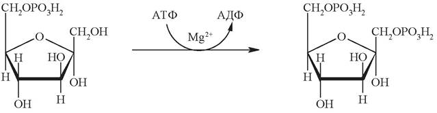
The enzyme Phosphofructokinase, which catalyzes this step, is a crucial key enzyme of glycolysis. Phosphofructokinase is classified as an allosteric enzyme; it is inhibited by ATP and stimulated by AMP. When the ATP/AMP ratio increases, phosphofructokinase activity is suppressed, leading to a slowdown in glycolysis. Conversely, when this ratio decreases, the rate of glycolysis increases. For instance, in resting muscle, the ATP concentration is relatively high, and consequently, phosphofructokinase activity is low. During Muscle contraction, ATP is consumed intensively, causing phosphofructokinase activity to rise and thereby accelerating the glycolytic process.
The Fourth Stage involves the cleavage of fructose-1,6-diphosphate precisely in half into two phosphotrioses. This reversible reaction is catalyzed by fructose-bisphosphate aldolase. The reaction products, 3-phosphoglyceraldehyde (PGA) and dihydroxyacetone phosphate (DHAP), are isomers of each other:
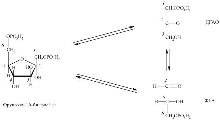
The fifth stage is mediated by the enzyme Triosephosphate isomerase, which accelerates The conversion of dihydroxyacetone phosphate into 3-phosphoglyceraldehyde. At equilibrium, the proportion of phosphoglyceraldehyde does not exceed 5%. However, subsequent reactions utilize precisely this isomer. Therefore, triosephosphate isomerase essentially ensures a sufficiently rapid replenishment of 3-phosphoglyceraldehyde as it is depleted (i.e., catalyzing the reaction, in effect, in a single direction). This concludes The first phase of glycolysis.
The sixth stage is The oxidation of the aldehyde group of 3-phosphoglyceraldehyde by the action of 3-phosphoglyceraldehyde dehydrogenase. Typically, the oxidation of an aldehyde group occurs via The addition of a Water molecule followed by the removal of 2 hydrogen atoms, whereby the aldehyde group is converted into a carboxyl group. However, during the oxidation of 3-phosphoglyceraldehyde, the Specificity of the NAD+-dependent dehydrogenase ensures The Use of phosphoric acid rather than water. As a result, the newly formed carboxyl group immediately becomes linked to a phosphate residue. The resulting mixed anhydride bond is high-energy (macroergic):

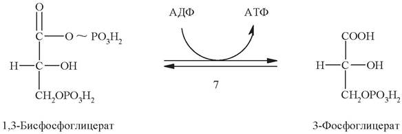
The seventh stage represents a substrate-level phosphorylation reaction. Driven by The energy released during the hydrolysis of the high-energy bond in 1,3-diphosphoglycerate, the cleaved phosphate residue is transferred to ADP, resulting in the formation of an ATP molecule. This reaction is reversible. The enzyme catalyzing this reaction is called phosphoglycerate kinase (named after the reverse reaction).
Next, under the action of the enzyme phosphoglycerate mutase, 3-phosphoglycerate is isomerized into 2-phosphoglycerate:
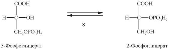
The ninth stage is the dehydration of 2-phosphoglycerate. This reversible reaction is catalyzed by the enzyme enolase:
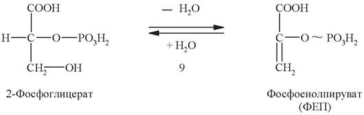
The result is the formation of a phosphoric acid ester of the enol form of pyruvate, known as phosphoenolpyruvate. Phosphoenolpyruvate contains a high-energy bond. The energy released upon the hydrolysis of this bond is utilized in the next stage for ATP synthesis.
The tenth stage is a substrate-level phosphorylation reaction. It is catalyzed by pyruvate kinase:
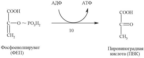
The transfer of the phosphate residue from the phosphoenolpyruvate molecule to ADP (yielding ATP) leads, however, to the release not of enolpyruvate, but of its more stable keto-isomer, pyruvate. This renders the pyruvate kinase reaction irreversible, providing it with strong thermodynamic backing.
Under aerobic conditions, pyruvate enters the mitochondria, where it is completely oxidized to CO2 and H2O. If oxygen availability is insufficient, as may occur in actively contracting muscle, pyruvate is converted into lactate:

Summarizing the chemistry of glycolysis, let us review its main features once again.
1. Regardless of whether it proceeds via an anaerobic or aerobic pathway, glycolysis can be divided into two main stages.
Reactions 1–5 constitute the first phase of glycolysis, The Essence of which is the conversion of a stable glucose molecule into two molecules of the more reactive phosphoglyceraldehyde. Two ATP molecules are consumed during this phase of glycolysis.
The second phase of glycolysis comprises reactions leading to the conversion of phosphoglyceraldehyde into pyruvate or lactate (reactions 6–10 or 6–11, respectively). These reactions are coupled with ATP synthesis;
2. Most glycolytic reactions are reversible, with the exception of three (reactions 1, 3, and 10);
3. All intermediates are in a phosphorylated form. The source of the phosphate group in phosphorylation reactions is ATP (reactions 1, 3) or H3PO4 (reaction 6);
4. Regeneration of NAD+, which is required for the oxidation of new phosphoglyceraldehyde molecules, occurs in aerobic glycolysis via the Respiratory Chain. In this process, hydrogen is transported from the cytosol into the mitochondria using a shuttle mechanism.
During anaerobic glycolysis, NAD+ is regenerated in the reduction of pyruvate to lactate, which is coupled with the oxidation of NADH2.
5. ATP formation during glycolysis can occur via two pathways: either by substrate-level phosphorylation, where the energy of a high-energy bond of a substrate is utilized to form ATP from ADP and H3PO4 (reactions 7, 9), or via Oxidative Phosphorylation driven by the energy of electron and proton transfer in the respiratory chain.
3.3.2. Calculation of ATP Yield in Anaerobic Glucose Oxidation
Cells with an insufficient oxygen supply can rely, partially or entirely, on the energy generated by glycolysis. Under anaerobic conditions, glycolysis
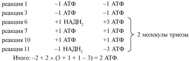
serves as the sole pathway for energy production to synthesize ATP from ADP and inorganic phosphate. Fig. 15 illustrates the steps of glycolysis where ATP is consumed and produced.
Fig. 15. Stages of glycolysis involving the consumption and production of ATP under anaerobic conditions
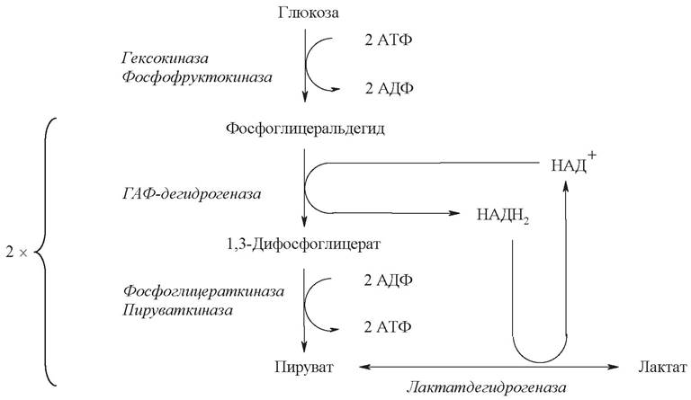
2 molecules of ATP are consumed to activate a single glucose molecule (reactions 1 and 3).
As a result of the conversion of each of the two C3 fragments during substrate-level phosphorylation (reactions 7 and 10), 2 molecules of ATP are produced.
NADH2 generated during the oxidation of phosphoglycéraldéhyde at the fifth stage is oxidized in the Lactate dehydrogenase reaction to form lactic acid and does not participate in ATP production.
Thus, the net energy yield under anaerobic conditions is 2 mol of ATP per mole of glucose:
3.3.3. Calculation of ATP Yield in Aerobic Oxidation
Most animal and plant cells normally function under aerobic conditions, resulting in the Complete oxidation of glucose to CO2 and H2O.
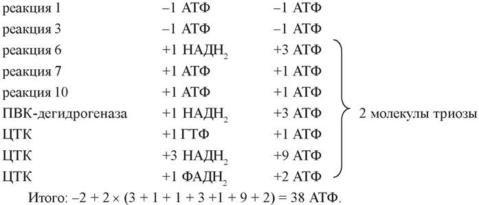
When oxygen is available in the cell, the NADH2 produced at the 6th stage is transported to the mitochondria for oxidative phosphorylation. There, its oxidation is coupled with the synthesis of 3 molecules of ATP.
Under aerobic conditions, the pyruvate generated during glycolysis is converted into acetyl-CoA by the pyruvate dehydrogenase complex, yielding 1 molecule of NADH2.
Acetyl-CoA enters the Krebs cycle and, upon oxidation, yields 3 molecules of NADH2, 1 molecule of FADH2, and 1 molecule of GTP. The subsequent oxidation of NADH2 and FADH2 in the respiratory chain generates another 11 molecules of ATP. Overall, the complete combustion of 1 acetyl residue yields 12 molecules of ATP.
Summing up the "glycolytic" ATP, the yields from both "glycolytic" and pyruvate dehydrogenase NADH2 oxidation, and the energy yield of the Krebs cycle, and multiplying the total by 2, we obtain 38 molecules of ATP:
Clearly, in energetic terms, the complete breakdown of glucose is far more efficient than anaerobic glycolysis (Fig. 16).
Fig. 16. Stages of AEROBIC GLUCOSE OXIDATION associated with the consumption and generation of ATP

However, the mitochondrial membrane is impermeable to NADH2; therefore, the transfer of hydrogen from cytosolic NADH2 into the mitochondria is mediated by shuttle systems. The core principle of this mechanism is that cytosolic NADH2 reduces a specific carrier compound capable of crossing the mitochondrial membrane. Inside the mitochondrion, this compound is oxidized—thereby reducing intramitochondrial NAD+—and then returns to the cytosol. This shuttle function is performed by either the glycerol phosphate shuttle or the malate-aspartate shuttle system (Figs. 17, 18).
Fig. 17. Glycerol phosphate shuttle: 1, 2 - oxidation-reduction reactions mediating the transport of hydrogen from the cytosol to the mitochondrial respiratory chain; 3 - FAD-dependent glycerol phosphate dehydrogenase

Fig. 18. Malate-aspartate shuttle: 1, 2 - oxidation-reduction reactions mediating the transport of hydrogen from the cytosol to the mitochondrial respiratory chain; 3, 4 - translocases facilitating the transport of malate, aspartate, and glutamate across the mitochondrial membrane

If hydrogen transfer from NADH2 occurs via the glycerol phosphate mechanism, the actual number of synthesized ATP molecules is lower, since energy is consumed to transport NADH2 from the cytosol across the mitochondrial membrane. Cytosolic NADH2 first reacts with cytosolic dihydroxyacetone phosphate to form glycerol-3-phosphate, which easily permeates the mitochondrial membrane. Inside the mitochondrion, glycerol-3-phosphate is oxidized to dihydroxyacetone phosphate with the participation of an FAD-dependent, rather than NAD-dependent, glycerol-3-phosphate dehydrogenase. The reduced flavoprotein (enzyme-FADH2) feeds the electrons it has acquired into the respiratory chain at the level of KoQ. Thus, a pair of electrons (from a single molecule of cytosolic NADH2) introduced into the respiratory chain via the glycerol phosphate shuttle mechanism yields 2 instead of 3 ATP, and the total energy yield is 36 instead of 38 ATP molecules.
Using this shuttle mechanism, the transfer of reduced equivalents from cytosolic NADH2 into the mitochondria takes place exclusively in skeletal Muscles and the brain.
In liver, Kidney, and Heart cells, a more complex malate-aspartate shuttle system operates (see Fig. 18).
The operation of this shuttle mechanism is made possible by the presence of malate dehydrogenase and aspartate aminotransferase in both the cytosol and the mitochondria. Cytosolic NADH2 reduces oxaloacetate to malate with the participation of the enzyme malate dehydrogenase.
Malate crosses The inner mitochondrial membrane into the matrix via a dicarboxylate transport system. Here, malate is oxidized to oxaloacetate by intra-mitochondrial NAD+, while the reduced NADH2 transfers its electrons to the respiratory enzyme chain.
In turn, the resulting oxaloacetate undergoes a Transamination reaction in the presence of glutamate and the enzyme AST. The resulting aspartate and α-ketoglutarate are able to cross the mitochondrial membrane via specialized transport systems. Transamination in the cytosol regenerates oxaloacetate, which triggers the next cycle.
Overall, the process involves readily reversible reactions, occurs without energy consumption, and as a result of the complete oxidation of a single glucose molecule, 38 rather than 36 ATP molecules may be formed.
During the transition from anaerobic to aerobic conditions, the accumulation of lactate in the cell ceases due to its oxidation to pyruvate. Louis Pasteur was the first to observe this phenomenon, formulating the thesis that with the onset of Respiration (i.e., oxygen consumption), Fermentation stops. He defined fermentation as life without access to oxygen.
The Mechanism of the Pasteur Effect (the inhibition of fermentation by respiration, accompanied by a sharp drop in the rate of glucose utilization) is explained by the fact that in the presence of oxygen, NADH2 and pyruvate are utilized by mitochondria immediately after their formation. Upon switching to aerobic oxidation, a decrease in previously accumulated lactate occurs. The utilization of lactate accumulated during the period of oxygen deficiency underlies the phenomenon known as "repayment of oxygen debt." The essence of this phenomenon is that after intense muscular activity, pulmonary respiration does not immediately return to normal, but remains elevated for some time. The excessive oxygen consumption during this period is determined by The amount of lactate accumulated during work insufficiently supplied with oxygen.
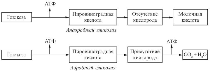
Thus, in the absence or deficiency of oxygen, lactate cannot fail to form, whereas with an adequate supply of oxygen, it cannot be formed (Fig. 19).
Fig. 19. Schemes of aerobic and anaerobic glycolysis
In A number of cases, lactate production can occur under aerobic conditions. In particular, it can be formed under aerobic conditions in malignant tumors. The reasons for the absence of the Pasteur effect in Cancer cells remain unclear to this day. Erythrocytes also produce lactate under normal aerobic conditions. This is due to the absence of mitochondria in these cells and, consequently, the inability to utilize reduced Coenzymes and pyruvate. Therefore, erythrocytes supply themselves with energy solely through the two ATP molecules generated in the substrate-level phosphorylation reaction (stage 7).
Questions for Comprehension Check
1. Carbohydrates: definition, Classification, biological significance. Monosaccharides: classification, major representatives, chemical properties. Isomerization of monosaccharides (using glucose and fructose as Examples), biological significance.
2. Disaccharides and Polysaccharides: major representatives, chemical properties, biological significance.
3. Pathways of glucose-6-phosphate metabolism in the cell: glycolysis, gluconeogenesis, The pentose phosphate pathway, synthesis of glycosaminoglycans, synthesis and breakdown of glycogen, essence, and biological significance.
4. Reactions of glycolysis, enzymes, regulation. Aerobic and anaerobic glycolysis, Energy balance. Fermentation: similarities to and differences from glycolysis.
5. Pathways of pyruvate and lactate metabolism in various tissues, the Cori cycle. The connection of carbohydrate metabolism with the Krebs cycle, Lipid Metabolism, and Amino acid metabolism. The Role of Hormones and The Nervous system in the REGULATION OF CARBOHYDRATE Metabolism.
Written Homework
Mandatory
1. Write the structural formulas for the 4 cyclic forms of D-ribose and its derivatives — the alcohol D-ribitol, D-ribonic acid, and D-2-deoxyribose. What
types of reactions lead to the formation of these compounds? What is the biological significance of ribose and deoxyribose?
2. Raffinose — α-D-galactopyranosyl-(1 —> 6)-α-D-glucopyranosyl-(1 —> 2)-β-D-fructofuranoside — is a reserve plant trisaccharide found in large quantities in legumes and sugar beets, yet it lacks a sweet taste. Upon hydrolysis catalyzed by the enzyme α-galactosidase, raffinose yields a sweet-tasting disaccharide. Write the structural formula of raffinose. Determine whether this trisaccharide is reducing or non-reducing. Write the equation for the hydrolysis reaction and name its products.
3. In the human body, Fructose Metabolism proceeds via phosphorylation at the expense of ATP, and the resulting fructose-6-phosphate enters glycolysis. Write out the reaction scheme and calculate the energy yield of the complete oxidation of fructose to CO2 and H2O.
4. A liver cell culture was supplemented with a glucose preparation radiolabeled with the 14C isotope at carbon atom 6. After some time, maximum accumulation of the radioactive isotope was detected in the Cell Cytoplasm. The experiment was repeated after adding a mitosis-stimulating substance to the cell culture. Under these conditions, the majority of the introduced radioactivity was concentrated in the cell nuclei. How can this observed pattern be explained, given the pathways of cellular glucose metabolism? In what other substances can labeled carbon atoms be detected?
Supplementary
1. The tubers of certain plants contain inulin, a polymer of β-D-fructopyranose. Hydrolysis of inulin yields raftilose, an oligosaccharide containing no more than 10 fructose units. Write the structural formula of a raftilose molecule comprising 3 monomers linked by β (2 —> 1)-glycosidic bonds, and provide its systematic name. Determine whether this trisaccharide is reducing or non-reducing.
2. A liver cell culture was supplemented with a lactic acid preparation radiolabeled with the 14C isotope at the carboxyl carbon atom. Upon Treatment of the culture with hormone No. 1, the radioactive label transiently concentrated in the mitochondrial fraction and then remained fixed in the cytosol for a prolonged period. Upon treatment of the culture with hormone No. 2, the label entered the mitochondria and left them exclusively as CO2. Which hormones were used in cases 1 and 2? How can the observed patterns be explained, given the pathways of lactate metabolism in the liver?
Sample Test Questions on the Topic "Carbohydrate Metabolism"
Instructions: Unless otherwise specified in the test question, select a single correct answer option.
1. Indicate the Structural Features of the monosaccharide:

a) ketose, pentose, D-isomer;
b) aldose, pentose, L-isomer;
c) ketose, hexose, L-isomer;
d) aldose, hexose, D-isomer;
e) ketose, pentose, L-isomer.
2. Select the reducing disaccharide composed of galactose and glucose:
a) maltose;
b) sucrose;
c) lactose;
d) cellobiose;
e) raffinose.
3. Indicate the biological role of starch:
a) structural plant polysaccharide;
b) animal reserve polysaccharide;
c) structural animal polysaccharide;
d) plant reserve polysaccharide;
d) structural polysaccharide of Arthropods.
4. Which chemical reaction occurs when glucose is boiled with Fehling's reagent:
a) oxidation;
b) reduction;
c) hydrolysis;
d) formation of a phosphate ester;
e) formation of a glycoside.
5. Choose the group of substances that serve as substrates for gluconeogenesis:
a) acetyl-CoA and ethanol;
b) glycerol and lactate;
c) ketogenic Amino Acids and cholesterol;
d) fatty acids and Ketone Bodies;
e) glucose and glycogen.
6. Name the enzyme that catalyzes the following reaction:

a) hexokinase;
b) phosphofructokinase;
c) phosphoglycerate mutase;
d) pyruvate kinase;
e) lactate dehydrogenase.
7. Which processes ensure the maintenance of physiological blood glucose concentrations during starvation:
a) insulin secretion decreases, which activates Glycogenolysis and gluconeogenesis;
b) insulin secretion increases, which activates glycolysis and glycogen synthesis;
c) insulin secretion decreases, which inhibits glycogenolysis and gluconeogenesis;
d) insulin secretion increases, which inhibits glycolysis and glycogen synthesis.
8. Specify the end Products of Anaerobic glycolysis per 1 mole of glucose:
a) 1 mole of lactate and 38 moles of ATP;
b) 2 moles of lactate and 19 moles of ATP;
c) 1 mole of lactate and 19 moles of ATP;
d) 2 moles of lactate and 2 moles of ATP;
e) 1 mole of lactate and 1 mole of ATP.
9. Insert the missing words (3 Answers) into the sentence: "The ... pathway of glucose metabolism provides the cell with ... for reduction reactions and ribose-5-phosphate for The Biosynthesis of ..."
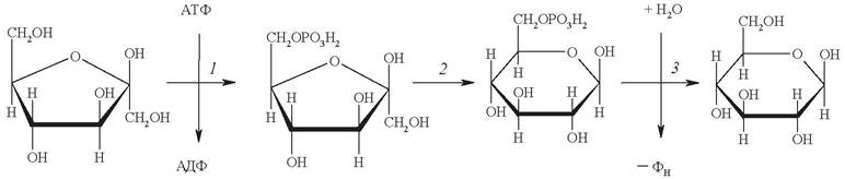
10. Provide the names and class numbers of the enzymes (6 answers) catalyzing reactions 1, 2, and 3:
Last update: 06/08/2026
Editorial and Educational Adaptation: This material has been compiled based on the primary/original source text. The project team performed an editorial review, corrected technical inaccuracies, structured sections, and adapted the content for an educational format.
What was processed:
- elimination of formatting defects (OCR errors, structural breaks, corrupted characters);
- editorial organization of content;
- standardization of terminology in accordance with academic sources;
- verification of factual statements against the original source text.
All mentions of the author, publication year, and origin of the primary text have been preserved in accordance with the source.