BOTANY: VOLUME 1 - CELL BIOLOGY. ANATOMY. MORPHOLOGY - 2007
3. PLANT TISSUES
3.2. Permanent Tissues
Permanent Tissues normally undergo no further Cell Division; their differentiated Cells are incapable of further growth and frequently even die, becoming filled with Water or air. The open Organization of plants means that large, long-lived plants contain a vast number of dead cells. For instance, the trunk of an old tree contains a minimal number of living cells, with the wood and bark consisting predominantly of dead cells (in contrast to animals, where aged or dead cells are normally rapidly eliminated). Meristematic cells adjoin one another tightly without intercellular spaces and often have the shape of irregular 14-hedron. Upon transitioning into permanent tissue, cells typically enlarge actively via postembryonic elongation: The Cell wall expands irreversibly (plastically) under the action of temporarily increasing turgor pressure. This results in a tendency toward cellular rounding. Adjacent cell walls, particularly at the corners and along cell junction lines, pull apart from one another along weaker middle lamellae, giving rise to air-filled intercellular spaces (Fig. 3.7; 3.8). These initially narrow slits widen, connect with one another, and ultimately form a continuous system of intercellular spaces. This system communicates with the external air via Stomata or lenticels (see 3.2.2.2) and facilitates gas exchange. Intercellular spaces arise either schizogenously (see Fig. 3.29) through the splitting of cell walls along the middle lamellae (from the Greek schizein – to split), or through the dissolution of cells or cell complexes (lysigenously – see Fig. 3.30, D, E), or finally through tissue tearing (rhexigenously) resulting from uneven growth (e.g., the stems of many plants with pith cavities; see Fig. 4.41). Depending on the relative volume of intercellular spaces, tissues are referred to as compact or loose. Examples of compact tissues include dermal and mechanical tissues, whereas chlorenchyma is a prime example of loose tissue.
Class="center">Fig. 3.7. Intercellular spaces (B, C – SEM micrographs)
A – parenchyma of the aerial ROOT of the orchid Vanda with narrow intercellular spaces between rounded cells (90x); B – spongy parenchyma (see 4.3.1.3) in a leaf of Parthenocissus tricuspidata, with large intercellular spaces between stellate cells (160x); C – "stellate parenchyma", the white pith tissue of the rush Juncus, with certain cell boundaries indicated by arrows; the intercellular spaces greatly exceed the very loose cellular tissue in volume (230x)

Fig. 3.8. Aerating tissue (aerenchyma, SEM micrographs).
A – air passages in the stem of the mare's tail Hippuris vulgaris, a plant rooted underwater and projecting above the water level (53x); B – aerating tissue in the petiole of the white water lily Nymphaea alba (55x)

3.2.1. Parenchyma
The ground tissue, or parenchyma (from the Greek para enchyma – a substance poured in between), is the least specialized tissue of the plant body. If one were to mentally remove all specialized tissues—such as vascular, dermal, and mechanical tissues—from a root, SHOOT, or leaf, the parenchyma would remain as the bulk mass (filling tissue) of these Organs. In herbaceous plants, it constitutes the main mass of the vegetative body; the loss of turgor in parenchyma due to water deficiency leads to the wilting of such plants. Parenchyma generally consists of large, isodiametric ("parenchymatous") and thin-walled cells. A significant portion of the ground tissue volume is accounted for by intercellular spaces (see Fig. 3.7).
Along with the view that parenchyma is poorly specialized, there is METABOLISM/2.html">THE CONCEPT OF the functional Diversity of the ground tissue. The following Functions are particularly noteworthy.
• Storage parenchyma serves for the accumulation of organic reserve substances (Polysaccharides: starch grains; Polypeptides: protein crystals; Lipids: fatty oils in oleosomes). Such parenchyma predominates in "fleshy" storage organs such as root crops, tubers, and bulbs, as well as in the storage tissue of seeds. Reserve substances are also frequently deposited in the pith parenchyma and cortex parenchyma. In the trunks of woody plants, the storage function is assumed by wood parenchyma (ray and axial parenchyma), which typically permeates the dead wood like a network.
✵ Hydrenchyma: plants of very dry habitats that remain active even during prolonged water deficits store water in the vacuoles of extremely large parenchymatous cells (up to 0.5 mm in diameter). The corresponding organs are externally noticeably swollen, their volume increased, and their surface area reduced. In extreme cases, they approach a spherical shape. This phenomenon is termed succulence (from the Latin succus – juice). Well-known examples include the leaves of stonecrop (Sedum – see Fig. 4.70) and the stems of cacti (see Box 4.1, Fig. A; Fig. 4.34; 4.35).
• In aerenchyma (aerating tissue; from the Greek aerios – airy), The system of intercellular spaces is strongly developed; up to 70% of the tissue volume consists of intercellular air spaces. In marsh and aquatic plants, it ensures Gas Exchange in submerged organs, as the intercellular space system reaches the stomata in floating or emergent leaves or shoots (see Fig. 3.8).
• Chlorenchyma (assimilation parenchyma) contains numerous Chloroplasts. This leaf tissue (mesophyll) is specialized for Photosynthesis. In the palisade layer of the mesophyll, the cells are elongated perpendicularly to the leaf surface (see Fig. 4.64). Spongy parenchyma functions simultaneously as both chlorenchyma and aerenchyma. The cells of this very loose tissue are irregularly stellate (see Fig. 3.7). The Abundance of large intercellular spaces creates conditions for substantial water loss by the spongy parenchyma, making this tissue the primary organ1 of Transpiration.
1 By definition, a tissue cannot be an organ. – Ed. note.
3.2.2. Dermal Tissues
The organs of herbaceous plants and the non-woody parts of woody plants are externally covered, as a rule, by a single-layered primary dermal tissue – the epidermis1
(from the Greek epi derma – outer Skin). When it ruptures during the secondary thickening of shoots and roots2 or due to damage, it is replaced by a multi-layered secondary dermal tissue, i.e., cork (phellem, Greek phellos – cork). Cork is produced by a specialized cambium – the cork cambium, or phellogen. Cork cells die off after the deposition of suberin layers in their walls; thus, cork is a dead tissue (see 3.2.2.2). In the trunks of trees, thick perennial branches, and roots, repeated ruptures of cork layers lead to the recurrent formation of cork cambium and cork layers, ultimately resulting in the buildup of thick dead cellular masses referred to as bark (see 4.2.8.9).
1 Shoots or thalli are covered by the epidermis. A specialized primary dermal tissue, the rhizodermis or epiblem, develops on roots, which lack an epidermis. – Ed. note.
2 See previous footnote. – Ed. note.
A common feature of dermal tissues is the tight arrangement of cells; intercellular spaces are absent. Lateral contacts between epidermal and cork cells are extremely strong, so that the leaf epidermis or cork layer can often be peeled away from the underlying tissues like a skin (Fig. 3.9). Vital gas exchange with the external environment is carried out in the epidermis via stomata, which are capable of regulating its intensity, and in cork through lenticels located within it.
Fig. 3.9. Single-layered epidermis of marsh marigold Caltha palustris, peeled from the lower side of the leaf (Interference contrast, 230x)
A—Cells are firmly connected to each other, as they are tightly interlocked by overlapping projections. The guard cells of stomata, containing numerous chloroplasts, are typical idioblasts within this homogeneous tissue (cf. also Fig. 3.13, E, F). B—Turgor-induced cushion-like Swelling of individual cells is clearly visible.

Plastids in the epidermis are mostly represented by leucoplasts or underdeveloped, grana-free chloroplasts. However, in perianth segments and fruits, the Cytoplasm of epidermal cells is often filled with chromoplasts, which serve to attract animals, thereby indirectly promoting pollination or the Dispersal of fruits and seeds. In other cases, a similar effect is achieved through coloring agents dissolved in the vacuolar sap (chumochromes: anthocyanins, betacyanins, Flavonoids). Often, both forms of pigmentation occur in combination.
A boundary tissue can also be found within the plant body, namely the single-layered endodermis. As a tissue barrier, it demarcates and physiologically isolates the Vascular Tissues from the parenchyma.
3.2.2.1. Epidermis and Cuticle
The molecular Structure OF THE cuticle (from Lat. cutis — skin) is perfectly adapted to reducing its permeability to water (see 2.2, 7.6). In plants of arid habitats
where water content is critical, water evaporation through the cuticle can be reduced to less than 0.01% of an equivalent free water surface area. Conversely, in locations where permeability is necessary, such as in glandular cells, the cuticles are porous. The surfaces of absorptive organs are generally devoid of a cuticle. In general, this is characteristic of the rhizodermis, the outermost layer of young roots.
The cuticle is capable of unlimited surface growth. Unlike insects, whose cuticle shares the function of protection against evaporation with that of plants, growing plant parts do not undergo molting. Along with the growing epidermis, the cuticle grows continuously and, in normal plants, never plays the additional role of a rigid shell. Extracellular cutinases render the molecularly reticulated cutin matrix plastically extensible, allowing for the deposition of new cutin and wax.
Frequently, the surface growth of the cuticle outpaces that of the epidermis, creating cuticular folds that extend across cell boundaries (Fig. 3.10). Cuticular striation reduces wettability: due to high surface tension, water droplets can only Touch the outer edges of the cuticular ridges and roll off the epidermis. This effect is often enhanced by The formation of bulging epidermal cells (papillose formations). During rain, the constantly rolling water droplets effectively clean the surface, washing away fungal spores that are usually loosely attached to the cuticular folds. (Many Fungi rank among the most dangerous enemies of plants and are often more harmful than insects.)
Fig. 3.10. Cuticular striation (SEM micrographs: A—W. Barthlott and N. Ehler; B, D—W. Barthlott): A—upper epidermis of petals of Anthemis tinctoria (140×); B—same in Viola tricolor, here the epidermal cells are even more convex than in Anthemis, "papillose" (95×); C—lower side of a leaf of Parthenocissus tricuspidata with a stoma (700×); D—seed surface of the cactus Neoporteria brevicylindrica (120×)

Alternatively, the same surface-cleaning function can be performed by epicuticular wax. Such superficial coatings of wax crystals are visible to the naked eye as a bluish-gray "bloom" (e.g., in "glaucous" varieties of cabbage, plums, and grapes; especially thick in the wax palm Copernicia, where rod-like wax crystals reach lengths of up to 20 µm and are used commercially as carnauba wax). Under the Electron microscope, epicuticular Waxes appear highly diverse (Fig. 3.11). Their Morphology correlates very well with their chemical composition, which is explained by self-organization in accordance with molecular parameters. The wax bloom does not occur simultaneously with cuticular striation. It renders the respective surfaces (such as the upper leaf surface of nasturtium or the lotus flower) completely non-wettable. Once abraded, the coating regenerates via the new secretion of wax through the cuticle. The wax molecules migrate to the outer surface of the epidermis, likely accompanied by water diffusing through the cuticle.
Fig. 3.11. Epicuticular wax (SEM micrographs by W. Barthlott)
A—lower side of a needle of Taxus baccata, exterior view; convex epidermal cells covered with a dense wax layer consisting of wax tubes (cf. F); stomata are sunken between 2–4 subsidiary cells (230×). B—upper side of a leaf of Hypericum buckleyi, partially aggregated wax platelets (1360×). C—long "wax hairs" are typical for many monocots (here Heliconia collinsiana), with a wax ridge surrounding the stoma (1280×). D—wax platelets in Lecythis chartacea (5400×). E—transversely cylindrical wax columns in Williamsonia quadrilocularis [Note: Wilhamodendron quadnlocelathum in original]; similar wax turrets are typical, for example, of Magnoliaceae, Lauraceae, and Aristolochiaceae (6200×). F—wax tubes (here in Lonicera tatarica) are formed when β-diketone or 10-nonacosanol are present as main components (23000×)

The secretion of cutin and wax monomers is stimulated, in a yet unknown manner, by the contact of cells with air not saturated with water vapor. Even the intercellular spaces of the mesophyll are lined with a very thin layer of cutin (inner cuticle), which can only be detected through special experiments and using an electron microscope. Extremely thick cuticles with additional cuticular layers are found on the long-lived leaves and stems of plants from very arid habitats, such as cacti and agaves. Such cuticles are very robust both chemically and mechanically, and can withstand the mouthparts of small animals. In other cases, the outer walls of epidermal cells become rigid due to calcification or, more frequently, silicification. Particularly pronounced silicification occurs, for example, in grasses and sedges. Because of this heavy silicification, horsetails were formerly used for polishing pewter tableware ("pewterwort").
The Cell walls of the epidermis in fruits and seeds are particularly diverse in Structure and Chemical composition. When dry, the aforementioned epidermises often have a horny consistency, but they swell strongly in water, becoming soft and mucilaginous in the process.
The epidermises of certain leaves assume the function of hydrenchyma; in this case, their cells are particularly large and can be arranged in multiple (up to 15) layers As a result of periclinal divisions of protodermal cells. In other instances, multi-layered epidermises perform a mechanical function, as seen in the leaves of sclerophyllous plants (see Fig. 4.71).
Stomatal complexes (stomata; from Greek stoma — Mouth) are characteristic of cutinized epidermises. They are mostly located on the lower side of leaves, though they are never entirely absent from stems and the perianth. Conversely, roots lack stomata altogether.
Each stoma consists of two elongated guard cells that are firmly connected to each other only at their ends, while their middle portions are separated by a schizogenously formed intercellular cleft—the aperture. The aperture forms a channel passing through the epidermis and cuticle, connecting the outside air with a particularly large intercellular cavity in the mesophyll or primary cortex, which—somewhat misleadingly—is designated
as the substomatal chamber1. The width of the aperture can be actively regulated by changing the shape of the guard cells. The cleft, bordered by the ventral walls of the guard cells, opens wider as their turgor increases (Figs. 3.12; 7.58, A; see 8.3.2.5). Stomata are the primary regulators of gas exchange, especially transpiration (see 6.3.4.1; 6.5.7).
1 This intercellular space is conventionally called the substomatal cavity. — Ed. note.
Fig. 3.12. Stomata of Commelina communis that have lost turgor (after K. Raschke)
A—in a 200 mM sucrose solution; B—in water in a turgescent state with widely opened stomatal clefts; in this state, the guard cells have elongated and the neighboring cells (*) have changed shape (400×)

On the lower surface of leaves, where typically 100 to 500 stomata per 1 mm2 are found, stomatal apertures account for only 0.5–2% of the total leaf surface area, even when fully open. However, stomatal transpiration can reach over two-thirds of The rate of evaporation (evaporation from a free water surface), while on the other hand, it can be reduced to almost zero.
Guard cells are typical epidermal idioblasts, differing from other epidermal cells in shape and size, as well as in the presence of chloroplasts containing assimilation starch. To a lesser extent, this is sometimes also true for cells immediately adjacent to the stoma, which are referred to as subsidiary (= accessory) cells. The stoma together with the subsidiary cells forms the stomatal apparatus. Morphogenetically, it corresponds to the terminal state of a meristemoid1 (Fig. 3.12; 3.13 B, C, E).
1 Only mesogenous stomatal apparatuses, in which both the stoma and all surrounding epidermal cells originate from a single meristemoid, can be considered terminal states of meristemoids — Ed. note.
Fig. 3.13. Stomatal development in iris (A), spiderwort (B), stonecrop (C), and corn (D) (A–D after E. Strasburger and A. de Bary)
Meristemoids and stomatal apparatuses are highlighted with stippling. E—as an example for C, the lower epidermis of a Sedum maximum leaf showing groups of stomata and subsidiary cells among partially papillate epidermal cells (60x). F—lower epidermis of a corn leaf (75x)

In grasses, guard cells are dumbbell-shaped. Their narrow middle portions, thickened due to strong cell wall deposition and stable in shape, are pushed apart as turgor increases because the thin-walled, bladder-like ends of the cells swell. The maximum opening of stomatal apertures in this grass type is small (wheat: 7 µm).
Alongside this type, there are others, among which the conifer type stands out for its exceptional complexity. The stomata on conifer needles are deeply sunken (see Fig. 4.65); subsidiary cells, typically with very unevenly thickened and partially lignified walls, actively participate in their turgor-driven movements.
Homologous to stomata, which function as gas exchange channels, are the water stomata, or hydathodes, found in certain plants, which serve to secrete liquid water (guttation; Lat. gutta — drop) (see 6.3.4.2). Their presence is indicated, for example, by apparent dew drops on nasturtium leaves. If the secreted water contains a high concentration of calcium bicarbonate, as in calcicolous saxifrage species (Saxibraga), white scales of calcium carbonate form on the hydathodes. Many nectaries secrete sugar-containing fluids through similar nectar stomata.
In many cases, the epidermis is covered with hairs. Often, individual epidermal cells grow directly into hairs or serve as meristemoid initial cells, giving rise to multicellular hairs. The Diversity of plant hairs (trichomes) is exceptionally vast; some examples are shown in Fig. 3.14. Their functions are correspondingly diverse. Through hairs, which by definition are idioblasts, the epidermis can extend far beyond its primary function as a protective tissue and participate, for example, in substance absorption or secretion.
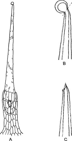
Papillate protrusions of epidermal cells act like lenses, focusing light and causing the surface to glisten, which in the perianth may serve to attract insects. Root hairs (see Fig. 4.75) function in nutrient and water absorption.1 Fruit and seed hairs can aid in wind dispersal. Seed hairs include commercially vital cotton, global production of which reached 18 million tons in 1999. Despite being unicellular, cotton fibers reach lengths of up to 5 cm and, before dying, develop quite thick secondary walls composed of nearly pure Cellulose with characteristic spiral striation. A dense woolly indumentum affects transpiration. Many plants inhabiting fog-prone regions absorb water from the surrounding mist using their pubescence; much like root hairs, such trichomes can be classified as absorptive organs. Dead, air-filled hairs reflect light, appearing snow-white, and can function as a shield against excessive illumination. In other cases, hooked climbing hairs develop to anchor climbing or scrambling shoots to a support, with well-known examples being hops and bedstraw (Galium aparine). Similar structures can also facilitate fruit and seed dispersal. Rough, often additionally branched bristle hairs with hard, silicon-impregnated cell walls serve to protect delicate leaves from being eaten by animals; even snails often ignore a plant if it presents a truly abrasive surface. A particularly sophisticated case is stinging hairs. The stinging Hair of the nettle (Urtica species, Fig. 3.15) is a single large cell with a polyploid nucleus that rises above the leaf and stem epidermis on a multicellular pedestal (an enation/emergence, see below). Its capitate-thickened tip breaks off at the base along a silicified, thin-walled zone. In this state, the stinging hair acts like a hypodermic needle: cell sap is expressed and can cause pain due to its content of formic acid, acetylcholine, and histamine. As in all large cells, intense cytoplasmic streaming can be observed in root hairs, growing cotton fibers, and even the stinging hairs of nettles (Urtica); this phenomenon was discovered in Urtica stinging hairs more than 300 years ago (R. Hooke, Micrographia, 1665). Finally, hairs can respond to stimuli as sensitive hairs, such as those of the Venus flytrap (Dionaea). Particularly common are glandular hairs, which almost invariably bear an enlarged terminal cell or a multicellular HEAD (see Fig. 3.14, D; 3.28, D, E; 9.18).
1 Root hairs are not homologous to shoot trichomes — Ed. note.
Fig. 3.14. Trichomes (SEM micrographs; C–F — W. Barthlott, G — C. Grünfelder)
A—unicellular hairs on the lower surface of a blackberry leaf (400x); B—hygroscopic hairs on the lower surface of mountain avens (Dryas octopetala) (350x); C—stellate hairs in Vitola surinamensis (Myristicaceae), a rainforest tree (papillate epidermal cells are covered with a wax layer, while hairs lack epicuticular wax) (285x); D—glandular emergences on a leaf of the sundew Drosera capensis (65x); E—hooked hairs on bean leaves ("bean straw" was formerly used as a remedy against lice and bedbugs) (220x); F—anchoring structures on the seed coat of hound's Tongue (Cynoglossum officinale) that also possess gripping properties (these structures should not be classified as trichomes — Ed. note) from the Boraginaceae family (60x). These are certainly not hairs, but emergences, since subepidermal tissue also participates in the formation of these multicellular structures; in other plants, similar structures are represented by hairs (some even unicellular); G—multicellular, concentric peltate hairs of sea buckthorn (Hippophae rhamnoides), which form a protective layer over the epidermis that reduces transpiration (160x); H—hair tufts on the floating leaves of the water fern Salvinia natans (50x). The hairs are covered with epicuticular wax, rendering the leaf surface non-wettable and trapping air bubbles upon forced submersion, which buoyancy pulls the leaf back to the water surface.
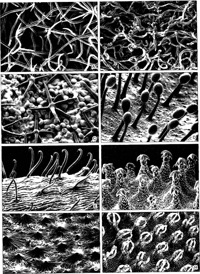
The term emergences refers to multicellular outgrowths in the formation of which subepidermal tissue also participates. Emergences largely correspond to trichomes in their Structural and functional diversity, but they are significantly larger. For example, hair-like glands are in many cases represented by macroscopic emergences (see Fig. 3.31). The pulp of citrus fruits (Citrus) is formed by "internal" emergences that grow as juice-filled tubes into the ovarian cavities. Furthermore, everyone knows from personal experience the prickles of roses and blackberries. These are also emergences and are therefore fundamentally different from spines, which are modified leaves (as in barberry and cacti) or shortened shoots (e.g., in blackthorn). Notably, common usage and botanical terminology diverge here: roses do not have thorns, but prickles, whereas cacti do not have prickles, but spines.
Fig. 3.15. Stinging hair of the stinging nettle Urtica dioica (A, 60x) (after D. von Denffer)
B—silicified tip with a predetermined breaking point (400x); C—poisonous cell sap being released after the tip has broken off (400x)
3.2.2.2. Cork (Phellem)
The periclinal cork cambium (phellogen), running parallel to the outer or wound surface, produces a thin layer of parenchymatous cells inward, known as the phelloderm, which often contains chloroplasts. This layer becomes visible as a green tissue layer when peeling, for example, elder branches or young beech stems. Outward, it produces the cork tissue, or phellum (phellem). The complete tissue complex—phellogen, phelloderm, and phelllem—is termed the periderm (see Fig. 3.17, C).
Cork often consists of only a few cell layers (potato periderm, the bark of young birch stems); however, the cork cambium can remain active for a long time and produce a cork layer more than a centimeter thick. The best-known and most practically significant example is the cork oak (Quercus suber). Fifteen-year-old trunks of this Mediterranean tree are already stripped, meaning the newly formed periderm is removed. A new, particularly active phellogen arises a few layers beneath the stripping surface, remaining functional for many years and producing "bottle cork." The resulting cork is harvested approximately every 10 years. Among European woody plants, spindle tree (Euonymus), as well as certain races of field elm and field maple, develop prominent cork ridges on young shoots (see Fig. 4.54, A). In beech, the cork cambium remains permanently active, forming a uniform, thick cork layer around trunks and branches.
Suberization of a cell involves the deposition of a waterproof suberin layer onto the cell wall (see 2.2.7.6). Upon completing the formation of its cell wall, the cork cell dies and fills with gas—an instance of apoptosis. As a result, cork tissue is very lightweight, elastic (acting as a cellular air cushion), and serves as an excellent thermal and radiation insulator. It can also be used for soundproofing.
The brown color of most types of cork is due to the deposition of Tannins, which protect against the penetration of parasites (insects, fungi).
Even a thin layer of cork reduces transpiration more effectively than a cutinized epidermis. Like any protective tissue, cork lacks intercellular spaces. This seems unexpected given that the phellogen arises as a secondary meristem within parenchyma (such as the primary cortical parenchyma of the stem) penetrated by an intercellular space system. However, during re-embryoniation, the intercellular spaces in the plane of the future cork cambium become sealed due to the localized growth of individual cells. Cells in the single-layered phellogen divide such that new cell walls are oriented exclusively periclinally. The orderly arrangement of cells in clear rows and layers, visible in cross-section, is driven by synchronous divisions of phellogen cells. Conversely, in tangential section, the outlines of the original parenchymatous cells remain recognizable (Fig. 3.16).
Fig. 3.16. Cork: bottle cork obtained from the bark of the cork oak Quercus súber (SEM micrographs by S. Grünfelder).
Left: transverse section showing rows of cells without intercellular spaces laid down by the phellogen; right: tangential section showing still recognizable outlines of initial parenchymatous Cells of the primary cortex that have transformed into phellogen initials (210×).

Total suberization of the stem surface would make the survival of cells inside trunks and branches impossible due to asphyxiation. Therefore, the cork layer is interrupted at intervals by lenticels (lenticula — a small lens) (Fig. 3.17; cf. also Fig. 4.55, A). Here, the phellogen lays down not tightly appressed cells, but rounded, loosely arranged cork cells that allow the Diffusion of Water vapor, oxygen, and carbon dioxide. The lenticel cells, which collectively form a mealy mass, are densely covered with wax crystals on their surface and are consequently non-wettable. Therefore, during prolonged rains, the lenticels do not become entirely waterlogged, remaining open for gas exchange. In bottle stoppers, the lenticels must be oriented transversely to hermetically seal the vessel.
Fig. 3.17. Lenticels (C — after K. Mägdefrau; D, E — SEM micrographs by S. Neinhuis and W. Barthlott).
A — two-year-old elderberry branch with lenticels (1.7×); B — cork on a potato tuber with numerous lenticels; C — Histology of cork and a lenticel (120×); D — lenticel of Akebia quinata (50×); E — cells of the complementary tissue of the lenticel in the same object, covered with wax (1640×); ep — epidermis, f — complementary tissue cells of the lenticel, kr — cork, kk — phellogen (cork cambium), kol — collenchyma.

The protective tissue composed of weakly suberized living cells is referred to as the cutis1. Sometimes this refers to the epidermis, but more frequently to a cell layer devoid of intercellular spaces located directly beneath the epidermis, known as the hypodermis, or — in the presence of Casparian strips — the exodermis. The cutis frequently forms also during wound healing, such as the formation of leaf scars following leaf fall, fruit abscission, etc. The programmed detachment of such organs is preceded by the formation of a thin-walled, cambium-like Separation tissue.
1 This term is currently obsolete. — Ed. note.
3.2.2.3. Endodermis
Wherever the endodermis develops (constantly in roots, and frequently in stems and leaves), it is distinctly differentiated from the tissues adjoining it externally and internally. This is related to its specific function, which will be examined using the root endodermis as an example. It demarcates the vascular tissue located in the center (the stele or central cylinder) from the surrounding parenchyma of the primary cortex (see Figs. 4.77 – 4.79).
In the endodermis at the primary developmental stage, the radial cell walls1 (anticlinal relative to the root surface and the parallel single-layered endodermis) are chemically modified such that a ribbon-like region encircles each cell; this is termed the Casparian strip after the botanist who discovered it (CS, Fig. 3.18). It is devoid of plasmodesmata. The Plasma Membrane is firmly attached to it and does not detach from the CS even during plasmolysis. The cell wall itself within the CS is impregnated with Lignin and lipophilic substances and is impermeable. This constitutes The Physiological Role of the CS and the entire endodermis in the root absorption zone: absorbed water with dissolved mineral nutrients can diffuse through the loose cell walls of the primary cortex parenchyma all the way to the endodermis, i.e., throughout the entire apoplast external to the endodermis. The total surface area of the primary cortex parenchymatous cells acts as a unified absorbing surface2. The extracellular, apoplastic diffusion pathway is blocked exclusively by the Casparian strips of the endodermis; water and ions enter the central cylinder solely via the symplastic pathway through plasmodesmata (see Fig. 6.22). In doing so, membrane translocators selectively regulate the passage of incoming ions. Endodermal cells actively transport ions into the central cylinder, where they subsequently re-enter the apoplast — into the dead Vessels of the central cylinder. The endodermis prevents the leakage of water and Mineral Substances out of the central cylinder.
1 Not only the radial but also the transverse walls are chemically modified. — Ed. note.
2 The absorbing surface is strictly represented by the outer walls of the rhizodermis and root Hair cells. An exodermal layer with similar Casparian strips develops beneath the rhizodermis, and the movement of aqueous solutions deeper into the root proceeds symplastically. The endodermis prevents water loss resulting from uncontrolled unloading of the xylem. — Ed. note.
Fig. 3.18. Endodermis with Casparian strips (CS) (C, D — light micrographs by I. Dörr; E — SEM micrograph by L. Schreiber and R. Guggenheim).
A, B — three-dimensional diagram (CS in black) and transverse sections showing primary, secondary, and tertiary structures (one cell is a passage cell); C, D — Introduction/19.html">Primary Structure of the root endodermis in Clivia nobilis (350×), transverse section of the root, with vascular tissue visible inside (interior to) the endodermis: C — lignified cell walls or parts thereof treated with phloroglucinol and Hydrochloric acid, stained dark (CS in the radial walls of the endodermis); D — incident-light fluorescence after staining with acridine orange; E — after enzymatic removal of all unlignified cell walls or parts thereof in the root of Clivia (longitudinal, external view), the xylem conducting elements and endodermal CS are preserved; the reticulate structure formed by the CS completely surrounds the xylem elements of the central cylinder (110×); E — endodermis; P — pericycle; Ph — phloem; R — primary cortex; X — xylem elements

This situation is analogous to that found in the epithelia and endothelia of higher animals. Here, the Casparian strips are functionally comparable to tight junctions, where closely apposed Plasma Membranes of adjacent cells are sealed together by specialized Proteins. Casparian strips and tight junctions represent structures that evolved completely independently across different biological kingdoms to fulfill similar functions and consequently differ structurally.
Beyond the absorption zone, in older Regions of the root, endodermal cells frequently undergo weak suberization similar to cutis cells (“secondary” structure of the endodermis). Finally, additional pronounced, often asymmetrical thickening of the cell walls may occur: the “tertiary” endodermis (see Fig. 4.78, B).
Secondary and tertiary endodermises feature passage cells opposite the xylem of the central cylinder, which retain their primary state.
3.2.3. Mechanical Tissues
Terrestrial plants require cells with tensile-strength and rigid walls (see 2.2.7.4). The limited mechanical strength of small herbaceous plants and delicate organs of larger plants (leaves, flowers, fleshy fruits) is maintained by the combined action of turgor and wall tension (turgescence), as becomes evident upon wilting. Tissue tensions, caused by slightly more vigorous growth of internal organ parts compared to their surfaces, can also contribute to the resilient state of berry-like fruits. However, such herbaceous or “fleshy” organs possess minimal actual strength; they can be easily bent, flattened, or crushed. In fact, such strength is insufficient for plants inhabiting drier environments and, even more so, for large, particularly perennial plants.
For instance, the tensile and compressive forces experienced by the roots and trunks of tall trees during gales far exceed the limits that parenchyma and protective tissues can withstand. Such plants utilize specialized mechanical tissues (stereome; from Greek sterigein — to support). These are dense, partially dead tissues whose cell walls are locally or entirely thickened through the deposition of wall layers particularly rich in cellulose. Due to impregnation with certain substances (mostly via lignification), cell walls can acquire additional rigidity and resistance to compression. This capability is also manifested in the seed coats and pericarp of fruits (nuts, drupes).
Incidentally, analogous strength-providing principles have also evolved in the animal kingdom. Hydraulic structures, tensile-strength wall layers kept under internal pressure, predominate in aquatic animals and Mollusks, but also occur, for example, in the intervertebral cartilaginous discs of vertebrates. In larger animals, tendons and skeletons can be formed by combining intercellular fibers (Collagen, Chitin) with or without impregnation by rigid substances (calcium carbonates, apatite, quinone-tanned proteins). Tensile and compressive loads are primarily borne by connective and Skeletal Tissues, and protective shells may also form (the chitinous exoskeleton of Arthropods, the vertebrate Skull).
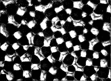
Collenchyma (from Greek kolla — glue) is the mechanical tissue of growing and herbaceous plant parts. Its prosenchymatous cells are living, capable of growth and even division. Wall thickenings are restricted to specific zones: along cell angles in angular collenchyma (Fig. 3.19), or along separate (mostly periclinal) longitudinal walls in lamellar collenchyma. This represents a primary cell wall, with its thickened regions composed of alternating lamellae consisting of cellulose and pectic substances. Wall strength is moderate, and no lignification occurs.
Fig. 3.19. Angular collenchyma in the stem of white dead-nettle (Lamium album), transverse section, cell wall thickenings appear light (420x) (light Microscopy by I. Dörr)
Sclerenchyma (from Greek skleros — hard, brittle) is a dead tissue composed of very thick-walled cells with narrow lumina, found exclusively in plant parts that have completed their growth. There are two MAIN TYPES OF sclerenchyma cells: prosenchymatous sclerenchyma fibers and isodiametric or palisade-like stone cells, known as sclereids.
Strands of stone cells (see Fig. 2.75) serve protective and supportive functions. Their thick, distinctly layered secondary walls are impregnated with lignin and perforated by branched pore canals. Sclereids occur in the hard seed coats of many fruits and in the bark of woody plants,
The functions of sclerenchyma fibers are more diverse (Fig. 3.20). In regions subject to tensile stress, the fibers generally remain unlignified (soft fibers), whereas in areas exposed to additional compressive forces, rigid, lignified fibers are formed. Sclerenchyma fibers are primarily found in stems and frequently in the large leaves of monocots. They are typically 1 — 2 mm long, although certain plants contain considerably longer fibers that are utilized in industrial manufacturing.
Fig. 3.20. Sclerenchyma fibers (A—after N. Fitting; B, C—after Eames and McDaniels): A—transverse section of a fiber strand in the leaf of New Zealand flax (Phormium tenax) (360x); B, C—development of a wood fiber in Robinia from a cambial initial (B) via bipolar apical tip growth, with the cell ends penetrating between neighboring cells (interposition) (150x); D—fibrous tracheids in pine wood with a spiral texture of the secondary walls (380x)


Since ancient times, bast fibers from fiber plants have been primarily used for manufacturing textiles, cords, and ropes. (Note the linguistic kinship of the following German and Latin terms: German binden — to bind, Band — ribbon, bandage, Bast — bast; correspondingly, Latin liber — bast, and libellus — a bound book.) The most important stem fibers are provided by flax (Linum, fiber length up to 7 cm), hemp (Cannabis), ramie (Boehmeria, belonging to the nettle family, with fiber cells exceeding 50 cm in length), and jute (Corchorus); sisal (from agave) and Manila hemp (from Musa textilis) represent fibers derived from leaves.
The length of sclerenchyma fibers always exceeds that of neighboring tissue cells. Young fiber cells grow at their tips, and their tapered ends wedge between adjacent cells (intrusive growth). Once established, the contacts between the fibers and their new surroundings are maintained subsequently (interpositional growth). Secondary plasmodesmata and, ultimately, pits may develop here. Due to the parallel arrangement of fibrils within the secondary fiber wall, its pits are slit-like. Their orientation reflects the direction of the microfibrils. The walls of most sclerenchyma fibers exhibit a spiral texture (which imparts additional elasticity), causing the slit-like pits to be oriented obliquely to the fiber axis (Fig. 3.20, B).
Not only sclerenchyma fibers but also the woody parts of vascular bundles contribute to the mechanical reinforcement of stems, leaves, and roots. The structural strength of tree trunks, older branches, and roots is provided entirely by wood. Numerous transitional forms exist between tracheids — the true conducting elements of wood — and fibers (fibrous tracheids — Fig. 3.20, D; 4.47, E).
The arrangement of mechanical tissues is of decisive importance for the biomechanics of individual organs and the plant as a whole (Fig. 3.21). This is particularly evident in erect stems, where bending stresses reach critical values even during minor gusts of wind. The resistance of a stem to bending is greater the closer the tissues with high flexural resistance (the so-called flexural elastic modulus) are located to the periphery, and the better these tissues are interconnected (composite construction). At the same time, evolution tends to optimize functionality while achieving maximum material economy. As a result of both factors, a hollow cylinder design is optimal for axial organs. This principle is realized primarily in the culms of grasses, which rank among the most remarkable natural structures in terms of their length-to-diameter ratio (up to 500:1). However, hollow cylindrical axes are characteristic only of relatively short, herbaceous plants, as they entail a risk of buckling (which is prevented in grasses by solid nodes) and preclude intensive branching (grass culms are largely unbranched, except for the inflorescences). Furthermore, the capacity to dampen mechanical stresses — which is crucial for trees and relies on the central region of the axis — would be diminished. Research has also shown that peripheral tissues in tree trunks exist in a state of tensile stress, which is counterbalanced by compressive stress in the core. Thus, the lower compressive strength of wood is compensated by its tensile strength — a feat that would be possible only to a limited extent in hollow cylindrical trunks. In lianas, whose older stems evidently do not require high bending stiffness but rather flexibility, a portion of the anti-flexural tissue is "relocated" back toward the periphery of the stem during secondary thickening. In roots, which require tensile rather than flexural strength, a rope-like design1 is realized from the outset, meaning that all mechanical elements are concentrated within a central cylinder surrounded by parenchyma (see Fig. 4.77).
1 Liana stems feature a rope-like construction in which individual strands of rigid tissue penetrate a mass of soft tissue. Roots embedded in the soil are not subjected to tensile stresses. — Ed. note.
Fig. 3.21. Functional arrangement of mechanical elements (F, G — after W. Rasdorski, H — L — after T. Speck)
A, B — stresses in a beam under deflection: tension on the convex side, compression on the concave side; the "neutral line" n deflects without changing its length. Enhanced bending strength is achieved primarily by reinforcing the convex and concave outer sides; C—E — schematic transverse sections of axial organs with differently arranged mechanical tissues (shown in black) at equal relative cross-sectional areas (11.1% of total cross-sectional area): C — central arrangement, e.g., root stele; D — intermediate arrangement, e.g., a sclerenchymatous ring with vascular bundles in eudicot stems; E — peripheral arrangement, e.g., a grass culm. Because mechanical tissues are approximately 100 times stiffer than parenchymal tissue, the bending strength at equal material expenditure stands in a ratio of 1:2.5:8; F — industrial chimney construction enabling material savings, reinforced by only 16 steel cables; G — for comparison, transverse section of the stem of deergrass (Trichophorum caespitosum); H, I — black alder (Alnus glutinosa), annual stem (H, 4 mm in diameter) with wide pith and cortex (stippled) is easily bent, whereas a multi-year branch (I, 37 mm in diameter) is substantially more resistant to bending due to its greatly expanded woody cylinder; K, L — in lianas (represented here by Aristolochia macrophylla), the opposite developmental trend can be observed: young axes (K, annual self-supporting "seeking shoot", 5 mm in diameter) are stiff against bending owing to peripheral collenchyma and an underlying continuous sclerenchyma ring (black), whereas older axes (L, 14-year-old, 30 mm in diameter) are flexible due to the fragmented arrangement of peripheral mechanical tissues and the formation of soft wood with wide rays and wide-lumen vessels (cf. Fig. 4.49, C)

Evolutionary solutions to biological problems are increasingly being applied in engineering (bionics). Indeed, this approach yields important technical innovations. However, one must account for the fact that biological structures are generally optimized for overall structure, whereas technical ones are optimized for material usage.
3.2.4. Conducting Tissues
To transport dissolved substances over cellular, microscopic distances within an Organism, diffusion driven by the thermal motion of particles in solution is entirely sufficient. However, the efficiency of diffusion decreases in proportion to the square of the distance (see 6.3.1.1 and 6.3.1.2). Even within particularly large cells, such as root hairs, internodal cells of charophyte Algae (see Fig. 5.9; 11.106, A), and the like, diffusion alone is insufficient and is supplemented by cytoplasmic streaming. Finally, in even larger Multicellular Organisms — both plants and animals — specialized conducting systems have evolved to maintain directed bulk flow of substances. Whereas in animals these transport fluids move through intercellular spaces (Body Cavities, Blood Vessels), higher plants develop specialized cells that channel liquids. These highly and terminally differentiated cells (i.e., incapable of any further development) are grouped into vascular bundles. Vascular bundles in leaves are visible to the naked eye as Veins or "nerves." In roots, the conducting tissues are assembled into a single central cylinder (stele).
All conducting systems comprise two distinct tissues. In the bast (phloem, leptome) (from Greek phloios — bast, bark; leptos — thin, soft), Organic compounds are transported through living, albeit enucleate cells with thin, unlignified walls. In the wood (xylem, hydrome) (from Greek xylon — wood; hadros — hard, rigid), water and dissolved mineral nutrients move from the absorbing root zones up to the leaves through dead, hollow, tube-like cells or multi-cellular tubes with robust, lignified walls, where the water is eventually released via transpiration or guttation (transpiration stream, see 6.3.5). In both phloem and xylem, the cells are prosenchymatous and longitudinally oriented within the vascular bundles. Consequently, longitudinally interconnected rows of cells form the conducting pathways.
3.2.4.1. Phloem
Figures 3.22 and 3.23 illustrate Various Forms of phloem conducting elements. Phylogenetically ancestral and less efficient are sieve cells. They possess narrow lumina and connect via strongly inclined end walls1 to adjacent sieve cells within a vertical cell row. These end walls (as well as lateral walls contacting other sieve cells) are perforated by enlarged plasmodesmata, referred to here as sieve pores2. These pores occur in groups known as sieve areas, a feature that inspired their name. In many angiosperms, this primitive conducting system has evolved into a system of continuous sieve tubes composed of longitudinally elongated cells of greater diameter, bearing sieve areas on oblique or transverse end walls — namely, sieve tube elements. In the most advanced types of phloem found in vines and climbing plants, the transverse end walls of the elements form simple sieve plates with exceptionally large sieve pores.
Fig. 3.22. Phloem elements (A—D — after W. Zimmermann, E — after N. Fitting, F—L — after A. Resch)
Phylogenetically ancestral are prosenchymatous cells lacking specialized cell wall structures (e.g., Rhynia, A). Primitive sieve areas arose in lycophytes (B); subsequent phylogeny led to sieve cells with sieve pores (e.g., nightshades, C) and, finally, sieve plates with sieve pores (e.g., in cucurbits, D). (All angiosperms possess sieve tubes with simple [cucurbits] or compound [nightshades] sieve plates rather than sieve cells. Sieve cells are characteristic of gymnosperms and vascular spore-producing plants, likely including Rhynia as well. — Ed. note.) E — Cucurbita pepo, transverse section of a sieve tube with a sieve plate and an associated companion cell g (600x). (The sieve plate and companion cell lie at different levels and cannot appear in the same section. — Ed. note.) F—L — development of a sieve tube element and companion cell in broad bean (Vicia faba) (F — unequal cell division, I—L — dissolution of The Nucleus and tonoplast in the sieve tube element).

Fig. 3.23. Sieve tube element with companion cells and phloem parenchyma in Passiflora coerulea (after R. Kollmann)
Left: compound sieve plate with five sieve areas (750x)

1 Sieve cells are spindle-shaped or flattened-spindle-shaped and lack end walls — Ed. note
2 A sieve pore is not simply a large plasmodesma, but the result of the regular transformation of an initial plasmodesma, involving complex reorganization of the adjacent cell wall region — Ed. note
Sieve cells and sieve tube elements possess living protoplasts with few Mitochondria and Plastids containing starch or proteins (see Fig. 11.167). The Cell Nucleus and tonoplast, dictyosomes, and Ribosomes break down early, and the cytoplasm and cell sap mix (one of the few exceptions to the compartmentalization rule, see Box 2.3). The Endoplasmic reticulum transforms into the so-called "sieve reticulum," consisting of branched tubules and smooth, folded cisternae. A characteristic component of mature sieve elements is filaments or tubules made of P-protein (phloem protein). As enucleate, delicate cells, sieve elements are short-lived; most of them degrade by the end of the vegetation period and are replaced by new ones in perennial plants. In perennial monocotyledons, such as palms, they can remain alive for years. Like plasmodesmata, sieve pores are closed by callose during the resting period. In cut or damaged sieve tubes of eudicots and some monocotyledons, sieve pores are found plugged with masses of P-protein or fragments of degenerated plastids.
In angiosperms, each sieve tube element is flanked by one (rarely more) smaller companion cell(s) containing a nucleus and numerous mitochondria (see Fig. 3.23). These glandular-looking cells are connected to the sieve tube elements by numerous plasmodesmata. They ensure the metabolism of the enucleate conducting elements. For example, P-protein (both the filament-forming PP1, 80–120 kDa, and its smaller accessory protein PP2) is synthesized in the companion cells and immediately transferred into the sieve tube elements, where highly ordered protein bodies are initially formed, which later break down into individual filaments. The second important function of companion cells is the controlled loading and unloading of sieve tubes. This is also reflected in their specific structural features: either they form a particularly large number of plasmodesmata with adjacent cells (symplastic type, predominantly in subtropical and tropical plants), or their cell surfaces are enlarged by a cell wall labyrinth (see Fig. 3.27; apoplastic type, in plants of temperate and cold zones).
In angiosperms, the complex consisting of a sieve tube element and companion cells arises from a single mother cell through its unequal division. Sieve cells of gymnosperms and ferns lack companion cells. However, these plants possess protein-rich parenchymal cells that are as closely associated with sieve cells as companion cells are with sieve tube elements, although, unlike the latter, they originate from different mother cells. These cells are called protein cells or Strasburger cells.
3.2.4.2. Xylem
The transpiration stream (see 6.3.5) moves through tube-like cells whose protoplasts die off and disappear upon reaching functional maturity due to self-dissolution (autolysis)—a striking example of programmed cell death (apoptosis) in plants. Only the lignified cell walls penetrated by bordered pits remain. There are Two Types of water-conducting "tracheal" elements: tracheids and vessels. Tracheids are elongated, narrow-lumen individual cells with very oblique end walls penetrated by numerous pits for communication with adjacent longitudinally arranged tracheids1 (see Fig. 2.75, C–F). Tracheid strands offer relatively high resistance to the flow of solution. This resistance is substantially lower in the wide-lumen, shortened vessel elements, in which the end walls are heavily perforated or completely lysed (Fig. 3.24). The larger diameter of the vessels (from 60 to over 700 µm)—they are usually distinguishable to the naked eye as "wood pores"—is due to the fact that young vessel elements grow in width as a result of the polyploidization of their nuclei (8–16 n) before their cell walls lose the capacity for growth due to the formation of thickened secondary walls.
1 Tracheids are spindle-shaped and lack end walls. — Ed. note
Fig. 3.24. Vessels (A–D — after E. W. Sinnott; E — SEM micrograph by W. Barthlott; G — SEM micrograph by S. G. Sombert):
A–D — development of a multi-celled vessel from a single row of cells via cell enlargement (polyploidization, vacuolization), formation of lignified wall thickenings, dissolution of transverse walls, and death of protoplasts (150x); E — based on the type of wall thickenings, pitted (left) and helical (right) vessels are distinguished; longitudinal section through a vascular bundle of pumpkin (360x); F — wide-lumen pits between vessel elements; pitted vessels in the wood of European elder Sambucus nigra (500x); G — scalariform perforation plate in a pitted vessel of birch wood (1300x)

Lignification of the walls of tracheids and vessel elements prevents the collapse of these tubular cells, in which negative pressure develops during intense transpiration. Therefore, when shoots are cut, air is sucked into the vessels. Since helical vessels are also structurally similar to the air-conducting tracheae of insects, the misleading term "tracheae" was formerly used for them (M. Malpighi, 1628–1694, one of the founders of plant anatomy) (from Greek trachelos — air tubes).
Particularly simple water-conducting elements are found in leafy mosses; their stems contain a central strand of longitudinally elongated, empty cells with thickened walls (hydroids). In ferns and gymnosperms, tracheids predominate; their diameter is larger, and the resistance of the end walls to flow is reduced due to their oblique arrangement and the presence of pits. The division of functions into conduction and support evolved later. In gymnosperms, predominantly tracheids still make up the supporting stem. Vessels arose evolutionarily multiple times and independently in various taxa. They already occur in some ferns and gymnosperms, but are particularly widespread in angiosperms. Vessels have retained solely the conducting function, while the supporting function is carried out by the mechanical tissue proper consisting of wood fibers (libriform fibers). Of course, in the wood of angiosperms—for example, in deciduous trees—tracheids still occur alongside vessels, and The Development of the vascular bundle generally repeats the evolution of water-conducting elements. Extremely sophisticated vessels are found in lianas. They have a very large diameter, and all transverse walls are lysed over lengths of up to 10 m along the stem, whereas normally individual transverse walls remain at intervals of a few centimeters to 1 m, probably to reduce the risk of massive embolism. The exceptionally high water conductivity of liana conducting tissues is teleonomically explained by the fact that, as climbing plants, they do not form an upright stem (relying on a host plant, rocks, or walls for support), yet must still supply a crown comparable in volume to a tree crown with water through their slender stems.
3.2.4.3. Vascular Bundle
In roots, stems, and leaves, conducting tissues are arranged in the form of vascular bundles (from Lat. fasciculi — little bundles). The conducting tissue itself is often externally strengthened by strands of sclerenchyma fibers and surrounded by an endodermis. In turn, vascular bundles in stems and leaves form a reticulate structure, whereas each root has a single radial vascular bundle in the central cylinder, which essentially represents a complex of bundles. Based on the arrangement of phloem and xylem, concentric and collateral bundles can be distinguished (Fig. 3.25). Concentric bundles with inner xylem are characteristic of ferns, whereas those with outer xylem are characteristic of underground stems of monocotyledons. Collateral bundles are much more common (horsetails, gymnosperms, and angiosperms) (Fig. 3.26; Lat. collateralis — side by side). In stems, the woody part is always located on the inside, and in horizontal leaves, on the upper side. A special type is the bicollateral vascular bundle with two strands of phloem; such bundles are found, for example, in Solanaceae and Cucurbitaceae. If the xylem and phloem directly border each other, it is termed a closed vascular bundle (Fig. 3.26, A). It consists entirely of permanent tissues. This bundle type is characteristic of monocotyledons, which has important consequences for the growth of these plants (see Box 4.3). In contrast, most bundles of gymnosperms and angiosperms are open, i.e., a layer of meristem—the fascicular cambium—is located between the phloem and xylem. In cross-section, it is noticeable due to the regular arrangement of very thin-walled cells (Fig. 3.26, B, C). The fascicular cambium plays a decisive role in secondary stem thickening.
Fig. 3.25. Types of vascular bundles
Arrangement of xylem (black), phloem (stippled), and cambium (white) in transverse sections: A — concentric vascular bundle with inner xylem (amphicribral bundle); B — same with outer xylem (amphivasal bundle); C — radial vascular bundle with inner xylem and, in this case, four xylem poles (tetrarch bundle) developed in the central cylinder of roots; on the left half "closed" (monocotyledons), on the right half "open" (magnoliids and eudicots); D–F — collateral vascular bundle: D — closed (monocotyledons); E — open (most eudicots); F — bicollateral open (e.g., in pumpkin)

Fig. 3.26. Collateral vascular bundle (A, B — after D. von Denffer, C — after K. Mägdefrau).
A — cross-section of a closed collateral bundle of maize Zea mays, showing an annular vessel (*) in the protoxylem, torn by the stretching of adjacent parenchyma (cf. Fig. 4.42); B — cross-section of an open collateral bundle of creeping buttercup Ranunculus repens; C — three-dimensional rendering of an open collateral bundle (all approx. 200x)

In a collateral bundle, the phloem generally develops from the outside inwards, and the xylem from the inner boundary of the bundle outwards to its middle. Consequently, the oldest conducting elements of the xylem—relatively little-differentiated initials and the protoxylem originating from them—are located at the inner edge of the xylem, whereas the phloem initials and protophloem are located, conversely, at the outer edge of the phloem. The later-formed, fully differentiated, and functionally mature conducting Tissues of the metaxylem and metaphloem are located in the middle of the bundle or adjacent to the fascicular cambium, respectively.
3.2.5. Glandular Cells and Tissue
Glandular cells produce specific substances known as secretions (Lat. secernere — to separate, to cast off), which they release to the outside. In other cases, Metabolic waste products, ballast substances, or harmful compounds are expelled as excretions (see 6.18). Generally, secretions are considered to be substances that are useful or essential to the organism itself, whereas excretions would be harmful if they were not removed. Secretions are formed within the cytoplasm of glandular cells, which are typically rich in Endoplasmic reticulum and/or Golgi bodies. The nuclei of glandular cells are comparatively large. Conversely, the vacuome is rather poorly developed, unless it serves as a storage site for the secretion or excretion. The products of glandular cells often accumulate in non-cytoplasmic spaces, primarily within vacuoles (intracellular secretion/excretion: "secretory cells," such as laticifers and oxalate-storing idioblasts; see below). More frequently, however, the secretion or excretion is released into the apoplast, a process often facilitated by a marked increase in surface area (Fig. 3.27). This can lead to the accumulation of the secretion or excretion within the plant (in secretion reservoirs or resin ducts) or its release into the environment (fragrant compounds, nectar).
Fig. 3.27. Cell wall protuberances and apical labyrinth in nectary cells (electron micrographs by E. Schnepf and R. Christ).
An increase in surface area at the site of substance secretion is typical for many glandular cells: A—cell wall labyrinth of a nectary cell on the perianth of Gasteria; numerous protuberances w originating from the apical cell wall W, where the secretion is released, almost reach the vacuole V. Mitochondria M within the cell wall labyrinth supply the energy required for The Active Transport of the secretion. Transfer cells (see 6.18) similarly increase their plasma membrane surface area via cell wall labyrinths; B—apical region of the nectary of Asclepias curassavica with countless folds of the plasma membrane, revealed by special carbohydrate-staining techniques for the secretion

Glandular cells in plants usually occur singly; less frequently, a few or many glandular cells are grouped together to form localized glandular tissue (see Figs. 3.28, 3.30). Plants lack large macroscopic glands comparable to those found in animals. On the other hand, the functional diversity of plant glands reflects the enormous scope of Secondary Metabolism. Ultimately, all of this is a consequence of the open ORGANIZATION OF THE plant body. The variety of secretions and excretions corresponds to the diversity of functions they fulfill. The most important examples include:
• Plant defense: many secretions are toxic (Alkaloids, steroidal Glycosides), bitter-tasting, or act as allergens. Fungal growth is inhibited by phenolic tannins and terpenoids, while plant secretions deter animals or disrupt their metabolism and development (see 6.16; 9.4.1). Through the latex, Gums, and resins released upon injury, plants can disinfect and rapidly seal wounds.
• Attraction of animals: Essential Oils and similar fragrant compounds, often produced in the glandular tissue of specialized osphores, play a role in pollination and seed dispersal. Nectar-producing glands (nectaries) reward animals that are beneficial to the plant. Nectaries are mostly located within flowers, but extrafloral nectaries also occur. Their sugar-rich secretions serve as food for insects, such as ants or termites, which act as biological enemies of pest insects. Some carnivorous plants (see Box 4.4 and 9.1.2) attract prey and capture them using glistening secretions of sticky mucus, subsequently digesting them chemically via digestive gland secretions and absorbing the breakdown products through osmotic uptake (see Fig. 3.31).
✵ Specialized excretory cells or tissues serve the purpose of excretion. The best-known example is cells containing oxalates, which bind calcium, remove it from active metabolism, and store it in their vacuoles in the form of calcium oxalate crystals1 (see Fig. 2.61). Halophytic plants of saline habitats, such as sea coasts, possess salt glands (similar to marine birds) to actively excrete the bound salts to the exterior.
1 The primary function of these cells is the removal of toxic oxalate from metabolism, not Ca. — Ed. note.
✵ A borderline case between glandular and non-glandular structures is represented by structures that carry out intensive long-distance transport of endogenous substances. Cells of this type are frequently found in boundary tissues: transfer cells of the endodermis (see Figs. 3.18 B, 4.78 B), bundle-sheath transfer cells, and the chlorophyll-poor "epithema" underlying hydathodes; companion cells of the angiosperm phloem serve a similar function. Unlike typical glandular cells, these cells often do not produce the transported substances independently; instead, they secrete them in a strictly polar (unidirectional) manner (though compare the aforementioned salt glands). Nevertheless, from a cytological standpoint, they exhibit hallmark features of glandular cells (large nuclei, dense cytoplasm, and an increased surface area generated by wall ingrowths).
A Selection of examples given below is intended to provide An Overview of the diverse STRUCTURE AND FUNCTIONS of plant glands.
3.2.5.1. Laticifers
When injured, certain plants exude a milky fluid known as latex. Well-known examples include species of spurge (Euphorbia), dandelion, ficus, celandine, and poppy. Latex corresponds to the cell sap or dilute plasma of an extensively branched network of tubes within the plant body. This system consists of specialized excretory cells. Their extraordinary size is partly due to the multinucleated (polyenergid) condition of these giant cells, which permeate the parenchyma as
non-articulated laticifers (see Fig. 3.28 A). Such plasmodial laticifers — which can be several meters long and rank among the largest plant cells overall — are found in many spurges, oleander, and rubber plants (Ficus elastica). Articulated laticifers, by contrast, are syncytia formed by the fusion of cells following the dissolution of their originally transverse walls. Laticifers of this type are widespread in the Asteraceae with ligulate flowers (Taraxacum, Scorzonera, Fig. 3.28 B, C; Lactuca derived its name from the Latin lac, milk) and many Euphorbiaceae (e.g., the rubber tree, Hevea brasiliensis).
Fig. 3.28. Glandular tissue and glandular hairs (D — after D. von Denffer; E — SEM micrograph by W. Barthlott)
A—non-articulated laticifers in the primary cortex parenchyma of Euphorbia (50x); B, C—articulated laticifers of Scorzonera purpurea in longitudinal and transverse cross-sections of the root (25x); D—glandular hair on the leaf petiole of Primula obconica, where the secretion accumulated between the cell wall and cuticle can cause itchy eczema (80x); E—glandular hair of Uncarina (Pedaliaceae) with a four-celled head (250x)

3.2.5.2. Resin Ducts and Secretory Cavities
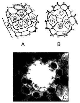
While latex is contained within laticifers, a viscous mixture of terpenoids (essential oils) known as resin or balsam accumulates in schizogenous intercellular spaces (Fig. 3.29). These resin ducts (canals) are lined with a Glandular Epithelium. Much like laticifers, resin ducts form a branched system of highly elongated tubes from which resin exudes upon wounding. Upon contact with the air, it hardens, disinfects, and seals the wound.
Fig. 3.29. Resin ducts (A, B — after W. H. Brown)
A, B—schizogenous formation of a resin duct with large-nucleated glandular epithelium in pine wood (250x); C—resin duct in a pine needle, with the glandular epithelium demarcated from the mesophyll by a sheath (all 250x)
Resin ducts are predominantly found in conifers. The resins of certain species have industrial Applications (turpentine and turpentine oil, Canada balsam). Amber is fossilized resin. In angiosperms, resin is a rare occurrence.
Essential oils are produced by the majority of higher plants; however, their quantity is often very small, or the volatile secretions are released so rapidly that no distinct accumulations are formed. For instance, the epidermal or mesophyll cells of many perianth segments contain droplets of essential oils that evaporate and diffuse into the surrounding air at appropriate temperatures (providing the fragrance of roses, violets, and jasmine). In many species, however, liquid essential oils accumulate in schizogenous or lysigenous cavities (Fig. 3.30). Examples include species of St. John's wort (Hypericum) and eucalyptus (Eucalyptus) (schizogenous cavities), as well as the lysigenous cavities found in the fruit peel of citrus fruits (Citrus).
Fig. 3.30. Oil reservoirs (B—after G. Haberlandt, D, E—after A. Tschirch)
A, C—schizogenous oil reservoirs in Hypericum perforatum, general view (A, 2x) and transverse section of the leaf (C, 50x); B, D, E—oil reservoirs in the outer layer of the orange fruit peel (B, 2x) and their lysigenous formation in Citrus limon (D, E, 25x)

3.2.5.3. Capitate hairs and glandular emergences
The ends of hairs and emergences often bear glandular cells or groups of them (see Fig. 3.28, D, E, 3.14, D). Since the glandular cells (or glandular tissue) are rounded and thicker than the stalk of the hair/emergence, it creates the impression of heads on slender necks, which is why they are called capitate. "Sessile" glands lack a stalk. The secretion (usually essential oil) accumulates between the cell wall and the cuticle and, due to its lipophilic nature, can evaporate through the distended cuticle. In other cases, the cuticle ruptures and releases a hydrophilic secretion (Fig. 3.31) (polysaccharide-containing trapping mucilage in sundew, Drosera, or protein-containing secretions of digestive glands in carnivorous plants).
Fig. 3.31. Accumulations of trapping mucilage on the heads of glandular emergences in the carnivorous plant sundew Drosera cuneifolia (see Box 4.4)
Bilaterally symmetrical dorsiventral leaf from above (A) and from the side (B) (2.5x)

Last update: 07/08/2026
Editorial and Educational Adaptation: This material has been compiled based on the primary/original source text. The project team performed an editorial review, corrected technical inaccuracies, structured sections, and adapted the content for an educational format.
What was processed:
- elimination of formatting defects (OCR errors, structural breaks, corrupted characters);
- editorial organization of content;
- standardization of terminology in accordance with academic sources;
- verification of factual statements against the original source text.
All mentions of the author, publication year, and origin of the primary text have been preserved in accordance with the source.