BOTANY VOLUME 1 - CELL BIOLOGY. ANATOMY. MORPHOLOGY - 2007
3. TISSUES OF VASCULAR PLANTS
A group of similar Cells is referred to as a tissue. This similarity is primarily evident in the external appearance of the cells, as well as in their functioning, given the general correspondence between Structure and function. Thus, Tissues can also be characterized by their role within the Organism; however, primarily, tissues remain morphological entities. Supra-cellular functional structures are designated as Organs; they are frequently composed of multiple tissues. The Study of tissues is the subject of Histology (from the Greek histos — tissue).
The initial, broad morphological Classification of the cells that make up tissues is based on their shape. More or less isodiametric cells, and the tissues they form, are termed parenchymatous, whereas longitudinally elongated cells and fibrous tissues are termed prosenchymatous. The properties of parenchymatous tissues are uniform in all directions (isotropy). Prosenchymatous tissues are characterized by a preferential direction—namely longitudinal, parallel to their parallel-arranged cells—for instance, regarding mechanical strength (anisotropy). Alongside these two basic forms—volumetrically polygonal and thread-like fibrous—there is also a flat, lamellar form, which is predominantly characteristic of protective tissues (the shape of epidermal cells).
Cells that deviate in appearance and function while located within a homogeneous tissue are termed idioblasts (see Fig. 3.9).
The richer the tissue composition of an organism, the higher the degree of differentiation it has achieved, i.e., the capacity for a division of labor among its cellular constituents. The level of Organization of an organism corresponds to the number of its Cell and tissue types. This forms the basis for the major division of the plant and fungal kingdoms. The evolutionary Development of the plant kingdom as a whole proceeded from simpler to increasingly highly organized, and frequently larger, forms.
Many Algae have attained a low degree of differentiation. In the simplest case, any cells of their body can perform all vital Functions, including reproduction. In more complexly organized algae and mosses, several distinct tissues can already be discerned. A greater diversity of tissues among plants is achieved by SHOOT-bearing forms1. Therefore, it is precisely using the latter as an example that plant tissues are examined in this chapter (Chapter 5 describes the highly aberrant tissue forms of algae and Fungi).
1 A shoot-based organization is characteristic of all mosses and many liverworts, as well as all vascular plants. Contrasting the latter with mosses is an error. — Editor's note.
Highly organized and evolutionarily advanced, historically younger shoot-bearing plants — seed plants (Spermatophyta) — are characterized by a clear Separation between meristematic tissues (Meristems) and permanent tissues. The function of a meristem (from the Greek merizein to divide) consists in producing the Cells of the body (from the Greek soma body). Cells of permanent tissues, by contrast, are incapable of division and are specialized for performing specific functions. Meristematic cells continuously undergo the Cell Cycle2 (see 2.2.3.5; 7.3.2), whereas cells of permanent tissues, conversely, normally arrest in the G1 phase (G0 phase). Furthermore, the meristematic cells of the shoot and ROOT tips are small, thin-walled, and devoid of a central vacuole. Permanent tissue cells are much larger. Frequently, they are even visible to the naked eye; their volume can exceed that of embryonic cells more than 1,000-fold. They possess a developed central vacuole, and their walls are considerably thicker. While meristematic cells grow through an increase in dry matter (embryonic or plasmatic growth), cell enlargement upon transition to the definitive state is driven by vacuolar expansion (post-embryonic growth or elongation; cf. 7.1.1). Thus, the embryonic cells of the shoot and root apical meristems (apical meristem = euhistogen) and their immediate derivatives in the primary meristem have yet to undergo elongation, whereas permanent tissue cells, by contrast, have already completed it. Elongation is typical of PLANT CELLS AND has no counterpart in animals. Since the phases of post-embryonic growth generally succeed one another rapidly, plants ("growers") can grow faster than animals with the same energy consumption.
2 Initials and meristematic cells usually divide at long intervals. — Editor's note.
During regeneration, as well as during normal development, permanent tissue cells can undergo re-embryonation and give rise to secondary (derived) meristems. Re-embryonation was previously referred to as dedifferentiation. However, this term is unfortunate because the meristematic state can also be the result of differentiation — as every re-embryonation clearly demonstrates.
3.1. Meristematic Tissue (Meristem)
The fertilized egg cell (zygote) of higher plants first develops into an embryo (Fig. 3.1; cf. also 7.4.1 and Fig. 7.27). Already During the first, typically unequal division of the zygote, the future polarity axis is established: derivatives of the smaller and optically denser apical cells later form the shoot, while the larger basal cells give rise to the primary root. Even earlier, a suspensor forms basally, through which the growing embryo is connected to the maternal plant (from the Latin suspendere to hang) and can draw upon the nutritional reserves of the seed's storage tissue, the endosperm.
As soon as the embryo enlarges, cell divisions become concentrated at the tips (apices) of the shoot and root (Latin apex tip, summit). Thus, shoots and roots exhibit apical growth, and the cells composing them are derivatives of their apical meristems. Lateral shoots and roots possess their own apical meristems. The immediate derivatives of the apical meristem frequently divide with particular vigor. They still retain a meristematic character, yet their position and external appearance often allow the future fate of their derivatives to be predicted. This apical formative tissue is designated as primary meristem and is subdivided into protoderm, which later gives rise to the protective tissue (epidermis), ground meristem, as the source of the fundamental tissue (parenchyma), and procambium, which will form the Vascular Tissues.
Class="center">Fig. 3.1. Embryo development in Arabidopsis thaliana (after U. Mayer and G. Jürgens)
A—zygote, B—two-cell stage following asymmetrical division of the zygote, C—"octant", from the upper half of the eight-celled spherical structure the shoot meristem and cotyledons later develop, while the lower half forms the hypocotyl and the main part of the embryonic root, D—dermatogen stage, with epidermal cell precursors having segregated at the periphery, E—globular stage, with the hypophysis adjoining a row of suspensor cells, from which the central part of the root meristem and the root cap will develop, F—triangle stage, establishment of bilateral Symmetry, which becomes even more pronounced at The Heart stage (G) and the subsequent torpedo stage (H) due to the Progressive development of both cotyledons

As they move further away from the primary meristem, its derivatives transform into permanent tissue cells. If, within a region that has already transitioned to permanent tissue, fairly large complexes of cells remain meristematic, they are distinguished from the apical meristem as residual meristems. A special case is represented by so-called intercalary meristems, located in the stem between already differentiated regions and capable of inducing localized longitudinal growth well below the apex. When small groups of actively dividing cells or single cells are present that eventually transition completely into permanent tissue, they are referred to as meristemoids.
Through cell Divisions of the apical and primary meristems, as well as subsequent histo- and morphogenetic processes, the primary plant body structure is formed. In herbaceous annual or biennial plants, this also corresponds to their final state. These plants die after seed production unless they propagate vegetatively via creeping shoots or other means. In perennial woody plants (shrubs, trees), by contrast, secondary thickening occurs, As a result of which stems transform into massive woody trunks, side branches into woody limbs, and roots into thick structures also consisting predominantly of wood. On the surface of perennial trunks, branches, and roots, secondary bark develops. Secondary thickening, in which the stem diameter may ultimately exceed the initial diameter by 10,000-fold, is driven by The activity of lateral meristems (secondary meristem, or cambium). This is a plate-like meristem running parallel to the organ surface. Such meristems are not located at the shoot or root apex like apical meristems, but instead form a lateral envelope around these axes (from the Latin lateralis lateral). There are two types of lateral meristems: stelar or root cambium (frequently called simply "cambium"), which produces the wood and bast of secondarily thickened stems and roots, and the cork cambium, or phellogen, which produces layers of cork tissue, which in turn lead to The formation of bark.
All apical meristems and cambia are distinguished by the presence of initials — stem cells. These cells divide unequally (see Fig. 4.43, A; 7.26, A, B): one daughter cell remains an initial, while the other transforms into a permanent tissue cell. Consequently, initial cells always remain within the meristem, thereby preserving the capacity for continued growth and the formation of new organs. The presence of apical meristems with initials characterizes the plant as an open system, which fundamentally distinguishes it from animals.
3.1.1. Apical Meristem and Primary Meristem
The meristematic cells of shoots and roots are isodiametric and small (diameter 10–20 µm). Their walls are very delicate and poor in Cellulose. All cells adjoin one another tightly, without intercellular spaces. The Cell is filled with a ribosome-rich Cytoplasm and a large, centrally located nucleus. A large central vacuole and reserve substances are absent, and Plastids are represented by proplastids.
In most higher plants, the complex of apical and primary meristems (growth point) at the shoot and root apices has a more or less conical shape (Fig. 3.2; 3.3; 3.5: apex); however, at shoot tips it may also be flattened or even concave, as in rosette plants and in the saucer-shaped large apices of many palms.
Cell number increases directly within the primary meristem, yet cell divisions within it occur for a limited time. Using microradioautography, it has been demonstrated that initial cells in the apical meristem divide relatively infrequently. In maize roots, the complete Cell Cycle of initial cells (central mother cells) lasts more than 7 days, i.e., almost 14 times longer than that of their actively dividing derivatives. In initial cells, which are more vacuolated than the others and possess smaller and denser nuclei, the G1 phase is prolonged. Consequently, the initial complex is frequently characterized as a quiescent centre.
The shoot and root apices differ substantially. Immediately behind the apex, the shoot apex (Fig. 3.2; 3.3) forms lateral outgrowths that develop into leaves or, under certain conditions, into lateral shoots. In this process, leaves and lateral shoots originate from superficial cell layers that retain a meristematic character, meaning they are exogenous. Initially, leaves grow faster than the apex; they envelop and protect it. However, unlike the apex, their growth is limited.
Fig. 3.2. Shoot apex of a horsetail (after E. Strasburger): A—longitudinal section; B—top view (180x). The apical cell cuts off segments S, S" with oblique walls. Subsequently, the segments divide further by additional walls m; f, f"—leaf primordia; g—initial cells of lateral buds; I—lateral wall of the segment
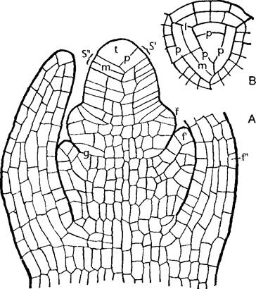
Fig. 3.3. Shoot apical meristem (A, B—light micrograph by H. Falk, D—SEM micrograph by W. Barthlott)
A—high-conical apex of Elodea densa with 2 tunica layers consisting exclusively of two cell layers; the leaves exceed the shoot apex, and a marked difference is visible between the embryonic cells in the apex and the differentiated, vacuolated leaf cells (140x). B—shoot apex of Coleus; the meristem of the terminal (*) and axillary apices stands out by its optical density (absence of vacuolization, ribosome-rich cytoplasm, large nuclei); in both leaves of the youngest node, the procambium and, correspondingly, the vascular tissue extending into the axis are already differentiated (85x). C—high-conical apex of Hippuris vulgaris (whorled phyllotaxis, cf. Fig. 4.12 A) (280x). D—shoot apex of Picea abies (spiral phyllotaxis, Fig. 4.12 D) (100x)

Roots do not bear leaves; therefore, the root apex is not protected by young leaves, but is covered by a root cap formed directly by the apical meristem. Lateral roots arise not exogenously, but endogenously, initially growing outward through the primary cortex and protective tissue. The lateral root primordium originates not in the apical region, but in a zone that has already undergone differentiation. This implies the de novo formation of an apical meristem, whereas the meristems of lateral shoots and leaf primordia are formed directly from the apical meristem (meristem fractionation).
3.1.1.1. Shoot Apical Meristem
In many marine algae, mosses, and horsetails, as well as in numerous ferns, the apical meristem contains a single, particularly large initial cell—the apical cell. It has the shape of a tetrahedron with a convex base facing outward. Cells are successively cut off from the other three sides (three-sided apical cell; see Figs. 3.2; 5.13). The resulting segments divide further, initially in a very regular manner. In ferns, leaf primordia also begin their development from wedge-shaped two-sided apical cells.
In higher ferns, especially lycophytes, and many gymnosperms, the apical cell is replaced by a group of equivalent initial cells, thereby increasing the number of stem cells. In this initial complex, cells can divide both anticlinally and periclinally (perpendicularly and parallel to the surface, respectively). In some highly developed gymnosperms and all angiosperms, the initials are arranged in layers. Only the cells of the innermost group divide periclinally and anticlinally, forming the bulk of the apex—the corpus. In the initial layers above it, cells divide only anticlinally. These cell layers form the tunica (from Lat. tunica — coat; Figs. 3.3, A; 3.4; 3.5). The total number of initial layers equals the number of tunica layers plus one.
The concepts of tunica and corpus are purely descriptive and do not indicate the subsequent developmental fate of the cells derived from them. The "tunica-corpus" concept replaced the older histogen concept, according to which the future fate of all arising cells was supposed to be predetermined already within the apical meristem. However, studies, particularly on mutants, have shown that other cells can assume The Role of missing cells in the meristem. The shoot apical meristem (in contrast to the sharply demarcated root meristem) acts as a dynamic, damage-correcting structural complex with significant regulatory potential, lacking rigid determination of subsequent cell fate.
Closest in properties to a histogen is the outermost tunica layer, designated as L1. It later develops into the epidermis and can accordingly be termed the dermatogen. The number of tunica layers varies (one in many gymnosperms, monocots, and cacti; two in most eudicots; more than two, for example, in Asteraceae). Their number can vary within the same species and often changes during ontogeny, such as during the transition to flowering. If the tunica is multilayered, each of its layers (from outside to inside L1, L2...) possesses its own initial cells. This was first demonstrated by polyploidizing Datura with colchicine. Polyploidy can manifest in either of the two tunica layers or in the corpus tissue. Nuclear and cell size correlate with the degree of ploidy, making such periclinal chimeras easily recognizable under a Microscope (Fig. 3.4). The same method can prove that each layer is formed by several initial cells. Often, polyploidy is restricted to a specific sector of the stem (sectoral chimeras).
Fig. 3.4. Shoot apex of Datura (80x) (after Satina, Blakeslee, and Avery).
A—normal diploid plant (n = 2); B—E—periclinal chimeras obtained by colchicine Treatment; B—outer tunica layer (protoderm) - 8n; C—second tunica layer - 8n, corpus = 4n; D—second tunica layer = 4n; E—corpus = 4n

The overall size of the shoot apical meristem mostly ranges between 50 and 150 µm. An exceptionally large apical meristem with a diameter of about one millimeter is found in cycads and in developing flower heads of sunflowers.
Normally, the shoot apical meristem (SAM) of angiosperms is structured as shown in Fig. 3.5. The central initial complex is surrounded by particularly actively dividing primary meristem cells: the ring-shaped peripheral meristem and the deeper pith (= medullary) meristem. The outermost layer of the peripheral meristem functions as the protoderm (dermatogen). Basipetally, the peripheral meristem gives rise to the peripheral meristem of the primary cortex and a hollow cylinder (often consisting of discrete longitudinal strands) of cells that begin to elongate along the axis. This procambium corresponds to that part of the primary meristem which remains meristematic longer than Other types of residual meristem. Later, it gives rise to the ring of stem vascular bundles, and the leaf trace primordia of young leaves—which subsequently become leaf trace bundles—connect into it. Together, the medullary and cortical meristems constitute the ground meristem.
Fig. 3.5. Shoot apex of a seed plant (longitudinal section): 1—apical initial group with stem cells (central mother cells, in gymnosperms often clearly visible as the "quiescent center"). The lateral peripheral meristem 2 includes the superficial dermatogen 3; it is represented here by the protoderm, from which the stem and leaf epidermis will develop, and the subprotoderm, from which leaf primordia arise via anticlinal divisions (7′–7"). In the adjoining histogenetic zone below, one can distinguish the medullary meristem 4, procambial strands 5, and primary cortex meristem 6. Vascular bundles will later develop from the procambial strands. The boundaries between these meristematic zones are rarely sharp, and developmental biology studies show that they can often mutually substitute for one another (e.g., in the absence of divisions)

However, before this tissue differentiation becomes distinct, leaf primordia appear on The surface of the apex as lateral bulges. Being sites of multiple anticlinal mitoses, they mark the aforementioned meristem fractionation. Cells of the leaf primordia enter the elongation phase earlier than stem cells, so that young leaves outgrow the apex. This reveals a gradient of increasing cell elongation (postembryonic growth!) that extends basipetally from the shoot apex toward the differentiation zones. The outer side of bud scales (their "abaxial" and later lower side) outpaces in elongation the "adaxial" (upper) side lying closer to the apex, causing the young leaves to curve over the apex, arching around it and thus forming a bud1 together with it.
1 This is how not only bud scales grow, but all leaves. — Editor's note.
In general, the distinction between the upper and lower sides of future leaves (their dorsoventral structure) is established already within the leaf primordium. This is manifested, for example, in asymmetric Gene Expression. The adaxial region of the primordium later gives rise to the upper epidermis, palisade parenchyma, and leaf xylem strands, whereas the abaxial region gives rise to the phloem, spongy parenchyma, and lower epidermis, respectively (see 4.3.1.1).
The formation and positioning of leaf primordia are determined by indole-3-acetic acid, a phytohormone that is transported strictly basipetally in the meristem region (see 7.6.13).
In addition to meristem subdivisions, three successive zones can be distinguished in the apex: the initial zone (10–50 µm in length); then the morphogenetic zone, or differentiation zone (20–80 µm), where leaf primordia are initiated and phyllotaxis is subsequently established; and,
finally, the histogenetic zone, where the transition to permanent Cells and Tissues takes place. It largely corresponds to the stem elongation zone.
The shoot apical meristem can also be formally subdivided according to geometrical features, which is often useful for descriptive purposes. Accordingly, one should distinguish mass meristem (in which cell divisions occur in all directions) from plate meristem (divisions in only one plane, cell walls anticlinal relative to this plane) and rib meristem (uniform, originating from cell rows via transverse divisions). In the apical and primary meristems, the corpus corresponds to mass meristem, the tunica to plate meristem, and the procambium to rib meristem.
3.1.1.2. Root Apical Meristem
The root apical meristem is covered by the root cap, or calyptra (derived from the Greek word for a covering or hood). The walls of the outermost, oldest cells of the root cap undergo mucilaginous degeneration due to abundant pectin secretion. As a result, root cap cells are short-lived; they are sloughed off every few days and constantly replenished by the root meristem. They undergo rapid terminal differentiation, a process frequently observed in plants (such as during secondary thickening; see 4.2.8.2, and in the periderm; see 3.2.2.2). The root cap facilitates the penetration of the delicate root apex through the soil, but its primary function is to act as a gravity-sensing organ (gravitropism; see 8.3.1.2). A crucial role in this process is played by prominent amyloplasts containing large starch grains. These develop primarily in longitudinal cell rows within the central region of the cap, known as the columella (Latin for small Column or pillar).
In most pteridophytes, the center of the root apex contains a tetrahedral apical cell, much like the shoot apex (Fig. 3.6, A). Being four-sided, it cuts off daughter cells along all four planes. The cells displaced outward undergo further divisions to form the root cap. In gymnosperms and angiosperms, by contrast, an apical cell is lacking. Instead, gymnosperms possess two groups of initial cells. The inner group gives rise to the bulk of the root through alternating anticlinal and periclinal divisions, whereas the outer group produces the primary cortex and the root cap, which is indistinctly delimited from it. Finally, in angiosperms, the tip of the root apex typically features a complex meristematic center composed of several independent groups of initials that give rise to the various permanent tissues (root cap, epidermis, primary cortex, and central cylinder) (Fig. 3.6, B; see also 4.4.2.1). Here, the developmental fate of the initial derivatives in the root is predetermined much more strictly than in the shoot, allowing us to speak of histogens with orderly cell lineages. The artificial elimination of specific initials through localized laser ablation often prevents the Formation of the corresponding tissue layers in the root entirely (see Fig. 7.26).
In individual cases, however, The structure of the initial zone varies. For instance, in the root apex of grasses (Poaceae), the outermost layer of the eumeristem (protoderm), which gives rise to the protective tissue of the root (rhizodermis), forms a single initial group together with the underlying meristem layer that produces the primary cortex. Lying external to this is the calyptrogen, a meristematic layer that generates the root cap. In most eudicots, however, the root cap is formed by anticlinal1 divisions from the same group of initials that also produces the protoderm (dermatocalyptrogen) (Fig. 3.6, B). Below this lies a second tier of initial cells, which form the primary cortex along with its internal barrier tissue, the endodermis. Finally, a third tier of initials, the plerome, produces the central cylinder containing the pericycle—a component of the vascular cylinder.
1 These are parallel periclinal surface divisions. — Ed. note.
Such "closed" root apices, which retain three distinct layers of initials acting as true histogens for the root cap, primary cortex, and central cylinder throughout their life (e.g., *Arabidopsis*) (see Fig. 7.26), contrast with the "open" root apices found in certain angiosperms (e.g., onion). In the latter, the initial segregation of histogens is soon lost due to the irregular proliferation of the initial complex, so that all permanent tissues develop from a unified group of meristematic cells, exhibiting a secondary resemblance to gymnosperms.
Fig. 3.6. Root apex and root cap (A — after E. Strasburger; B — after L. Kny): A — Cytology/practical/54.html">Longitudinal section of the root tip of the fern *Pteris cretica*. The four-sided apical cell is highlighted in gray (160×); B — longitudinal section of the root tip of *Brassica napus*, a eudicot. The outermost of the three initial layers (dermatocalyptrogen, in) produces the dermatogen, from which the rhizodermis develops, and the root cap (wh), whose cells contain readily distinguishable statolith starch grains (for gravity perception; see 8.3.1.2). The second initial layer lying above it produces the cells of the primary cortex with the endodermis. Finally, the third initial layer produces the central cylinder with the pericycle (50×) (cf. also Fig. 7.26); e — endodermis; in — initial zone; p — pericycle; pr — protoderm or rhizodermis, respectively; r — primary cortex; st — statolith starch grains; wh — root cap; z — central cylinder

3.1.2. Lateral Meristems (Cambia)
Cambial initials differ from the corresponding cells of the apical meri
stem by their larger size and pronounced vacuolation. In the prosenchymatous, so-called fusiform initials of the shoot or root cambium (see 4.2.8.2), which give rise to the cells of the secondary vascular tissues, this vacuolation dictates a specific mode of Cell Division: The Nucleus divides within a cytoplasmic strand traversing the longitudinally elongated vacuole, and the phragmoplast expands centrifugally within it (see Fig. 2.32). This process is unusually time-consuming, as fusiform initials can exceed a millimeter in length.
In most cases, cambial initials do not originate directly from the apical or primary meristem, but instead arise via the dedifferentiation (re-embryonicization) of cells in permanent tissues.
This is characteristic of the cork cambium (phellogen) (see 3.2.2.2) and, for the most part, also the so-called interfascicular cambium of shoots (see Fig. 4.44).
The structure and function of cambia can only be understood against the backdrop of morphological and anatomical data; therefore, they will be discussed in detail below (see 4.2.8.2; 4.4.2.3).
Box 3.1. Residual Meristems and Meristemoids
Posterior to the histogenetic zone, i.e., well back from the apex, remnants of the meristem in the form of restricted cell layers, groups, or strands retain their capacity to divide for some time. For example, in many monocots, the basal regions of internodes remain meristematic for a prolonged period as intercalary growth zones. Cambial strands within the vascular bundles of dicots later facilitate secondary stem thickening (see 4.2.8.2). Similarly, the root pericycle gives rise to lateral roots (see 4.4.2.2). In many monocots, leaves continue to grow at their base long after the leaf tip has fully differentiated. An extreme manifestation of this growth pattern is found in the southwestern African gymnosperm *Welwitschia* (see Fig. 11.211, A), in which two ribbon-like leaves grow indefinitely at the base while their distal zones continuously wear away and die off.
In the differentiation zones of shoots and leaves, one frequently encounters small nests of actively dividing cells that lack initials. Consequently, all the cells of such meristemoids1 ultimately differentiate into permanent cells that stand out visually and functionally from the surrounding tissue (idioblasts). Meristemoids give rise, for instance, to the stomatal apparatus and multicellular trichomes (hairs) (see Figs. 3.13, 3.14). Leaf primordia at the shoot apex are likewise ultimately meristemoids2, which accounts for the determinate growth of leaves (the aforementioned *Welwitschia* being an exception).
Meristemoids often originate from single cells produced by asymmetric division: a mother cell divides to form one larger, highly vacuolated cell that ceases dividing, and one smaller, cytoplasm-rich cell that undergoes limited further divisions to form a meristemoid3. From a developmental biology perspective, the spatial arrangement of meristemoids and the structures derived from them is fascinating: they form regular patterns (see Fig. 3.13, Box 4.1). This is the result of overlapping inhibitory fields that an established meristemoid generates around itself, within which the formation of subsequent meristemoids is suppressed. This principle underlies, for example, the regular patterns of phyllotaxis (see 4.2.2, 7.4.2).
1 Meristemoids are not groups ("nests") of cells, but rather the individual cells themselves. — Ed. note.
2 This is a specific view of the textbook authors, not shared by the majority of botanists. — Ed. note.
3 The small cell itself already functions as a (primary) meristemoid. — Ed. note.
Last update: 07/08/2026
Editorial and Educational Adaptation: This material has been compiled based on the primary/original source text. The project team performed an editorial review, corrected technical inaccuracies, structured sections, and adapted the content for an educational format.
What was processed:
- elimination of formatting defects (OCR errors, structural breaks, corrupted characters);
- editorial organization of content;
- standardization of terminology in accordance with academic sources;
- verification of factual statements against the original source text.
All mentions of the author, publication year, and origin of the primary text have been preserved in accordance with the source.