Human Anatomy - A Course of Lectures - Kostylenko Yu.P. 2015
Introduction to Anatomy. General Osteology. Doctrine of Bone Connections
Lecture outline:
1.1 Anatomy as a science and its place among medical disciplines.
1.2 Main Research Methods in anatomy.
1.4 Universal concepts and terms in anatomy.
1.5 General Osteology.
1.6 The bone as an organ.
1.7 The Doctrine of bone connections (Arthrology).
1.1 Anatomy as a science and its place among medical disciplines
Anatomy is one of the oldest natural sciences. It studies the shape and Structure OF THE human body. Its name comes from the Greek word "anatemno", which means "to dissect", and originated in those distant times when dissection of corpses was the only method for studying The Human Body.
Anatomy constitutes a branch of biology (the science of living things) and belongs to the group of morphological disciplines that study the structural patterns of living organisms. Morphology, in turn, is subdivided into several disciplines that examine different Levels of Organization in living systems.
Currently, the following Structural levels of the Organism are distinguished:
1. Level of the intact organism.
2. Level of morphofunctional systems (systemic level).
3. Level of individual body Organs (organ level).
4. Level of tissues that make up organs (tissue level).
5. Cellular level.
6. Level of organic macromolecules, molecular complexes, and subcellular structures (subcellular level).
Anatomy studies The structure of the Human Body at the first three of the aforementioned levels. The Study of the body and its parts with the unaided eye falls under The Scope of macroscopic anatomy. Microscopic anatomy investigates the structure of organs using a Light Microscope. Histology studies the development, structure, and function of tissues that compose the organism. Research at THE CELLULAR LEVEL forms the subject of Cytology. The Study of the organism at the level of molecules and subcellular structures belongs to the field of molecular biology.
Historically, several branches have branched off from anatomy, associated with the Practical Application of anatomical data in medicine and other fields of human activity. These primarily include: topographical or surgical anatomy, which studies the layered structure of body parts, the mutual arrangement of organs, and the spatial relationships of anatomical formations by body regions; and pathological anatomy, which studies the changes occurring in organs and tissues during various diseases.
1.2 Main research methods in anatomy
Like any science, anatomy has its own research methods, its own ways of cognizing the object of study and grasping scientific truth. The research methods used in anatomy allow us to study both the external and Internal Structure of a human. The main methods include:
1. Somatoscopy (body inspection) provides an idea of the shape of the body and its parts.
2. Somatometry (measuring the body and its parts) complements the inspection data. The main body dimensions—total length (height), chest circumference, shoulder width, and limb length—are used to determine human body type (constitution) and evaluate physical development.
3. Autopsy and dissection are Traditional Methods that have not lost their significance. The Development of anatomy as a science is primarily associated with these two methods. From antiquity to the present day, dissection remains an integral part of the educational process in the department of human anatomy.
4. Maceration is also one of the oldest methods in anatomy. It involves soaking soft tissues to soften and separate them, and is used, in particular, for studying bones.
5. Injection method involves filling cavities, fissures, lumens, and tubular structures within the human body with a colored or colorless Setting mass. This method is used to obtain a cast of the studied cavity or vessel, as well as to facilitate the Separation of a vessel from surrounding tissues.
6. Corrosion method consists in removing tissues that are difficult to dissect by etching them with acids. Tubular organs or an organ cavity are pre-filled with a substance that resists acid degradation.
7. The method of serial sectioning of frozen cadavers was proposed in the 19th century by the great Russian surgeon N.I. Pirogov to study topographical relationships within the body. The utility of this method lies in preserving the life-like relationships between various anatomical structures in a specific body region.
8. Method of multi-layered plastic or graphic reconstructions. The Essence of this method is that a series of histological sections and histotopograms can provide a visual representation of the shape of anatomical structures and their spatial arrangement.
9. Clearing method for anatomical preparations. This method involves Processing organs or parts thereof so that the studied object becomes clearly visible against the Background of cleared tissues. It is most frequently used to study the nervous and vascular systems.
10. Method of macro-microscopic research. This method was developed in the early 20th century by the prominent anatomist V.P. Vorobyov. It involves fine dissection of stained objects (small vessels, nerves) followed by examination under a binocular magnifying Glass.
11. X-ray method (radiological anatomy), the study of individual organs or body parts using X-rays. The advantage of the X-ray method is that it allows for the study of the structure of a living person, observation of functioning organs, and dynamic investigation of age-related changes. Radiological anatomy has emerged as a distinct branch of anatomy essential for clinical practice. Currently, alongside traditional methods—fluoroscopy (direct examination of an organ or body part using X-rays) and radiography (recording the X-ray image on film)—modern, specialized X-ray techniques are employed. The latter include: stereoradiography, which provides three-dimensional images of body parts and organs; cineradiography, which allows for the study of organ movements, Heart contractions, and the passage of contrast media through vessels; and tomography, a sectional X-ray imaging technique that yields a clear image of anatomical structures located in a specifically designated layer, free from extraneous overlays. Modern tomographic methods are implemented using computer technology. This technique, known as computed tomography (CT), enables the acquisition of a three-dimensional image of the studied object.
12. Endoscopic methods refer to the observation of the internal surface of hollow organs using specialized optical instruments: the Larynx via laryngoscopy, the Bronchi via bronchoscopy, The Stomach via gastroscopy, and others.
13. Ultrasound echolocation (echography, ultrasound scanning) is an examination based on differences in the acoustic properties of organs and tissues, enabling the visualization of certain organs that are difficult to assess via X-ray Examination, such as The Liver and Spleen.
14. Magnetic Resonance imaging (MRI) is the most modern and advanced visualization method utilizing the physical phenomenon of nuclear magnetic resonance, widely used in radiology for detailed imaging of internal body structures. This method produces high-contrast images of body tissues and is therefore extensively applied in neuroimaging and the visualization of The Heart and Muscles, unlike other research methods (such as computed tomography or radiography). It also allows for obtaining three-dimensional images of the study object.
1.3 Anatomical Terminology
Every science has its own professional language—a system of specialized terms designating the objects and processes with which that science deals. Anatomical terminology, which includes the names of body parts, organs, Blood Vessels, nerves, and other anatomical structures, constitutes a major part of medical language. The names of diseases and surgical interventions are derived from organ names. Anatomical terms originate from Ancient Greek, Latin, and Arabic. Today, new terms are constructed on a Latin basis.
Efforts to standardize anatomical nomenclature have been underway since the late 19th century. In 1895, at the Congress of Anatomists in Basel, the first (Basel) anatomical nomenclature was adopted. This was followed by the British Revision list (1933) and the Jena Anatomical Nomenclature (1936). Finally, in 1955, at the Congress of Anatomists in Paris, the International (Paris) Anatomical Nomenclature was approved. Amendments and additions have been incorporated into the International Anatomical Nomenclature at subsequent anatomical congresses in New York (1960), Wiesbaden (1965), Leningrad (1970), Tokyo (1975), Mexico City (1980), London (1985), and São Paulo (1997).
1.4 Universal Concepts and Terms in Anatomy
The study of human anatomy typically begins with familiarization with General Concepts and terms that serve as the foundation for forming a comprehensive understanding of initial coordinates, THE POSITION OF the human body in space, the relationships between its parts, and their movement during motor acts.
First of all, to avoid discrepancies, it should be agreed that the human body, as the object of study, must be placed before us in a deliberately defined position known as the anatomical position. In this position, the human (regardless of their actual posture) stands vertically before us, facing forward, with arms hanging down and turned outward (palms facing forward), i.e., in a state of supination.
It is easy to imagine that through any point in or on the human body, three mutually perpendicular lines, or trajectories, known as axes can be drawn. One of them is oriented vertically (perpendicular to the horizon) — the vertical axis (axis verticalis), while the other two, perpendicular to it, lie in the horizontal plane.
The axis passing through the human body (in its initial position) from front to back or vice versa is designated as the sagittal, or arrow-like, axis (axis sagittalis), as it corresponds to the trajectory of an arrow shot straight forward by an archer. The second axis, perpendicular to the first and parallel to the plane of the forehead, is termed the frontal axis (axis frontalis).
All three axes (vertical, sagittal, and frontal), being mutually perpendicular, are considered the geometric centers of movements of body parts performed within a given joint (Fig. 1.1).
Class="center">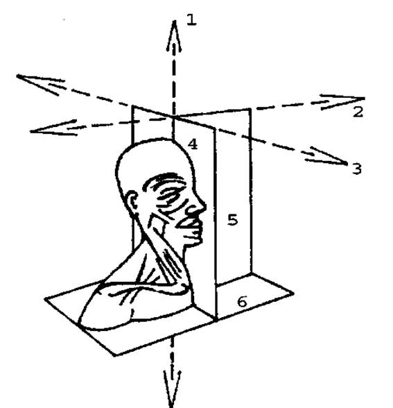
Fig. 1.1 Axes and their corresponding planes:
1 - vertical axis; 2 - frontal axis; 3 - sagittal axis; 4 - sagittal plane; 5 - frontal plane; 6 - horizontal plane.
An isolated movement centered around the vertical axis is called rotation (rotatio). Examples of this movement include turning the HEAD to the right and left.
The sagittal axis serves as the center for two opposing movements: one is called abduction (abductio) and the other is adduction (adductio). Abduction refers to a movement around the sagittal axis whereby a body part moves away from the median plane, whereas adduction brings it closer to the initial position. For instance, this occurs when moving the upper limb sideways away from the body and during the reverse movement. In this case, the center of movement is the sagittal axis passing through the geometric center of the shoulder joint.
Finally, the horizontal axis is used to analyze movements known as flexion (flexio) and extension (extensio). Flexion is a movement in which the angle between the related body parts decreases, whereas extension leads to its increase. A movement in the elbow joint serves as a good illustration of this.
Mastering the above material will also help you understand more complex concepts related to the spatial characteristics used in dissecting the human body. The body has an extremely complex shape and occupies a specific volume in three-dimensional space, which can be virtually or physically sectioned across three mutually perpendicular planes. Each of these planes is easily determined using two of the three known axes.
Let us begin with the plane formed by the vertical and sagittal axes. This plane cuts through the human body from front to back (or back to front) and from top to bottom (or bottom to top), corresponding to the sagittal axis that defines it.
If this plane is used as the basis for anatomical dissection, the human body is divided into A number of parts located from right to left or vice versa. One of these cutting planes may divide the body into two similar, yet unequal halves: the right (dexter) and the left (sinister). This unique sagittal plane is referred to as the median plane (mediana).
Using the sagittal plane makes it possible to perform a sectional Analysis of the right and left halves of the human body. The resulting parts will lie either to the right or to the left of the median plane. Two concepts are used to describe them: medial (medialis) and lateral (lateralis), the meanings of which become clear only when comparing certain parts or structures. Thus, the term medial typically designates a body part that lies closer to the median plane compared to another similar part, while lateral refers to a part that lies farther from the median plane. In descriptive anatomy, the term "lateral" is sometimes used as a synonym for "side" and "medial" for "middle", which is not entirely accurate in terms of meaning. Closely related to these concepts is the term intermediate (intermedius).
If the objects of analysis are corresponding PARTS OF THE two halves (right and left), they are referred to as opposite or contralateral.
To describe anatomical structures located in the anteroposterior direction, a second vertical plane is used—the frontal plane, which cuts the human body from top to bottom and from right to left, or vice versa. Primarily, two concepts are associated with it: anterior and posterior. Synonyms for these terms include abdominal or ventral (from venter — belly, abdomen) and dorsal (from dorsum — back).
Finally, the horizontal plane is used for the anatomical description and analysis of body parts and individual structures that change their shape or structure along the vertical axis. This plane cuts the body from front to back and from right to left, or vice versa. To denote these variations, terms such as superior and inferior are used. Within the human trunk (up to the level of the Pelvic Girdle), these are often replaced by the terms cranial (from cranium — Skull) and caudal (from cauda — tail). Naturally, these terms also have a conventional and relative meaning.
At the same time, when describing the structure of the limbs, instead of the terms "upper" and "lower", more universal concepts are used. These allow for describing the relationship between the two opposite ends of the limbs or their individual long bones, regardless of their specific position. For example, when the upper limb is abducted, the terms upper and lower (applied to the shoulder region and bones, respectively) lose their strict meaning. Therefore, it is more convenient to use designations that indicate the relative distance of these ends from the median plane. Obviously, the shoulder region relative to the hand (regardless of the position of the upper limb) is closer to the trunk, whereas the hand occupies the position farthest from it. Accordingly, the terms proximal (proximalis), meaning located closer to the trunk, and distal (distalis), meaning located farther from the trunk, are adopted.
From an anatomical perspective, the human body is generally divided into two parts: the soma (body) and the Internal Organs or viscera, which are located within the Body Cavities. The external shape and configuration of the body constitute its exterior. If the internal organs are removed from the cavities during dissection, its interior is revealed. Consequently, when describing the structure of the walls of these cavities, terms such as external (externus) and internal (internus) are used. These terms are equally applicable when studying the structure of hollow internal organs and blood vessels.
Because many structures (blood vessels, nerves, etc.) are located at varying depths within the body, anatomy also employs concepts such as superficial (superficialis) and deep (profundus).
1.5 General Osteology
The aggregate of bones forming the framework of the human organism is called the Skeleton. The term "skeleton" originates from the Greek word *skeleton*, which translates from Ancient Greek as "dried up", and emerged in ancient times when skeletons were prepared by drying corpses in the sand.
The exact number of bones making up the human skeleton cannot be specified, as it varies with age. Throughout life, approximately 806 separate bone elements are formed: 270 appear during the prenatal period, and the rest after birth. Some of these elements fuse together, so that in adulthood the skeleton contains about 206 individual bones, while in elderly people their number drops below 200. Additionally, non-constant (accessory) bones may occur, representing individual variations in the Skeletal System's structure.
The skeleton as a whole and the bones comprising it perform various Functions in the body. They provide support for the musculature and viscera, and counteract the force of gravity. Bones protect vital organs, vascular trunks, and nerve pathways from potential injury. For instance, the cranium forms a robust case that protects the Brain tissue from trauma. The rib cage shields the heart, Lungs, and major blood vessels. The pelvic bones safeguard the organs located within the pelvic cavity.
The skeleton represents the passive part of The Musculoskeletal System, with most bones acting as levers influenced by Muscle pull.
Bones serve as a reservoir for certain substances contained within the body, participate in Calcium and phosphorus METABOLISM, and house the Bone Marrow, which is The Organ of hematopoiesis and The Immune System.
Based on their shape, size, and structural features, the following types of bones are distinguished (According to the Classification by A. I. Svyridov):
1. Tubular (long) bones:
A — long bones (humerus, Femur); B — short bones (metacarpals).
2. Spongy bones:
A — long bones (Ribs); B — short bones (carpals); C — sesamoid bones (pisiform bone).
3. Flat (broad) bones (cranial vault bones, pelvic bones, scapulae).
4. Pneumatic bones (frontal, Maxilla, sphenoid, ethmoid, temporal).
5. Irregular (mixed) bones (vertebrae: body is spongy, arch is flat).
Long bones have a shaft closely resembling a cylinder in shape (diaphysis), two ends (epiphyses), and metaphyses located between them.
The wall of the long bone shaft is formed primarily by compact Bone tissue, which surrounds the medullary cavity like a tube containing yellow bone marrow. The epiphyses of long bones are formed by spongy bone tissue, the spaces of which are filled with Red bone marrow. In humans, long bones form the Skeleton of the free limbs and predominantly perform weight-bearing and locomotor functions (Fig. 1.2).
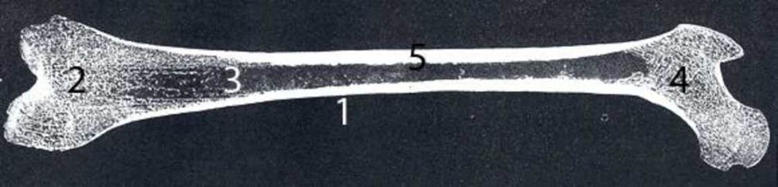
Fig. 1.2 Structure of a long bone (femur):
1 - diaphysis; 2 - epiphysis; 3 - medullary cavity; 4 - spongy substance; 5 - compact substance.
Spongy bones consist of spongy bone tissue covered by a thin layer of compact bone. The spaces of the spongy tissue contain red bone marrow. Spongy bones primarily perform supportive and protective functions.
Sesamoid bones, a variant of spongy bones, develop as additional structures within muscle tendons near various joints and are not directly connected to the skeletal bones. Typically, sesamoid bones increase the leverage of specific muscles. Examples of sesamoid bones include the Patella and the pisiform bone of the wrist.
Flat bones include the cranial vault bones (parietal bones) and the limb girdle bones (pelvic bones, scapulae). They consist of external and internal plates of compact bone tissue with spongy bone tissue sandwiched between them. Flat bones perform a protective function for the brain and several internal organs, while limb girdle bones perform a supportive function.
Pneumatic bones are part of the skull (frontal, sphenoid), vary in shape and size, and contain air-filled cavities lined with mucous membrane that communicate with the external environment.
Mixed bones include bones that exhibit great diversity in origin, shape, structure, and functions (example).
During ontogenesis, most human skeletal bones pass through Connective Tissue, cartilaginous, and bony stages of differentiation; based on their mode of ossification, such bones are called secondary. Some bones bypass the Cartilage stage in their development; based on their mode of ossification, such bones are called primary (BONES OF THE face, cranial vault, middle part of the clavicle).
1.6 The Bone as an Organ
Skeletal bones are built of bone tissue and covered by a membrane—the periosteum, *periosteum*, which separates them from surrounding tissues. The periosteum plays a major role in the development and Nutrition of bones. Its inner osteogenic layer is the site where bone matrix is formed. Blood Vessels and nerves penetrate deep into the bone from the periosteum. Almost all bones have articular surfaces covered with hyaline cartilage, where the periosteum is absent. Inside many bones, There is a marrow cavity lined with a membrane called the endosteum, *endostum*.
A living bone contains 50% Water, 15.75% fat, 12.25% protein-based organic substances, and 22% inorganic Mineral Substances. Bone tissue plays the main role in bone composition. Dried and degreased bone contains approximately 2/3 inorganic matter and 1/3 organic matter.
Bone tissue consists of bone Cells—osteocytes—and intercellular substance, or bone matrix, which includes organic and Inorganic Components. The former includes the protein ossein, which forms the ground substance of bone tissue. The inorganic components of bone are represented primarily by calcium salts in the form of submicroscopic hydroxyapatite crystals. Mineral fibers are formed from these crystals.
Bone strength exceeds that of many building Materials. In experiments by P.F. Lesgaft, a human femur withstood 5500 N/cm2 under tension and 7787 N/cm2 under compression. This equals the elasticity of oak and the strength of cast iron.
Organic and inorganic components of bone, taken separately, have much lower elasticity and strength. In decalcified bone, the modulus of elasticity decreases by about 20 times. A decalcified vertebra can be compressed by hand like a sponge, and a rib can be tied into a knot. Collagen isolated from bone has significantly lower tensile strength than an intact bone. When organic substances of bone are destroyed by incineration, the remaining mineral skeleton becomes extremely brittle, and the bone crumbles under slight pressure.
Bone Structure is studied using ground sections and histological preparations. As an example, let us consider a cross-section of the diaphysis of a long bone (Fig. 1.3). At the periphery of the bone diaphysis, there are several rows of bone lamellae parallel to the diaphysis surface. This is the layer of outer circumferential lamellae, through which perforating canals containing blood vessels pass. Deeper lies the osteon layer. Here, bone lamellae are arranged concentrically, forming systems called osteons, or Haversian systems. This is the structural unit of bone.
Several cylindrical tubes of varying diameters nested one inside the other can serve as a model of an osteon (Fig. 1.4). A single osteon may contain from 4 to 20 bone lamellae, with an average of 10. In the center of each system runs the osteon canal (Haversian canal) with a diameter ranging from 22 to 110 µm. These canals run primarily along the length of the diaphysis and interconnect. Small blood vessels and nerve trunks are located within the osteon canals. Interstitial lamellae, which run in various directions, are located between osteons; they are remnants of old, resorbed osteons. The layer of inner circumferential lamellae at the border with the medullary cavity is not continuous because numerous vascular canals pass through it. Nutrient canals, which begin on the bone surface with small openings, penetrate the entire thickness of the diaphysis. Vessels and nerves pass through the nutrient canals.
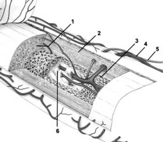
Fig. 1.3 Diagram of the structure of a long bone:
1 — osteon layer; 2 — compact bone; 3 — spongy bone; 4 — artery; 5 — vein; 6 — medullary canal
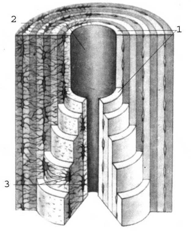
Fig. 1.4 Structure of an osteon, diagram:
1 - osteon lamella; 2 - central canal (osteon canal); 3 - bone cells (osteocytes).
The surrounding bone lamellae and osteons are also present in the compact substance of the epiphyses of tubular bones and the cortical layer of spongy bones. The trabeculae of the spongy substance are built of osteons. The latter are not arranged randomly, but are oriented in a specific direction corresponding to the main stress trajectories within the bone. Bone material concentrates along the lines of greatest tension and compression, thereby achieving minimal material expenditure. Consequently, the pattern of trabecular arrangement in the spongy substance corresponds to the mechanical conditions to which the given bone is subjected.
1.7 Doctrine of Bone Connections
The study of bone connections is termed arthrology. Along with osteology and Myology, arthrology constitutes a branch of anatomy that examines the musculoskeletal system. All bone connections are divided into two main groups: continuous and discontinuous.
Continuous connections. Continuous connections, or synarthroses, are phylogenetically older and have a simpler structure. Depending on the type of tissue participating in the bone connection, they are subdivided into fibrous, cartilaginous, and bony.
Fibrous connections, articulationes fibrosae, are formed by regular dense Fibrous connective tissue. These connections, in turn, are subdivided into syndesmoses and sutures.
Syndesmoses include ligaments and membranes. Both consist of bundles of collagen fibers and a small amount of elastic fibers.
Sutures represent thin plates of fibrous connective tissue located between the edges of the skull bones. Depending on the configuration of the bone margins, sutures are subdivided into serrated, squamous, and plane.
A special type of fibrous connection is dentoalveolar syndesmosis, or gomphosis (gomphosis, articulationes dentoalveolares); the roots of the Teeth are attached to the dental alveolus by means of connective tissue fibers.
Cartilaginous connections, articulationes cartilagineae, are also called synchondroses. Cartilaginous connections include the synchondroses of the skull located between the bones of the cranial base, as well as the sternal synchondroses connecting the manubrium and xiphoid process to the body of the Sternum. Most synchondroses are temporary. They exist only up to a certain age, after which the cartilaginous tissue is replaced by bone tissue, thus forming a synostosis.
Symphysis, symphysis, is distinguished as a special type of cartilaginous connection. It differs from synchondrosis in that the cartilage contains a small slit-like cavity. Symphysis represents, as it were, a transitional form from continuous to discontinuous connections. The Pubic Symphysis belongs to this category. It is believed that the cavity in the cartilage forms As a result of its stretching. Small cavities are also present in intervertebral discs; therefore, they are currently referred to as intervertebral symphyses.
Discontinuous connections. Discontinuous bone connections, or diarthroses, differ not only in their greater structural complexity but also in their functional properties. In contrast to slightly movable or completely immobile continuous connections, diarthroses allow for diverse and directed movements of individual skeletal segments. The capability for differentiated Movements of the head and limbs in terrestrial vertebrates is determined by the degree of development of discontinuous connections in their skeleton.
Diarthroses include synovial connections, articulationes synoviales, commonly referred to as joints (articulatio). A joint is an organ whose structure involves cartilaginous, osseous, and Connective Tissues proper. The structure of a joint comprises main and auxiliary elements.
The main elements invariably present in any synovial connection include articular surfaces, articular cartilage, joint cavity, Joint Capsule, and synovial fluid (Fig. 1.5).
Articular surfaces, facies articulares, are located on the bones participating in The formation of the joint. Each joint contains at least one pair of articulating surfaces. One of them is typically convex, known as the articular head, while the other is concave, known as the articular fossa. The convex surface is always larger in extent than the concave one.
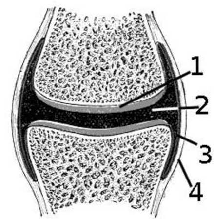
Fig. 1.5 Basic structure of a synovial connection:
1 - articular cartilage; 2 - joint cavity; 3 - synovial membrane of the joint capsule; 4 - fibrous membrane of the joint capsule.
The articular surfaces are covered by articular cartilage, cartilago articularis. Most articular surfaces are covered with hyaline cartilage, and only in a few joints, such as the temporomandibular and sternoclavicular joints, is fibrocartilage present. Along the margin of the articular cartilage, the fibrous layer of the periosteum continues directly into the superficial fiber layer of the cartilage itself. Thus, the entire bone, together with the articular cartilage, is surrounded by a single fibrous sheath. The outer surface of the cartilage is smooth, which allows the articular surfaces to glide easily relative to one another. Due to its elasticity, articular cartilage protects the ends of bones from damage during impacts and jolts.
The joint capsule, capsula articularis, encloses the parts of the bones that form the joint. The capsule attaches along the margins of the articular surfaces or at a short distance from them, hermetically sealing the joint. The joint capsule consists of two membranes: fibrous and synovial. The fibrous membrane forms the outer layer. The synovial membrane constitutes the inner layer of the joint capsule. It lines all structures within the joint, with the exception of the articular cartilages. This membrane is thin, loosely connected to the fibrous layer and therefore movable, containing collagen and elastic fibers. In some joints,
the synovial membrane forms folds, plicae synoviales, which contain adipose tissue and project into the joint cavity, filling its free spaces. Smaller projections—synovial villi, villi synoviales—increase the surface area of the synovial membrane, which is important for metabolic processes within the joint. The synovial membrane is richly supplied with blood vessels, Lymphatic vessels, and nerves.
The synovial membrane and articular surfaces bound the joint cavity, cavitas articularis, which under normal conditions appears as a narrow slit, and only in pathological conditions accompanied by the accumulation of a large amount of fluid in the joint (bursitis) does the volume of the joint cavity increase to such an extent that the articular surfaces may separate.
Synovial fluid, or synovia (synovia), is produced by the synovial membrane. Under normal conditions, it is present in the joint cavity in a small amount. Synovia acts as a lubricant in joints. It also performs protective and metabolic functions, ensuring the trophic supply of the avascular articular cartilage. Synovial fluid participates in the exchange of substances between the joint contents and the vascular bed of the synovial membrane. When various substances are introduced into the joint cavity, they are absorbed into the Vessels of the synovial membrane, with joint movements significantly accelerating this absorption.
Auxiliary elements of the joint include articular discs, menisci, labra, ligaments, and synovial bursae.
The articular disc, discus articularis, is a fibrocartilaginous plate covered by a synovial membrane, located within the joint cavity between the articulating bone surfaces and fused with the joint capsule. Similar structures are found in the temporomandibular, sternoclavicular, and radiocarpal joints.
A variation of the disc is represented by the articular menisci, menisci articulares, located in the knee joint. They are curved cartilaginous plates of semilunar and sickle-like shape, secured within the joint by special ligaments. Due to their elasticity, articular discs and menisci cushion shocks and jolts transmitted to the joint. They also play a specific role in the mechanics of movement.
The articular lip, labrum articulare, is a ring-shaped fibrocartilaginous structure attached to the margin of the articular cavity, deepening it and increasing its surface area. Articular Lips are found in the shoulder, hip, and certain other joints.
Ligaments play a major role in joint stabilization and movement. Together, they form the Ligamentous apparatus of the joint. Based on their relation to the articular capsule, ligaments are divided into 3 types:
1. Extracapsular ligaments, ligamenta extracapsularia — located outside the joint capsule, though they frequently blend into it.
2. Capsular ligaments, ligamenta capsularia — thickenings of the joint capsule.
3. Intracapsular ligaments, ligamenta intracapsularia — located within the joint cavity and covered by the synovial membrane.
Along with the joint capsule and muscles, ligaments ensure joint stability and maintain contact between the articulating bone surfaces. Ligaments restrict and limit joint movements. Some act as guiding ligaments that influence the trajectory of movement while interacting with other joint components, such as the ulnar collateral ligament of the elbow joint. In a number of ligaments, these functions are combined. For instance, the medial (deltoid) ligament of the ankle joint performs stabilizing, restraining, and guiding functions. Certain patterns are observed in the arrangement of ligaments:
1. Ligaments are distributed in each joint according to the number and position of its axes of rotation.
2. Ligaments are positioned perpendicular to a given axis of rotation, predominantly at its ends.
3. Ligaments lie within the plane of the joint's movement.
Synovial bursae, bursae synoviales, are protrusions of the synovial membrane through thinned areas of the fibrous layer of the joint capsule.
The size and shape of synovial bursae vary. As a rule, synovial bursae are located between the bone surface and moving muscle tendons. Bursae eliminate friction between contacting surfaces of tendons and bones.
It should be noted that due to joint airtightness, a negative pressure of 60-120 mm of water Column is maintained within the joint cavity. As a result, atmospheric pressure presses the articular surfaces against each other with a force that in the hip joint reaches 25 kg. If a joint is isolated and all muscles and ligaments are removed, the connection of the articular surfaces is still preserved. For the articular surfaces to separate, it is necessary to incise the joint capsule or introduce gas under pressure into the joint cavity.
Principles of joint classification. Joint classification is based on anatomical and functional features. Depending on the number of articular surfaces, joints are classified as simple or complex.
A simple joint, articulatio simplex, has only one pair of articular surfaces. Most human joints are simple, such as the interphalangeal joints.
A complex joint, articulatio composita, includes two or more pairs of articular surfaces, such as the elbow joint.
Functionally, joints are classified into complex (composite) and combined joints.
A complex joint, articulatio complexa, is a joint whose cavity is completely or partially divided into two parts by an articular disc or meniscus, such as the temporomandibular joint.
Combined joints, articulationes combinatae, are anatomically isolated joints that always function together in movements, such as the right and left temporomandibular joints.
1. Plane joint, articulatio plana — its articular surfaces can be regarded as segments of a sphere with a large radius. Plane joints include the zygapophysial joints, carpometacarpal joints of digits II-V, sacroiliac joint, and tibiofibular joint.
2. Spheroidal (ball-and-socket) joint, articulatio spheroidea — this also does not strictly correspond to its name, as its surfaces have varying curvature in different regions. Spheroidal joints include the joint of the rib head, shoulder joint, and humeroradial joint.
3. Cotyloid (cup-shaped) joint, articulatio cotylica — a specialized variant of the ball-and-socket joint. There is only one such joint: the hip joint.
4. Ellipsoid joint, articulatio ellipsoidea — its surfaces can be compared to a piece of eggshell. This type includes the acromioclavicular and radiocarpal joints. Ellipsoid surfaces, much like spheroidal ones, are either concave or convex in all directions.
5. Saddle joint, articulatio sellaris — possesses the opposite property: the articular surfaces are convex in one direction and concave in the opposite direction. Saddle joints include the sternoclavicular joint, the carpometacarpal joint of the thumb (digit I), and the calcaneocuboid joint.
6. Condylar joint, articulatio condylaris — an articulation in which one bone articulates with another via two separate surfaces. Each of these articular surfaces is termed a condyle, regardless of whether it is convex or concave. Condylar joints include the knee joint.
7. Hinge joint, ginglymus — a joint with cylindrical articular surfaces. The axis of the convex articular surface is perpendicular to the axis of the bone itself, and the trochlea features a small ridge that guides its movement. Examples include the interphalangeal joints, humeroulnar joint, and ankle joint.
8. Trochoid joint, articulatio trochoidea — the axis of the convex articular surface runs along the longitudinal axis of the bone itself, rather than perpendicularly to it, as in a ginglymus (hinge) joint. These include the atlanto-axial, costotransverse, radioulnar, and subtalar joints.
Depending on the number of axes around which movements can occur, joints are classified into:
- uniaxial joint — movements occur around 1 axis (hinge or pivot in shape);
- biaxial joint — movements occur around 2 axes (ellipsoid, saddle, or condylar in shape);
- triaxial (multiaxial) joint — movements occur around 3 axes (plane, spheroid/ball-and-socket, or cotyloid in shape).
Outline for studying a joint during a practical class:
1. Name of the joint (in Ukrainian and Latin).
2. Primary Components of the joint:
a) demonstrate the articular surfaces on the skeleton;
b) demonstrate the articular cartilage and joint capsule on wet specimens.
3. Accessory (auxiliary) structures of the joint:
a) trace the direction of the ligaments between bony prominences on the skeleton;
b) demonstrate the articular disc, meniscus, labrum, and ligaments on wet specimens.
4. Type of joint.
5. Shape of joint.
6. Function of the joint (demonstrate on the skeleton and on a living person (e.g., yourself) which movements occur around which axes and in which planes).
Possible movements: flexion (flexio) and extension (extensio) — primarily around the frontal axis in the sagittal plane; abduction (abductio) and adduction (adductio) — primarily around the sagittal axis in the frontal plane; rotation (rotаtio) — primarily around the vertical axis in the horizontal plane.
Temporomandibular joint (articulatio temporomandibularis).
Features of primary components: the articular surfaces are the mandibular fossa and the head of the Mandible (Fig. 1.6). Accessory structures:
— articular disc, which divides the joint cavity into two compartments (upper and lower);
— intracapsular ligaments (discotemporal and discomandibular);
— extracapsular ligaments:
a) lateral ligament (ligamentum laterale) — extends from the zygomatic arch to the neck of the mandible;
b) stylomandibular ligament (ligamentum stylomandibulare) — between the styloid process and the angle of the mandible;
c) sphenomandibular ligament (ligamentum sphenomandibulare) — between the greater wing of the Sphenoid bone and the angle of the mandible.
The joint type is combined. In shape, it is a condylar joint. Function:
— vertical movements occur in the frontal plane (depression and elevation of the mandible);
— sagittal movements occur in the sagittal plane (forward and backward Displacement of the mandible);
— transversal movements occur in the horizontal plane (right and left displacement of the mandible).
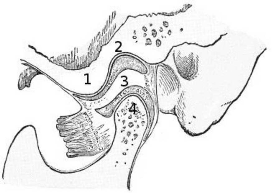
Fig. 1.6 Structure of the temporomandibular joint:
1 - articular tubercle; 2 - mandibular fossa; 3 - articular disc; 4 - mandibular head.
Age-related Structural Features of the temporomandibular joint. In newborns, the head of the mandible is rounded, with nearly equal mediolateral and anteroposterior dimensions. With age, it gradually elongates transversely. From the eruption of deciduous teeth up to 2 years of age, the head enlarges, after which its dimensions stabilize. This stabilization lasts until 6 years of age: with the appearance of the first permanent tooth, the dimensions of the head increase again.
In newborns, forward inclination of the head is not pronounced; with age, the head tilts forward relative to the neck of the articular process. In infancy, the mandible assumes a distal position. With the eruption of deciduous molars and the increase in bite height, further anterior displacement of the articular head occurs.
In newborns, the head is covered with a thick layer of fibrous connective tissue, whereas in adults, it is covered with fibrocartilage, which thins with age.
In newborns, the articular tubercle is absent; it only begins to form anterior to the mandibular fossa. Posteriorly, there is a well-defined retroarticular process that protects the tympanic part of the Middle ear from the pressure of the articular head. With the eruption of deciduous teeth, the dimensions of the articular tubercle increase. By 6–7 years of age, it is already well developed. As the articular tubercle develops, the retroarticular process atrophies.
In newborns, the mandibular fossa is virtually flat and rounded in shape. It functions fully, and its bone thickness slightly exceeds 2 mm. Subsequently, the depth of the mandibular fossa increases, and it elongates transversely, acquiring an elliptical shape that corresponds to the Changes in the mandibular head. The articular surface is covered with fibrocartilage.
The articular disc in newborns is a soft, rounded layer, concave inferiorly and convex superiorly, with barely visible anterior and posterior thickenings. It consists of collagen fibers. Parallel to the Formation of the bony structures of the joint, the disc also forms, gradually acquiring anterior and posterior thickenings and a thin central part, which serves to ensure the congruence of the articular surfaces.
There are 4 Zones of the disc: the anterior pole, intermediate zone, posterior pole, and bilaminar zone (located between the posterior pole of the disc and the joint capsule, represented by two ligaments enclosing a neurovascular zone). In the area of the bilaminar zone, the synovial membrane of the joint capsule forms villi, which serve as sites of interoception. In newborns, villi are absent; a small number of them appear at 1–2 years and increase up to 3–6 years of age. By 16–18 years, they are numerous. As the body ages, involution of the villi occurs.
Last update: 08/08/2026
Editorial and Educational Adaptation: This material has been compiled based on the primary/original source text. The project team performed an editorial review, corrected technical inaccuracies, structured sections, and adapted the content for an educational format.
What was processed:
- elimination of formatting defects (OCR errors, structural breaks, corrupted characters);
- editorial organization of content;
- standardization of terminology in accordance with academic sources;
- verification of factual statements against the original source text.
All mentions of the author, publication year, and origin of the primary text have been preserved in accordance with the source.