Human Anatomy (with the Fundamentals of Dynamic and Sports Morphology) - Ivanitsky M. F. 2008
The Doctrine of Internal Organs
Digestive System
The Digestive System comprises a complex of Organs responsible for the mechanical and chemical Processing of food products. It is the site where nutrients, derived from food breakdown, are absorbed into the lymphatic and Blood Vessels. Unabsorbed food residues, which are subsequently eliminated to the exterior, are also formed here. The digestive system consists of an 8–10 meter long digestive tract (with localized dilations) and digestive glands whose ducts open into the lumen of the tract.
The largest of these are the Salivary Glands (parotid, sublingual, and submandibular), as well as The Liver and Pancreas. The digestive tract is divided into the following sections: Oral Cavity, Pharynx, Esophagus, Stomach, Small Intestine, and Large Intestine.
Oral cavity
The oral cavity is subdivided into two compartments: the oral vestibule and the oral cavity proper.
Oral vestibule
The oral vestibule refers to the space situated between the Lips and Cheeks externally, and the Teeth and Gums internally. The vestibule communicates with the external environment via the oral fissure, and with the oral cavity proper through the cleft behind the ramus of the Mandible, which occurs not only when the jaws are parted, but also when they are clenched.
The oral fissure is bounded by the upper and lower lips. The Structure of the lips and cheeks is similar; their bulk is formed by Muscles. The inner surface of the lips and cheeks is lined with mucous membrane, while the exterior is covered by Skin. Between the skin and the mucous membrane, the lips possess an intermediate zone covered with epithelium, beneath which lies a rich network of blood vessels.
The mucous membrane of the lips and cheeks extends onto the alveolar margins of the upper and lower jaws, forming the gums, which firmly adhere to the alveolar margins of the jaws and the necks of the teeth.
Teeth. Teeth are located along the alveolar margins of the upper and lower jaws at the boundary between the oral vestibule and the oral cavity proper.
It is customary to distinguish between deciduous and permanent teeth. Deciduous teeth (20 in total: on each half of the upper and lower jaw there are 2 incisors, 1 canine, and 2 molars) exist only during childhood. They are shed and replaced by permanent teeth (30–32 in total): 2 incisors, 1 canine, 2 premolars, and 3 molars.
Class="center">
Fig. 72. Permanent teeth of the right side (medial view):
1 - medial incisor; 2 — lateral incisor; 3 - canine; 4 - first premolar; 5 - second premolar; 6 - first molar; 7 - second molar; 8 - third molar (wisdom tooth) (after Braus)
The arrangement of teeth is conventionally designated by a so-called dental formula. For deciduous teeth, it appears as follows:

(premolars are absent). For permanent teeth, this formula takes the form:

Eruption of deciduous teeth typically begins at 6 months of age. Tooth replacement begins after 6 years. Usually, the eruption of permanent teeth starts with a molar or an incisor, concluding by 12–14 years of age. An exception is the 3rd molar, known as the wisdom tooth, which erupts after the age of 18.
A tooth consists of a crown, a neck, and a ROOT. The crown is the external part of the tooth visible upon inspection. The neck serves as the site of attachment for the gum. The root is embedded in the dental alveolus. Inside the tooth is a pulp cavity that extends into the root, opens at its apex, and contains blood Vessels and nerves making up the so-called dental pulp. The tooth root fuses quite firmly with The surface of the dental alveoli.
The external shape of teeth varies (Fig. 72). A tooth presents four surfaces: occlusal or masticatory, lingual, vestibular or facial, and contact. The masticatory surface faces the tooth of the opposite jaw, the lingual surface faces the Tongue, the vestibular surface faces the lips and cheeks, and the contact surface faces the adjacent tooth.
A tooth is constructed of dentin, which forms the bulk of the tooth; enamel, which covers the exterior of the tooth in the crown region; and cementum, which covers the root of the tooth.
Each tooth possesses its own characteristics. Incisors resemble a chisel in shape. Their root is compressed transversely and lacks bifurcation at the tip. Upper incisors are larger than lower ones. Canines also have a single root, laterally compressed. The cone-shaped crown of a canine features two cutting edges converging toward the apex. Premolars have a single root. In upper teeth, the root is flattened in the anteroposterior direction. The crown of premolars is also somewhat flattened anteroposteriorly and bears two cusps on its masticatory surface. Molars have an approximately quadrangular crown. Upper molars typically have four cusps, the 1st lower molar has five, and the 2nd and 3rd lower molars have four. Upper molars possess three roots, while lower ones have two. The roots of wisdom teeth frequently fuse into a single common root. The size of molars decreases posteriorly.
When the mandible is in a relaxed position, even with the Mouth closed, a slight free space exists between the upper and lower teeth because the lower jaw hangs down slightly. The occlusion of the teeth is termed the bite. In a normal bite, the teeth of the upper jaw project slightly anteriorly relative to the lower jaw teeth due to the alveolar margin of the upper jaw being somewhat larger than that of the lower jaw.
Oral cavity proper
The oral cavity proper is bounded superiorly by the hard and partly soft palate, inferiorly by the floor of the mouth formed by the mylohyoid Muscle, and anteriorly and laterally by the teeth and gums. For the most part, it is occupied by the tongue.
The mucous membrane of the hard palate is tightly fused with the bone periosteum. Posteriorly, the hard palate transitions into the soft palate, which is formed of muscles covered by a mucous membrane. In its middle, There is a downward-directed projection known as the uvula. Posteriorly, the oral cavity communicates with the pharyngeal cavity via the fauces—an unpaired aperture bounded superiorly by the soft palate (its hanging inferior part, termed the palatine velum) and the uvula, inferiorly by the root of the tongue, and laterally by the palatine arches.
The tongue. The tongue is divided into the body, the tip or apex, and the root. Its upper surface is called the dorsum. The dorsum is free, whereas the lower surface is free only in its anterior portion. The lateral margins of the tongue are also free.
The tongue comprises an anterior part, located entirely within the oral cavity, and a posterior or pharyngeal part, positioned almost vertically with its dorsum facing the pharynx.
On the mucous membrane of the posterior part, there is an accumulation of lymphoid tissue known as the lingual tonsil.
The mucous membrane of the tongue forms papillae (Fig. 73) that vary in shape and function, primarily on its dorsum and lateral surfaces. The anterior part of the tongue features filiform, conical, fungiform, vallate (circumvallate), and foliate papillae. In the filiform and conical papillae, receptors perceive pain and Temperature stimuli. Fungiform papillae are relatively few in number; they contain taste buds that perceive gustatory stimuli. A large number of taste buds are located within the vallate papillae, which are the largest papillae. They are arranged in a V-shaped formation at the border between the anterior and posterior PARTS OF THE tongue, numbering up to 12. The central part of these papillae is cylindrical in shape, surrounded by a sulcus, outside of which lies a wall (vallum). Foliate papillae are found exclusively on the margins of the tongue and are more pronounced in its posterior part than in the anterior.
The tongue is constructed of striated muscles. Some muscles originate and terminate within the tongue, altering its shape upon contraction, while others originate from bones, the soft palate, or the pharynx, terminating within the tongue to facilitate its movement within the oral cavity. The intrinsic Muscles of the tongue possess fibers running in three directions: transverse, anteroposterior, and vertical. Upon their contraction, the tongue correspondingly shortens, decreases in transverse diameter, and flattens.
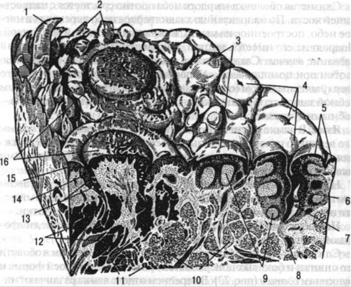
Fig. 73. STRUCTURE OF THE tongue:
1 — conical papilla; 2 — filiform papillae; 3 — fungiform papillae; 4 — accumulation of lymphoid tissue; 5 — epithelium; 6 — leukocytes within the epithelium; 7 — crypt; 8 — duct of a gland; 9 — individual follicles; 10 — mucous Glands of the mucous membrane; 11 — striated m.m.; 12 — serous glands; 13 — taste buds; 14 — circular sulcus; 15 — vallum; 16 — vallate papillae (after Braus)
Muscles originating from adjacent bones include: the genioglossus (displaces the tongue forward), the hyoglossus (displaces the tongue backward and downward), and the styloglossus (displaces the tongue backward and upward).
Laterally, the soft palate directly transitions into the anterior (palatoglossal) and posterior (palatopharyngeal) arches, which are formed of muscles and covered by mucous membrane. The anterior arch extends from the palate to the lateral surface of the tongue, whereas the posterior arch extends into the pharynx. Between them lies a depression containing the palatine tonsil, which represents an accumulation of lymphoid tissue. It varies greatly in size and shape, sometimes projecting significantly beyond the arches.

Fig. 74. Diagram of the soft palate musculature:
1 — tensor veli palatini m.; 2 — levator veli palatini m.; 3 — pterygoid hamulus of the Sphenoid bone; 4 — palatoglossus m.; 5 — uvular m.; 6 — palatopharyngeus m. (after G. F. Ivanov)
The soft palate is formed by the following paired muscles: the palatopharyngeus, levator veli palatini, tensor veli palatini, palatoglossus, and the unpaired musculus uvulae (Fig. 74). Upon contraction, all of them alter the shape of the soft palate by elevating, shortening, and tensioning the palatine velum. The palatopharyngeus muscle participates in the act of swallowing by elevating the pharynx. Contraction of the palatoglossus muscle results in the constriction of the fauces.
Salivary glands. The ducts of the salivary glands—both minor and major—open into the oral cavity (Fig. 75). Minor salivary glands are named according to their Location: palatine, labial, buccal, and lingual. There are three pairs of major salivary glands: the parotid, submandibular, and sublingual. Based on The Nature of their secreted product (saliva), compound glands are divided into protein (serous), mucous, and mixed. Saliva contains Enzymes that perform the primary breakdown of dietary CARBOHYDRATES.
The parotid gland produces a serous (protein-rich) secretion containing enzymes; it is located slightly anterior and inferior to the auricle upon the masseter muscle and partially fills the retromandibular fossa. The weight of the gland is 20–30 g. It is covered by the parotid fascia and exhibits a lobular structure.
The parotid gland is directly adjacent to the mandible, which is of great significance during mastication. During Movements of the mandible, this gland is massaged, facilitating the expulsion of its secretion into the oral cavity. The excretory duct of the parotid gland runs anteriorly, parallel to the zygomatic arch; curving over the anterior margin of the masseter muscle, it pierces the buccinator muscle and opens into the oral vestibule at the level of the upper second molar.

Fig. 75. Salivary glands
1 — parotid duct; 2 — zygomaticus major m.; 3 — buccal glands; 4 — levator anguli oris m. (cut); 5 — labial glands; 6 — orbicularis oris m.; 7 — anterior lingual gland; 8 — opening of the sublingual duct; 9 — mandible (sectioned); 10 — genioglossus m.; 11 — geniohyoid m.; 12 — mylohyoid m.; 13 — sublingual gland with minor excretory ducts; 14 — digastric m. (anterior belly); 15 — Hyoid bone; 16 — submandibular duct; 17 — submandibular gland; 18 — facial artery; 19 — masseter m.; 20 — parotid gland; 21 — superficial temporal artery and vein (after Braus)
The submandibular gland belongs to the mixed group and weighs 10–15 g. It lies against the inner surface of the body of the mandible and the medial pterygoid muscle, filling the submandibular fossa. The excretory duct of the submandibular gland, curving over the posterior margin of the mylohyoid muscle, opens onto an elevation—the sublingual caruncle—located on the floor of the oral cavity lateral to the lingual frenulum.
The sublingual gland weighs approximately 4 g. It is situated beneath the mucous membrane of the floor of the Oral Cavity and secretes mucus. Its 18–20 excretory ducts open into the oral cavity along the sublingual fold. A larger duct opens into the oral cavity together with the submandibular duct.
Pharynx
The pharynx serves as a passageway for food and air. It represents the upper portion of the digestive tract and is attached to the external Base of the Skull, the pterygoid processes of the sphenoid bone, as well as to the hyoid bone and the cartilages of the Larynx.
The pharynx is conventionally divided into three parts: the nasal, oral, and laryngeal parts. The nasal part communicates with the Nasal cavity through the choanae and with the Middle ear cavity via the auditory tubes; the oral part communicates with the oral cavity through the fauces; and the laryngeal part directly continues into the esophagus at the level of the 6th–7th cervical vertebrae.
The wall of the pharynx is composed of muscles lined internally by a mucous membrane and covered externally by an adventitial layer. Between the mucous and muscular membranes lies a fibrous coat, which anchors the pharynx to the external base of the skull. Posterosuperiorly on the pharyngeal mucosa lies an accumulation of lymphoid tissue known as the pharyngeal tonsil (adenoid), while anterior to the pharyngeal opening of the auditory tube is the tubal tonsil. The lingual, palatine, pharyngeal, and tubal Tonsils form a nearly continuous ring of lymphoid tissue in the upper digestive tract—the so-called Waldeyer's ring (Pirogov-Waldeyer ring).
Of the seven openings of the pharynx (two auditory tube openings, two choanae, and one opening each for the fauces, larynx, and esophagus), six close during the act of swallowing. The choanae leading to the nasal cavity and the openings of the auditory tubes are closed because the soft palate is elevated by the contraction of its striated muscles. The fauces are closed by the dorsum of the tongue, and the laryngeal inlet is closed by the epiglottis, which is pushed downward and backward by the pressure of the tongue's dorsum. During swallowing, the only opening that remains patent is the one leading from the pharynx into the esophagus.
The pharyngeal muscles are striated (voluntary) and feature a rather complex architecture. It is customary to distinguish three pharyngeal constrictors—superior, middle, and inferior—with transversely oriented fibers, alongside several smaller muscles (the stylopharyngeus and palatopharyngeus) that run vertically. Upon contraction, these vertical muscles primarily elevate the lower part of the pharynx, helping to shorten it vertically.
The pharyngeal constrictors are significantly better developed than the longitudinal muscle group. Where the constrictors meet posteriorly, they form a midline raphe, which is a Connective Tissue structure. A structural feature of the pharyngeal constrictors is their funnel-like shape, with each superior funnel nesting inside the one below it, which facilitates the propulsion of food.
Esophagus
The esophagus is a direct continuation of the pharynx. It is a muscular tube connecting the pharynx to The Stomach. The esophagus begins at the level of the 6th–7th cervical vertebrae and descends anterior to THE Vertebral Column along the midline of the body down to the level of the 11th thoracic vertebra. Its total length reaches 25–30 cm. The esophagus is divided into three parts: the cervical, thoracic, and abdominal parts, the latter of which transitions into the stomach.
The longest portion of the esophagus is the thoracic part, and the shortest is the abdominal part (approximately 1 cm). Throughout its course, the esophagus exhibits three narrowings: at the junction of the pharynx with the esophagus (upper), at the level where the Trachea bifurcates into the Bronchi (middle), and at the transition of the esophagus into the stomach (lower).
The upper part of the esophagus lies posterior to the trachea, and in the cervical region, slightly to the left of it. In the thoracic cavity, it is located in the posterior Mediastinum alongside the descending aorta, azygos, and hemiazygos Veins. Initially, it runs to the right of the descending aorta, and further down, curving around it, comes to lie anterior to the aorta at the level of approximately the 9th thoracic vertebra, subsequently passing through the Diaphragm also anterior to it. At the level of the 4th–5th thoracic vertebrae, the esophagus passes posterior to the left bronchus. In its lower half, it deviates slightly to the left of the body's midline before passing through the diaphragm into the Abdominal cavity.
The wall of the esophagus is approximately 4 mm thick. Its muscular coat in the upper section is composed of Striated Muscle tissue, whereas in the thoracic and abdominal sections it consists of Cytology/cytology/32.html">Smooth muscle tissue. The gradual replacement of one type of muscle tissue by the other occurs progressively along the length of the esophagus.
The luminal surface of the esophagus is lined with a mucous membrane, while its exterior is covered by a fascia. The serous coat of the esophagus is absent in the cervical and thoracic regions. The mucous membrane features longitudinal folds, which facilitate the expansion of the lumen during the passage of a large food bolus. Alongside intrinsic esophageal glands, gastric glands are occasionally found in the human esophagus.
Stomach
The stomach is a dilation of the digestive tube that varies considerably in shape and position. It is located in the upper left region of the abdominal cavity behind the costal margin (Fig. 76). The stomach presents anterior and posterior walls, as well as greater and lesser curvatures; the former faces left and downward, while the latter faces right and upward. The omenta attach to the curvatures of the stomach. Anatomically, the stomach comprises the cardiac part (cardia) where the esophagus opens, the body of the stomach, the pyloric part communicating with the duodenum, and the fundus, which lies to the left of the esophageal opening. The stomach wall consists of three layers: mucous, muscular, and serous. The mucous membrane forms numerous folds and contains a large number of glands that secrete gastric juice. These glands consist of chief and parietal Cells. Chief cells secrete gastric enzymes, whereas parietal cells secrete Hydrochloric acid, which acts as a catalyst. The muscular coat is subdivided into three layers: circular, longitudinal, and oblique. The serous coat is formed by the visceral Peritoneum and covers the stomach on all sides.
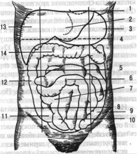
Fig. 76. PROJECTION OF THE BOUNDARIES OF THE liver, stomach, and intestines onto the anterior abdominal wall:
1 — xiphoid process of the Sternum; 2 — left lobe of the liver; 3 — stomach; 4 — level of the pylorus of the stomach; 5 — level of the lower Ribs; 6 — loops of the jejunum; 7, 10 — loops of the ileum; 8 — level of the anterior superior iliac spines; 9 — descending colon; 11 — level of the cecum and vermiform Appendix; 12 — ascending colon transitioning into the transverse colon; 13 — right lobe of the liver; 14 — transverse colon (after G. F. Ivanov)
Small Intestine
The small intestine is the direct continuation of the stomach. It has a length of 4.18–8.80 m and is divided into three parts: the duodenum, the jejunum, and the ileum.
The duodenum is the proximal part of the small intestine and is shorter than the other sections (25–30 cm). It lies deep within the abdominal cavity, predominantly to the right of the midline, abutting the posterior abdominal wall. The duodenum is C-shaped (horseshoe-shaped) and is subdivided into superior, descending, and horizontal parts. It begins at the stomach at the level of the 12th thoracic to 1st lumbar vertebrae and descends down to the level of the 3rd lumbar vertebra. Its horizontal part runs from right to left, ascending to the body of the 2nd lumbar vertebra, where it transitions into the jejunum. The upward-directed segment of the duodenum is sometimes described as its fourth or ascending part. The mobility of the duodenum is relatively limited.
Of the three main parts of the duodenum, only the superior part possesses a mesentery in the form of the hepatoduodenal ligament, which extends from the porta hepatis. The peritoneum covers the duodenum only anteriorly. The mucous membrane of the duodenum features circular folds (plicae circulares). These folds bear numerous villi—microscopic projections of the mucosa that play a crucial role in nutrient absorption—as well as accumulations of lymphoid tissue in the form of solitary lymphatic follicles that perform a protective function. On the mucosa of the descending part of the duodenum, there is a longitudinal fold and a papilla where the ducts of the pancreas and liver open. In addition to these, a large number of minor glands secrete their products into the lumen of the duodenum. Some of these glands exhibit endocrine activity; hence, the duodenum is sometimes figuratively referred to as the "hypophysis of the abdominal cavity."
The junction between the duodenum and the jejunum is located at the level of the 2nd lumbar vertebra, to the left of the body's midline. The transition from the jejunum to the ileum is gradual, without a sharp demarcation. Approximately the upper two-fifths of their combined length constitute the jejunum, while the lower three-fifths constitute the ileum. Thanks to its well-developed mesentery, the jejunum is quite mobile and can change its position. Individual loops of the jejunum may partially lie in the left hypochondriac region or be covered by the transverse colon. However, the largest portion of the jejunum is located in the umbilical region (see Fig. 76).
The ileum, as the continuation of the jejunum, extends from the umbilical region into the right half of the abdominal cavity, where it terminates at the cecum near the right iliac fossa.
Thus, the small intestine occupies a significant portion of the ABDOMINAL CAVITY AND is situated across all its regions. It has a relatively uniform diameter (unlike the large intestine). Most of its extent is covered by the serous coat. The muscular coat consists of two layers (circular and longitudinal) that completely encircle the intestine (unlike the large intestine, where the longitudinal muscle layer does not form a continuous sheet but rather runs in three distinct bands). The mucous membrane features numerous folds, villi, intestinal juice-producing glands, and lymphoid tissue aggregates (solitary and aggregated follicles). The size and number of these lymphoid aggregates increase toward the large intestine.
The folds in the small intestine (circular folds, or plicae circulares) are formed by the mucous membrane and submucosa. These folds do not flatten out when the intestine is distended. They increase in height at the beginning of the jejunum, whereas in the ileum they become flatter and sparser.
Intestinal villi are mucosal evaginations devoid of a submucosal core. They vastly increase the absorptive surface area of the intestine. They are extremely numerous (4–5 million). In the duodenum and jejunum, there are about 30 to 40 villi per 1 mm2. The length of the villi ranges from 1 to 1.5 mm. Muscle cells within the villi allow them to function somewhat like pumps, facilitating the expulsion of secretions from the glands and the absorption of digested food products. Each villus contains nerves, blood capillaries, and lymphatic capillaries that form dense capillary networks within it.
The movement of the villi is regulated by the hormone villikinin, while their blood filling is mediated by arteriovenous anastomoses located within them (see p. 283). Proteins and carbohydrates, broken down by the action of intestinal juice, are absorbed into blood capillaries, whereas fat breakdown products enter the lymphatic capillaries.
The surface of intestinal epithelial cells features cytoplasmic outgrowths known as microvilli. Each Cell contains up to 3,000 microvilli, which further increase the absorptive surface area of the intestine. Among the epithelial Cells of the small intestine are numerous goblet cells that secrete intestinal juice. In addition to villi, the mucous membrane contains crypts, which are tubular invaginations of the epithelium into the mucosa. Their number can reach up to 100 per 1 mm2, with a higher concentration in the duodenum and jejunum.
The muscular coat, consisting of longitudinal and circular layers of muscle fibers, is characterized by a helical arrangement of these fibers with varying degrees of pitch in their coils.
The visceral layer of the serous membrane covers the jejunum and ileum from all sides and forms the mesentery, which consists of two layers enclosing fat, blood vessels, Lymphatic vessels, Lymph Nodes, and nerves.
Large Intestine
The large intestine is the continuation of the small intestine and is subdivided into the cecum with the vermiform appendix, the colon, and the rectum. In turn, the colon is divided into the ascending colon, transverse colon, descending colon, and sigmoid colon. These parts of the large intestine are located in the abdominal cavity to the right, to the left, and partially below the small intestine, framing it accordingly (see Fig. 76).
The total length of the large intestine reaches 2 m, though it can be shorter (1.5 m). The diameter of the large intestine is significantly greater than that of the small intestine, ranging from 4 to 7 cm. The cecum is the widest part of the large intestine.
The large intestine differs from the small intestine both in appearance and internal structure. The first distinctive feature is that three bands, or taeniae coli (free, mesocolic, and omental), run from the vermiform appendix to the rectum. These are bands of longitudinal smooth muscle which, as previously mentioned, are distributed unevenly in the large intestine.
The second feature of the large intestine is the sacculations of its walls, known as haustra, which are clearly visible both from the mucosal side and on the external surface of the gut. They are formed because the longitudinal bands are shorter than the intestine itself.
The third feature of the large intestine is the presence of omental Appendices (appendices epiploicae), which are fat-containing outpouchings of the serous layer of the intestinal wall. They protect the wall from being pinched between segments of the intestine containing dense contents.
The mucous membrane of the large intestine lacks villi; it is smooth and features semilunar folds, intestinal crypts, glands, and solitary lymphatic follicles (Aggregated lymphoid follicles are absent).
The muscular coat consists of two layers: a longitudinal layer running as three muscular bands, and a circular layer.
The serous membrane is absent in certain parts of the large intestine wall. The lower two-thirds of the rectum lack this covering, while the ascending and descending colons are covered by the peritoneum on three sides. The remaining parts of the large intestine are covered by the serous membrane on all sides and possess a mesentery (except for the cecum), which accounts for their considerable mobility.
The cecum is the initial segment of the large intestine. It is located in the right part of the abdominal cavity, inferior to the junction of the ileum with the large intestine; its upper boundary lies approximately at the level of the midpoint between the umbilicus and the anterior superior iliac spine (see Fig. 76). At this junction lies the ileocecal valve, which permits contents to flow in only one direction—from the small intestine into the large intestine. The length of the cecum is approximately 6 cm, and its width is about 7 cm.
The vermiform appendix (appendix) extends from the posteromedial surface of the cecum, with an average length of about 8 cm. It is covered by the serous membrane and has a mesentery. Clusters of lymphoid tissue are located within the mucous membrane of the appendix. Its position varies greatly depending on the LOCATION OF THE cecum. It may drape over the iliac vessels and descend into the pelvic cavity, or curve upward and lie posterior to the cecum.
The ascending colon serves as the continuation of the cecum. It is located in the right region of the abdominal cavity, lying directly against the abdominal wall. Ascending upward, it reaches the liver, beneath which it forms the right colic (hepatic) flexure and transitions into the transverse colon (see Fig. 76).
The transverse colon is the longest part of the large intestine, measuring 25–50 cm. It comes into contact with the liver, gall bladder, stomach, pancreas, and reaches the Spleen with its left end. Here it forms the left colic (splenic) flexure and continues as the descending colon (see Fig. 76).
The descending colon is located in the left side of the abdominal cavity. Compared to the preceding segment of the large intestine, it has a smaller diameter.
The sigmoid colon projects at the level extending from the iliac crest to the 3rd sacral vertebra. It is usually somewhat shorter than the transverse colon, but in individual cases may be abnormally long (megasigmoid), which is one of the causes of prolonged and habitual constipation.
The rectum, the terminal segment of the digestive tube, extends from the 3rd sacral vertebra to the anus. It forms the sacral and perineal flexures. The sacral flexure conforms to the curvature of the anterior surface of the sacrum, while the perineal flexure faces forward, corresponding to THE POSITION OF the coccyx. In its upper part, the rectum is relatively narrow. Inferiorly, it dilates to form the ampulla, with a transverse diameter of 7.5 cm. The rectum terminates at the anus. Its total length is 14–18 cm.
The muscular layer of the rectal wall contains longitudinally and circularly arranged fibers. The longitudinal fibers are distributed evenly and do not form taeniae. Their layer is thicker anteriorly and posteriorly than laterally. The circular muscle fibers run throughout the entire length of the rectum. They form two thickenings—the internal and external anal sphincters. The former is composed of smooth muscle tissue (involuntary); the latter is composed of striated muscle tissue (voluntary).
In men, the lower part of the rectum lies anteriorly adjacent to the Seminal Vesicles and Ductus Deferentes, a small area of the base of the Urinary Bladder, and the posterior surface of the Prostate Gland. A well-defined fascial layer is situated between all these structures and the rectum. In women, the rectum is anteriorly adjacent to the Vagina, and posteriorly to the sacrum and coccyx.
Liver
The liver is located primarily in the upper right region of the abdominal cavity (see Fig. 76). It is the largest gland in The Human Body, with its weight reaching 1.5 kg. The liver presents diaphragmatic (superior) and visceral (inferior) surfaces, as well as an inferior (anterior) margin.
The diaphragmatic surface faces upward as well as slightly forward and lies in contact with the Inferior surface of the diaphragm. The sagitally positioned falciform ligament divides the upper surface of the liver into two lobes, of which the right is considerably larger than the left.
The visceral surface faces not only downward, but also slightly backward (Fig. 77). It features three sulci, two of which run sagittally, while the third connects them transversely. These sulci delineate 4 lobes: the right, left, quadrate, and caudate, the first two of which are further subdivided into segments. The quadrate lobe is located anterior to the transverse sulcus, and the caudate lobe is situated posterior to it. The porta hepatis is located within the transverse sulcus, serving as the site of entry for blood vessels and nerves, and the exit for Lymphatic vessels and the common hepatic duct.
The anterior section of the right longitudinal sulcus expands to form a fossa that houses the Gallbladder. In the posterior section of this sulcus, there is an expansion for the INFERIOR VENA CAVA. The left longitudinal sulcus serves as the passageway for the ligamentum teres hepatis (round ligament of the liver), which represents the obliterated umbilical vein that functioned during the fetal period. In the posterior section of the left longitudinal sulcus lies the ligamentum venosum (venous ligament), which extends from the round ligament to the inferior vena cava.
In the fetus, this ligament Functions as a duct through which blood from the umbilical vein passes directly into the inferior vena cava.
The inferior (anterior) border of the liver is sharp. It features notches that accommodate the Fundus of the gallbladder and the round ligament of the liver. Sometimes the left and right borders of the liver are also distinguished, with the former being sharp and the latter blunt. The liver is in contact with numerous organs: superiorly with the diaphragm, and through it with The Heart and Lungs; to the left with the stomach; posteriorly with the esophagus; inferiorly and to the right with the colon; and posteriorly and to the right with the right Kidney, right Adrenal gland, duodenum, and inferior vena cava.

Fig. 77. The liver (inferior and posterior view), showing the areas of contact with the esophagus, stomach, and duodenum:
1 — caudate lobe; 2 — inferior vena cava; 3 — bare area of the liver (extraperitoneal field); 4 — right triangular ligament; 5 — renal impression; 6 — duodenal impression; 7 — colic impression; 8 — gallbladder; 9 — quadrate lobe; 10 — round ligament of the liver; 11 — porta hepatis with the portal v., hepatic a., and Bile duct passing through it (after Braus)
The entire liver is covered by the peritoneum, with the exception of the posterior border, where it fuses directly with the diaphragm, the porta hepatis, and the fossa formed by the gallbladder. A fibrous capsule lies beneath the serous membrane.
It also covers the entire liver and is particularly well-developed in areas where the serous membrane is absent.
The position of the liver is maintained by ligaments (the falciform and coronary ligaments, which extend to the diaphragm) and blood vessels, notably the inferior vena cava. In addition, intra-abdominal pressure and the partial direct adhesion of the liver to the inferior surface of the diaphragm play a major role.
The liver is divided into segments: the anterior and posterior segments in the right lobe, and the medial and lateral segments in the left lobe.
The structural unit of the liver is the lobule (Fig. 78), an approximately prismatic structure measuring about 1–2 mm across. Each lobule, in turn, consists of the so-called hepatic cords, or trabeculae, which radiate relative to the central vein between the blood capillaries (sinusoids) that drain into it. The hepatic cords are formed by two rows of epithelial cells (hepatocytes), between which runs a bile canaliculus.
The hepatic cords function as a type of tubular glands that make up the liver. The secretion (bile), released through the bile canaliculi into the interlobular ductules, subsequently flows into the common hepatic duct exiting the liver.
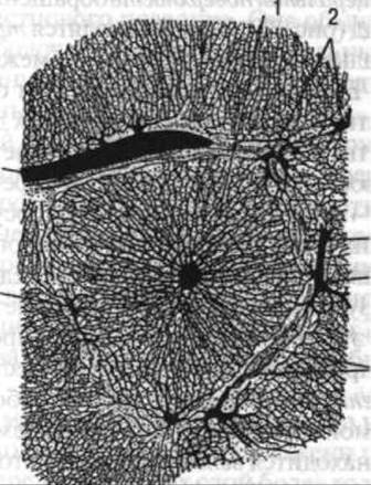
Fig. 78. Liver lobules (veins filled with a colored mass):
1 — liver lobule shown entirely in cross-section (the central vein is in the middle of the lobule); 2, 4 — interlobular v.v.; 3 — interlobular bile ductule
The liver receives blood from the hepatic artery proper and the portal vein. Blood returning from the stomach, pancreas, intestines, and spleen via the portal vein is cleansed of harmful chemical impurities within the liver lobules. The presence of fenestrations (gaps) in the walls of the sinusoids ensures contact between the blood and the hepatocytes, which absorb certain substances from the blood and release others into it. The blood, having altered its composition, collects in the central veins and flows through the hepatic veins into the inferior vena cava.
The structural complexity of the liver corresponds to The Diversity of functions performed by this organ. First and foremost, the liver carries out a detoxicating function, neutralizing toxic substances that are formed in the intestines, absorbed into the blood, and transported to the liver. As a gland of the digestive system, the liver produces bile, which enters the duodenum, where it emulsifies fats, preparing them for the action of enzymes. In addition, the liver synthesizes urea, which is highly soluble in Water and excreted from the body; synthesizes proteins (albumin, globulin, and prothrombin) and Phospholipids that constitute Nervous Tissue; and converts glucose into Glycogen, which is stored in the liver. The reticuloendothelial System of the liver participates in the phagocytosis of dead erythrocytes originating from the spleen, as well as microorganisms and other cells. The liver also serves as a major blood reservoir (depot).
The gallbladder is pear-shaped, with its fundus directed forward and downward, projecting slightly from beneath the anterior border of the liver. The gallbladder is divided into a fundus, body, and neck. It continues into the cystic duct, which joins the hepatic duct to form a single common duct that empties into the duodenum, running within the hepatoduodenal ligament.
The length of the gallbladder is 8–12 cm, its width is 3–5 cm, and its capacity is 40–60 cm3. The wall of the bladder consists of mucous and muscular membranes, and its inferior surface is covered by a serous membrane (peritoneum). Superiorly, the gallbladder is attached directly to the inferior surface of the liver.
Depending on the phase of Digestion, the cystic duct conducts bile in two directions: from the liver into the gallbladder, and from the gallbladder into the common bile duct. Here, bile is not only accumulated and stored, but also changes its composition: it becomes thicker and more viscous due to the reabsorption of water and chlorides and mixing with mucus.
The Pancreas
The pancreas is the second large gland that secretes its product into the duodenum (Fig. 79). Like the liver, it develops from the epithelium of the mucous membrane of the duodenum. The pancreas weighs approximately 70–80 g, has a soft consistency, and in its internal structure somewhat resembles the salivary glands. It lies retroperitoneally, covered by the peritoneum only anteriorly and inferiorly. The pancreas is divided into a HEAD, body, and tail. The head of the pancreas is directed to the right and surrounded by the duodenum; the body lies transversely at the level of the 1st lumbar vertebra and is somewhat flattened in the anteroposterior direction; and the tail reaches the left kidney and spleen.
The anterior, posterior, and inferior surfaces of the body of the gland are distinguished. The anterior surface is in contact with the body of the stomach and its pyloric part; the posterior surface is adjacent to the body of the 1st lumbar vertebra and the Abdominal Aorta and inferior vena cava located anterior to it; and the inferior surface faces downward and anteriorly, contacting the duodenum, jejunum, and transverse colon. The splenic artery and splenic vein run along the upper margin of the pancreas.

Fig. 79. The pancreas and adjacent organs:
1 — aorta; 2 — left gastric a.; 3 — celiac trunk; 4 — left suprarenal gland; 5 — spleen; 6 — splenic artery; 7 — pancreas; 8 — left kidney; 9 — ascending part of duodenum; 10 — superior mesenteric a.; 11 — superior mesenteric v.; 12 — horizontal (inferior) part of duodenum; 13 — head of pancreas; 14 — descending part of duodenum; 15 — right kidney; 16 — common bile duct; 17 — cystic duct; 18 — common hepatic duct; 19 — common hepatic a.; 20 — right suprarenal gland; 21 — gallbladder; 22 — hepatic parenchyma; 23 — inferior vena cava (after Sobotta)
The pancreas is classified as a compound alveolar gland possessing both exocrine and endocrine functions. As an exocrine gland, it secretes pancreatic juice containing Enzymes for the digestion of proteins, fats, and carbohydrates. The main excretory duct traverses the entire gland and empties into the duodenum. Occasionally, it merges with the common bile duct into a single channel. In such cases, a dilation is present within the duodenal papilla, which serves as the common opening for both ducts. Sometimes the pancreas features an accessory excretory duct that opens independently into the duodenal lumen.
The Endocrine portion of the pancreas is formed by specialized cell clusters scattered throughout the gland as individual islets, particularly within its tail. These islets lack ducts. Insulin, the hormone produced by the endocrine cells of the pancreas, is released directly into the bloodstream and regulates Carbohydrate METABOLISM. In addition, the islet cells secrete Glucagon, a hormone antagonistic to insulin that stimulates not the storage, but The breakdown of glycogen in the liver and fat in adipose tissue.
Peritoneum
The peritoneum consists of two layers: the parietal layer and the visceral layer (Fig. 80). The parietal layer lines the walls of the abdominal cavity, specifically the inferior surface of the diaphragm, the inner surfaces of the anterior, lateral, and posterior abdominal walls, and extends down into the pelvis to cover its superior aspect. The visceral layer invests the abdominal organs. The degree of peritoneal coverage varies among organs: some are invested entirely (jejunum and ileum), others on one side only (Kidneys, Ureters), and still others on three sides (ascending and descending colons).
Passing from one organ to another, as well as from the abdominal walls to the organs, the peritoneum forms folds, ligaments, and mesenteries.
The potential space between the parietal and visceral peritoneum, as well as between individual folds of the visceral peritoneum, is termed the peritoneal cavity. It is a complex, slit-like compartment containing a small amount of serous fluid. In males, the peritoneal cavity is a completely closed sac, whereas in females it communicates with the exterior via two openings leading into the uterine tubes.
From the diaphragm, the peritoneum reflects onto the liver, forming ligaments: the falciform ligament and two coronary ligaments that run along the posterior margin of the liver to the right and left. The latter terminate in expansions known as the right and left triangular ligaments.
From the inferior surface of the liver, at its porta hepatis, the peritoneum extends to the stomach and duodenum as a double fold known as the lesser omentum. It comprises the hepatoduodenal and hepatogastric ligaments. The former contains the portal vein, hepatic artery, and common bile duct, while the latter encloses blood vessels running along the lesser curvature of the stomach. Additionally, lymphatic vessels and nerves pass through these ligaments.
The stomach is covered by the peritoneum anteriorly and posteriorly. The greater omentum arises from its greater curvature, extends inferiorly to cover the small intestine anteriorly, and then turns upward to attach to the transverse colon. The greater omentum is a double fold of peritoneum, meaning it consists of four layers. The segment passing from the stomach to the transverse colon is called the gastrophocolic ligament. To the left, the greater omentum continues as a ligament extending from the stomach to the spleen, known as the gastrosplenic ligament.

Fig. 80. Course of the peritoneal layers:
1 — caudate lobe of liver; 2 — pancreas; 3 — duodenum (inferior part); 4 — retroperitoneal space; 5 — peritoneal cavity; 6 — rectovesical pouch; 7 — rectum; 8 — Testis; 9 — tunicae of testis; 10 — Pubic Symphysis; 11 — urinary bladder; 12 — small intestines possessing a mesentery; 13 — greater omentum; 14 — transverse colon; 15 — transverse mesocolon; 16 — omental bursa; 17 — stomach; 18 — lesser omentum (after Spalteholz)
The spleen (see p. 325) is invested by the peritoneum on all sides, forming ligaments that pass to the serous coat of the diaphragm and stomach. As it reflects onto the transverse colon, the peritoneum forms the mesocolon of this segment, which lies horizontally and is directed from behind and above forward and downward. This mesentery divides the peritoneal cavity into superior and inferior compartments (floors).
Inferior to the transverse colon and its mesocolon lies the region of the abdominal cavity containing primarily the small intestine, as well as the ascending and descending colons. The mesentery of the small intestine attaches to the posterior abdominal wall, forming a root partition that extends from the left side of the 1st and 2nd lumbar vertebrae to the right sacroiliac joint. Toward the small intestine, the mesentery broadens and assumes a pleated, fan-like shape. To the right and left of the mesentery, the peritoneum forms recesses known as the right and left mesenteric sinuses, which house the small intestine.
The ascending and descending colons are covered by the peritoneum on three sides, the cecum on all sides, and its vermiform appendix possesses a well-defined mesentery.
The sigmoid colon also features a well-defined mesentery that continues into the lesser pelvis down to the Origin of the rectum.
In the male lesser pelvis, the peritoneum reflects from the rectum onto the urinary bladder, forming the rectovesical pouch, which is occupied by the sigmoid colon and partly by the small intestine. In females, the peritoneum passes from the rectum first onto the posterior surface of the Uterus and then onto its anterior surface. The lateral continuations of these visceral peritoneal folds away from the uterus form the broad ligaments of the uterus, which extend from the lateral pelvic walls. The uterine tubes are enclosed within the free margins of the broad ligaments. The broad ligament of the uterus is lax and therefore does not restrict uterine mobility in anteroposterior, vertical, or transverse directions. From the anterior surface of the uterus, the peritoneum continues onto the posterior wall of the urinary bladder. Between the rectum and the uterus posteriorly, and the uterus and the urinary bladder anteriorly, the peritoneum forms pouches termed the rectouterine and vesicouterine pouches, respectively. The urinary bladder is covered by the peritoneum only on its superior and posterior aspects.
On the anterior abdominal wall, the parietal peritoneum forms the median and lateral umbilical folds, overlying the ligaments of the same names. Furthermore, the peritoneum forms a fold along the course of the inferior epigastric artery. Between these folds, the parietal peritoneum exhibits supravesical, medial inguinal, lateral inguinal, and femoral fossae. The supravesical fossa represents a potential space that diminishes as the urinary bladder fills and ascends. The medial inguinal fossa lies between the lateral umbilical fold and the epigastric fold, corresponding to the superficial (subcutaneous) ring of the Inguinal Canal. The lateral inguinal fossa corresponds to the deep (abdominal) ring of the inguinal canal. These two fossae are located superior to the inguinal ligament. The femoral fossa lies inferior to this ligament, corresponding to the depression bounded by the pubic bone and inguinal ligament on one side, and the femoral vein on the other.
Last update: 08/08/2026
Editorial and Educational Adaptation: This material has been compiled based on the primary/original source text. The project team performed an editorial review, corrected technical inaccuracies, structured sections, and adapted the content for an educational format.
What was processed:
- elimination of formatting defects (OCR errors, structural breaks, corrupted characters);
- editorial organization of content;
- standardization of terminology in accordance with academic sources;
- verification of factual statements against the original source text.
All mentions of the author, publication year, and origin of the primary text have been preserved in accordance with the source.