Human Anatomy (with the Fundamentals of Dynamic and Sports Morphology) - Ivanitsky M. F. 2008
The Doctrine of Internal Organs
Respiratory System
The Respiratory system comprises the Lungs and the respiratory tract, through which air flows into and out of the lungs. The respiratory tract includes the Nasal cavity (see p. 85), Pharynx (see p. 212), Larynx, Trachea, and Bronchi. Normally, a person breathes through the Nose with the Mouth closed, although oral breathing is also possible (for a Description of the Oral Cavity, see pp. 85, 234).
A characteristic feature of the respiratory tract is the presence of a rigid structural framework in its walls: bone (in the nasal cavity) and Cartilage (in the larynx and bronchi). This architecture prevents the respiratory tract from collapsing, thereby ensuring continuous airflow during respiration.
Larynx
The larynx serves not only as a passage for airflow but also Functions as a vocal organ. Compared to all other PARTS OF THE respiratory apparatus, the larynx has the most complex Structure. It features a well-developed cartilaginous Skeleton and numerous Muscles that actuate it. The larynx is located on the anterior aspect of the neck, protruding between the cervical muscles at the level of the 4th to 6th cervical vertebrae. By means of the hyothyroid membrane, the larynx connects to the Hyoid bone and thus follows its movements, elevating and depressing during actions such as swallowing.
The cartilages of the larynx consist of three unpaired and three paired elements. The unpaired cartilages are the thyroid cartilage, cricoid cartilage, and epiglottis; the paired ones are the arytenoid, corniculate, and cuneiform cartilages (Fig. 81). Each has a distinct structure. The epiglottis, as well as the smaller cartilages (corniculate and cuneiform), is composed of elastic cartilage, whereas the thyroid, cricoid, and arytenoid cartilages are made of hyaline cartilage.
The thyroid cartilage is the largest cartilage of the larynx. It consists of two quadrilateral plates that meet anteriorly at an almost right angle to form a prominent laryngeal prominence (which is easily palpable beneath the Skin just below the hyoid bone). The thyroid cartilage features superior and inferior horns: the superior horns connect via ligaments to the hyoid bone, while the inferior horns articulate with the cricoid cartilage via joints. The thyroid cartilage serves as an origin and insertion site for several muscles. It gives rise to the inferior pharyngeal constrictor, the vocalis Muscle, and muscles extending to the cricoid cartilage and epiglottis. On the external surface of the thyroid cartilage plate, There is a rough, oblique line that serves as the attachment site for muscles running from the Sternum to the thyroid cartilage, and from the thyroid cartilage to the hyoid bone.
Class="center">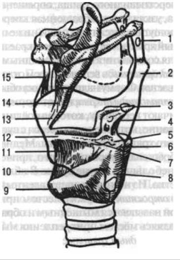
Fig. 81. Cartilages and ligaments of the larynx (lateral view; the thyroid cartilage is outlined):
1 — triticeal cartilage (in the lateral thyrohyoid ligament); 2 — superior horn of thyroid cartilage; 3 — corniculate cartilage; 4 — arytenoid cartilage; 5 — muscular process of arytenoid cartilage; 6 — posterior cricoarytenoid ligament; 7 — vocal process of arytenoid cartilage; 8 — inferior horn of thyroid cartilage; 9 — cricoid cartilage; 10 — cricothyroid ligament; 11 — elastic cone; 12 — vocal ligament; 13 — thyroepiglottic ligament; 14 — stalk of epiglottis; 15 — median thyrohyoid ligament (after G. F. Ivanov)
As a rule, the thyroid cartilage is more developed in men than in women, which accounts for the greater length of the vocal cords in males.
The cricoid cartilage articulates movably with the arytenoid cartilages and the thyroid cartilage, and connects via the cricotracheal ligament to the first tracheal ring below. The expanded posterior portion of the cricoid cartilage is called the lamina, while the narrow anterior portion is termed the arch (which is readily palpable beneath the skin inferior to the thyroid cartilage). The upper margin of the lamina bears articular surfaces for connection with the arytenoid cartilages.
The epiglottis expands in its upper section and tapers inferiorly. The shape of the epiglottis is commonly compared to that of a leaf.
The arytenoid cartilage has the shape of a triangular pyramid. It presents an apex, which articulates with the corniculate cartilage; a base, which articulates with the articular surface of the cricoid cartilage; and three surfaces. The Medial surface of one cartilage faces the other, with a small space between them. Near its base, the cartilage features two processes: one directed anteriorly, termed the vocal process, serves for the attachment of the vocal ligament; the other, directed laterally and slightly posteriorly, is the muscular process, which serves for muscle attachment.
The corniculate cartilage is conical in shape and sits by its base upon the apex of the arytenoid cartilage.
The cuneiform cartilage is located within the fold of the mucous membrane extending from the arytenoid cartilage to the epiglottis.
The larynx is anchored to the hyoid bone. The median thyrohyoid ligament extends between the posterior edge of the hyoid bone body and the superior margin of the thyroid cartilage. The lateral thyrohyoid ligament stretches from the tip of the greater horn of the hyoid bone toward the superior horn of the thyroid cartilage and frequently contains a small triticeal cartilage. The remaining space between the hyoid bone and the upper edge of the thyroid cartilage is enclosed by the thyrohyoid membrane, which is composed of Connective Tissue containing elastic fibers. Additionally, the laryngeal region features ligaments running from the hyoid bone to the epiglottis, as well as between the thyroid and cricoid cartilages. The epiglottis connects to the thyroid cartilage via the thyroepiglottic ligament.
The most important JOINTS OF THE larynx are the cricoarytenoid joints (between the cricoid and arytenoid cartilages). The cricoarytenoid joint permits rotation around a vertical axis passing through the joint, as well as limited gliding of the arytenoid cartilage forwards and backwards, laterally and medially. The cricothyroid joint allows only a slight tilting movement of the thyroid cartilage relative to the cricoid cartilage.
The internal surface of the larynx is lined with a mucous membrane containing small clusters of lymphoid tissue and mucus-secreting glands. The Superficial layer of the mucosa is covered by pseudostratified ciliated epithelium, whose Ciliary movement traps dust particles, mixes them with mucus, and expels them via coughing. Beneath the mucosa lies a layer of elastic tissue that, in the sagittal plane extending from the angle of the thyroid cartilage to the vocal process of the arytenoid cartilage, forms a dense accumulation known as the elastic cone.
The connective tissue bundles extending from the thyroid cartilage to the vocal process of the arytenoid cartilage constitute the vocal ligament. It is covered by mucous membrane, which here forms the vocal fold. Superior to it lies the vestibular fold, the mucosa of which covers the vestibular ligament. The latter contains a relatively small number of elastic fibers. A recess known as the laryngeal ventricle is situated between these folds. The rima glottidis lies between the right and left vocal folds.
The musculature of the larynx is composed of Striated Muscle tissue. Anatomically, all laryngeal muscles are divided into extrinsic and intrinsic groups. The extrinsic muscles include the cricothyroid muscle, which runs from the upper margin of the anterior cricoid cartilage to the lower margin of the lateral plate of the thyroid cartilage. All intrinsic laryngeal muscles are classified into adductors (which narrow the glottis) and abductors (which widen the glottis). These two groups are numerically unequal: the glottis is widened by a single posterior cricoarytenoid muscle (Fig. 82) and narrowed by all the other intrinsic muscles (lateral cricoarytenoid, transverse and oblique arytenoids, and thyroarytenoid).
Regarding The Mechanism of phonation, the larynx can be compared simultaneously to a stringed and a wind musical instrument. The vocal cord functions analogously to the vibrating string that produces sound in stringed instruments. Pitch is determined by the length of the vibrating segment of the cord and its tension. Elongation of this segment occurs when the thyroid cartilage shifts relative to the arytenoid cartilage (due to contraction of the cricothyroid muscles). The degree of tension is determined by the contraction of fibers within the vocalis muscle, which is interwoven into the vocal ligament. Like a wind instrument, the larynx modifies the sounds generated within it, imparting a specific timbre through resonating structures (the laryngeal ventricles, Upper Respiratory Tract, and Paranasal Sinuses). Sound volume is determined by the width of the glottis, which depends on the tone of the intrinsic laryngeal muscles.
The larynx also participates in the act of swallowing, during which it is displaced upward (driven by the contraction of muscles that elevate the hyoid bone). The laryngeal inlet is closed as the epiglottis is deflected posteriorly by the pressure of the Tongue and, in part, by the contraction of the aryepiglottic muscles.

Fig. 82. Muscles of the larynx (posterior view):
1, 2 — epiglottis; 3, 15 — cuneiform tubercles; 4 — aryepiglottic fold; 5, 13, 14 — oblique arytenoid muscles; 6 — transverse muscle; 7 — trachea; 8 — membranous wall of trachea; 9 — cricothyroid ligament; 10 — lamina of cricoid cartilage; 11 — inferior horn of thyroid cartilage; 12 — posterior cricoarytenoid muscle; 16 — greater horn of hyoid bone (after G. F. Ivanov)
Trachea
The trachea is a tube 11–13 cm long, composed of 16–20 C-shaped hyaline cartilage rings that are open posteriorly and joined by a fibroelastic membrane. This anatomical arrangement is of great functional significance: the Esophagus lies directly behind the trachea and can expand into the tracheal lumen as a food bolus passes. At its upper end, the trachea is attached to the cricoid cartilage by a ligament at the level of the intervertebral disc between the 6th and 7th cervical vertebrae. At the level of the 4th–5th thoracic vertebrae, the trachea bifurcates into the bronchi. Because the trachea lies within both the neck and the thoracic cavity, it is divided into two parts: the cervical and thoracic regions.
The inner surface of the trachea is lined with a mucous membrane containing scattered lymphoid nodules and lined with ciliated epithelium. In the cervical region, the sternohyoid and sternothyroid muscles lie anterior to the trachea, and the isthmus of The Thyroid Gland is located at the level of the 2nd to 4th tracheal rings. The groove between the trachea and the esophagus serves as a pathway for nerves and Blood Vessels. Within the thoracic cavity, the trachea is located in the Mediastinum, where it is bordered anteriorly by the Thymus gland, the aorta, and the left brachiocephalic vein.
Bronchi
The trachea divides into two main bronchi: the right and the left. The right main bronchus is shorter, wider, and runs more vertically than the left. Consequently, foreign bodies are more likely to enter the right bronchus. In structure, the main bronchi resemble the trachea, also consisting of C-shaped hyaline cartilage rings. The arch of the aorta passes over the left bronchus, while the azygos vein arches over the right. The bronchial mucosa contains small aggregates of lymphoid tissue.
The main bronchi divide into lobar bronchi, which enter the lung tissue.
Lungs
The right and left lungs are located in the thoracic cavity on either side of the mediastinum (Fig. 83). In shape, they resemble a truncated cone. The right lung is shorter and wider than the left. Each lung has three surfaces: the costal surface facing the Ribs, the medial surface facing the mediastinum, and the diaphragmatic surface facing downward. The lung has an apex and a base. On the medial surface of the lung is the hilum, a depression through which the bronchus, Blood and Lymphatic vessels, and nerves pass. Together, these structures form the ROOT of the lung. Lymph Nodes are also located here. In the hilum of the right lung, the bronchus is situated superiorly, with the pulmonary artery and Veins below it; in the hilum of the left lung, the artery lies superiorly, with the bronchus and Pulmonary veins located inferiorly and slightly posteriorly.

Fig. 83. Lungs and anterior mediastinum of a child (anterior view):
1 — esophagus; 2 — trachea; 3 — left common carotid a.; 4 — left subclavian a.; 5 — thymus gland; 6 — left brachiocephalic v.; 7 — superior lobe of the left lung; 8 — mediastinal Pleura; 9 — inferior lobe of the left lung; 10 — subpleural fat; 11 — Pericardium; 12 — epicardial fat; 13 — middle lobe of the right lung; 14 — inferior lobe of the right lung; 15 — superior lobe of the right lung; 16 — right brachiocephalic v.; 17 — right subclavian a.; 18 — right common carotid a. (after G. F. Ivanov)
The specific gravity of the lungs is less than 1 because they contain air. Naturally, in a fetus (which has no air in its lungs), the specific gravity of the lungs is greater than 1.
The lungs are divided into lobes by fissures: the right lung is divided by oblique and horizontal fissures into superior, middle, and inferior lobes, while the left lung is divided by an oblique fissure into superior and inferior lobes. The lobes are further subdivided into bronchopulmonary segments (10 on the right and 9 on the left), each of which receives a segmental bronchus (a branch of the lobar bronchus) and a corresponding branch of the pulmonary artery. The bronchi branch dichotomously into smaller and smaller airways, forming the bronchial tree. Pulmonary segments consist of lobules (primary lobules).
Lobules bordering the outer surface of the lung are pyramidal in shape, with their apex pointing inward; those located deeper are polyhedral. The pulmonary lobules are separated from one another by connective tissue septa. Upon entering a lobule, the intralobular Branches of the segmental bronchi branch repeatedly and transition into terminal bronchioles (Fig. 84), each of which gives rise to an acinus (secondary lobule).
Within the acinus, the terminal bronchiole branches into respiratory bronchioles, which have alveoli budding from their walls. Finally, the respiratory bronchioles branch into alveolar ducts, each of which leads into two alveolar sacs. The walls of these sacs contain the vast majority of the pulmonary alveoli, the total surface area of which spans several dozen square meters.

Fig. 84. Diagram of The structure of a pulmonary acinus:
1 — intralobular bronchus (branches of the segmental bronchus); 2 — terminal bronchiole; 3 — respiratory bronchiole; 4 — alveolus; 5 — alveolar duct (after Stöhr-Möllendorff)
Like the trachea, the bronchi are lined with a mucous membrane covered by pseudostratified ciliated epithelium, possess a cartilaginous skeleton, and contain elastic fibers and Cytology/cytology/32.html">Smooth muscle tissue.
The walls of the respiratory bronchioles contain bundles of smooth muscle tissue located between the openings of the alveoli that branch off them. Cartilage is absent here. From the lumen side, the bronchioles are lined with ciliated epithelium. The presence of ciliated epithelium in the airways is essential for clearing inhaled air of suspended particles such as dust.
The structure of pulmonary alveoli is quite complex and corresponds to their function in gas exchange. The foundation of the alveolar wall is a very thin basement membrane lined with simple squamous epithelium. Elastic fibers within the alveolar wall facilitate its recoil during exhalation. Alveolar expansion follows the expansion of the thoracic cavity during inhalation. A dense, fine-meshed network of blood capillaries surrounding each alveolus is closely apposed to its wall. Through the walls of the alveoli and blood capillaries, oxygen from the air enters the blood, while carbon dioxide passes from the blood into the alveolar cavity to be expelled from the lungs during exhalation.
The lung is covered by the visceral pleura (except at the hilum), which is a serous membrane. The walls of the thoracic cavity are lined internally by the parietal pleura. Between these two layers lies the pleural cavity. Extending downward from the hilum of the lung to the Diaphragm is the pulmonary ligament, a pleural fold representing the transition zone from visceral to parietal pleura. The parietal pleura is divided into costal, diaphragmatic, and mediastinal parts. The costal pleura covers the ribs, the diaphragmatic pleura covers the diaphragm, and the mediastinal pleura covers the mediastinum. At the junction where the costal pleura meets the diaphragmatic pleura, the costodiaphragmatic recess is formed, serving as a reserve space for the lungs to expand.
The mediastinum is a complex of Organs located between the right and left sheets of the mediastinal pleura. By an imaginary transverse plane passing through the bifurcation of the trachea, the mediastinum is divided into anterior and posterior parts. The anterior mediastinum consists of superior and inferior divisions. The superior division is formed by the thymus gland, major vessels (SUPERIOR VENA CAVA, ascending aorta, and aortic arch), and nerves, while the inferior division contains The Heart. The posterior mediastinum is composed of the esophagus, its accompanying vagus nerves, the Thoracic Aorta, the Thoracic duct, the Sympathetic trunk with the greater and lesser splanchnic nerves arising from it, and the azygos and hemiazygos veins. Lymph nodes are also located here.
Last update: 08/08/2026
Editorial and Educational Adaptation: This material has been compiled based on the primary/original source text. The project team performed an editorial review, corrected technical inaccuracies, structured sections, and adapted the content for an educational format.
What was processed:
- elimination of formatting defects (OCR errors, structural breaks, corrupted characters);
- editorial organization of content;
- standardization of terminology in accordance with academic sources;
- verification of factual statements against the original source text.
All mentions of the author, publication year, and origin of the primary text have been preserved in accordance with the source.