BOTANY VOLUME 2 - PLANT PHYSIOLOGY - 2007
6. PHYSIOLOGY OF METABOLISM
6.10. Energy Generation via Carbohydrate Breakdown
Since ATP synthesized through Photophosphorylation (or via electron transport during The oxidation of an inorganic substrate in chemoautotrophs) is typically utilized for CO2 reduction (carbon dioxide assimilation), the ATP required for other cellular processes in autotrophs must be generated through alternative pathways (see Fig. 6.2). Photoautotrophs therefore had to evolve the capacity for ATP Synthesis in the dark. All Cells of heterotrophic and autotrophic organisms rely exclusively on reduced carbon compounds as starting Materials for synthesizing their cellular organic components and as Energy Sources; ultimately, these compounds originate from Photosynthesis or METABOLISM/21.html">Chemosynthesis.
Energy generation through The breakdown of reduced carbon compounds (dissimilation) proceeds continuously via redox reactions, i.e., through The transfer of electrons from a donor to an acceptor. Depending on the terminal electron acceptor in the energy-yielding breakdown reactions (catabolic reactions), two MAIN TYPES OF dissimilation are distinguished: in the first type, oxygen serves as the terminal electron acceptor (aerobic dissimilation, or cellular Respiration; see Fig. 6.2), whereas In the second type, an organic molecule derived directly from the breakdown process acts as the acceptor (anaerobic dissimilation, or Fermentation). Fermentation does not involve the complete net oxidation of the substrate; rather, it relies on an internal redox reaction, i.e., electron transfer within the substrate or exchange between products of substrate breakdown.
Organisms completely incapable of utilizing oxygen—obligate anaerobes (which rely exclusively on fermentation)—are extremely rare, restricted to a small number of Bacteria and invertebrates inhabiting environments such as organic-rich aquatic sediments and animal intestines. Most living cells are facultative anaerobes that generate energy via fermentation under oxygen-limiting conditions. However, the efficiency of anaerobic dissimilation (along with sensitivity to oxygen deficiency) and the biochemical pathways of fermentation vary. Most Yeasts, for example, can survive under anaerobic conditions by means of fermentation, yet they reproduce exclusively under aerobic conditions, i.e., when respiration is operational. The metabolic shift from aerobic to anaerobic Catabolism is facilitated by the fact that both biochemical pathways share many identical steps, with aerobic breakdown utilizing intermediates generated during the anaerobic pathway.
Hexoses, primarily glucose, generally serve as substrates for fermentation. Specialized bacteria are also capable of fermenting pentoses, Amino Acids, and Fatty acids. Respiration likewise predominantly utilizes glucose as a substrate. The shared biochemical pathway of glucose fermentation and glucose respiration leads to The formation of Pyruvate and is known as Glycolysis (see 6.10.1). It takes place in the Cytoplasm of cells.
Glucose is supplied to the plant either directly from The Calvin Cycle (see 6.5, Fig. 6.70) or through the breakdown of transport and reserve CARBOHYDRATES (see Fig. 6.72, and also 6.17.1.2), thereby sustaining photosynthetically inactive Cells and Tissues. Additionally, plants are capable of converting reserve Lipids into carbohydrates (see 6.12). Finally, the breakdown products of amino acid carbon skeletons (e.g., degradation of Reserve Proteins) can also feed into ATP synthesis.
Glucose is produced via sucrose Cleavage by invertases, as well as starch breakdown by amylases and maltases (see 6.17.1.2). Glucose-1-phosphate is generated directly either through sucrose cleavage by sucrose synthase or via phosphorolytic degradation of starch (see Fig. 6.72, and also
6.17.1.2). Glucose is converted into glucose-6-phosphate with the involvement of ATP, phosphoglucomutase, and hexokinase as an intermediate step, yielding glucose-1-phosphate; glucose-6-phosphate exists in equilibrium with fructose-6-phosphate, the initial metabolite of glycolysis (Fig. 6.92, phosphoglucose isomerase reaction).
Following its conversion into fructose-1,6-bisphosphate via aldolase, fructose-6-phosphate is cleaved into one molecule of glyceraldehyde 3-phosphate and one molecule of dihydroxyacetone phosphate, which remain in equilibrium through a reaction catalyzed by Triosephosphate isomerase.
The energy-yielding reactions of glycolysis proceed via the formation of pyruvate from glyceraldehyde 3-phosphate. The reaction sequence illustrated in Fig. 6.92 runs twice per hexose molecule (once for each of the two triosephosphates), yielding 2 ATP per triosephosphate, or a total of 4 ATP per hexose. At the same time, it must be taken into account that 2 ATP molecules are consumed for the synthesis of fructose-1,6-bisphosphate from glucose. The net yield of glycolysis is therefore 2 ATP per glucose molecule.
The reactions converting fructose-1,6-bisphosphate into 3-phosphoglycerate mirror those of the Calvin cycle, but operate in reverse (see Fig. 6.70). The participating Isoenzymes differ slightly in their molecular Structure: chloroplast glyceraldehyde-3-phosphate dehydrogenase is NADP-dependent, whereas the isoform involved in glycolysis is NAD-dependent.
ATP synthesis during glycolysis occurs via substrate-level phosphorylation. Both synthetic reactions proceed with a release of Free energy. The standard molar Gibbs Free energy of Hydrolysis for phosphoenolpyruvate to pyruvate and inorganic phosphate is ∆G0' = -61.9 kJ • mol-1 (see Fig. 6.3). The pyruvate kinase-catalyzed reaction, in which a phosphate group is transferred to ADP to yield ATP (∆G0' = +30.5 kJ • mol-1), is exergonic overall (∆G0' = -31.4 kJ • mol-1) and practically irreversible. Under standard conditions, approximately 50% of the energy stored in the enol phosphate bond is conserved in the form of ATP.
The standard molar Gibbs free energy of hydrolysis for 1,3-bisphosphoglycerate to 3-phosphoglycerate and phosphate is also sufficiently high to drive ATP synthesis (∆G0' = -49.4 kJ • mol-1; see Table 6.3). The phosphoglycerate kinase reaction is likewise exergonic (∆G0' = -49.4 + 30.5 kJ • mol-1 = -18.9 kJ • mol-1); the removal of 1,3-bisphosphoglycerate shifts the equilibrium and drives the mildly endergonic oxidation of glyceraldehyde 3-phosphate during the glyceraldehyde-3-phosphate dehydrogenase reaction (∆G0' = +6.3 kJ • mol-1). Thus, the overall oxidation of glyceraldehyde 3-phosphate to 3-phosphoglycerate is exergonic (∆G0' = -18.9 + 6.3 kJ • mol-1 = -12.6 kJ • mol-1).
Class="center">Fig. 6.92. Glycolytic breakdown of glucose to pyruvate and (on a gray Background) fermentation reactions for the reoxidation of NADH + H+ produced during glycolysis under oxygen-deficient conditions. Reactions enclosed within the rectangle occur twice, since the aldolase reaction yields two triosephosphate molecules from a single glucose molecule

6.10.2. Types of fermentation
In the presence of oxygen, pyruvate is ultimately oxidized to CO2, accompanied by the reoxidation of NADH generated during
glycolysis. Under oxygen deficiency, these pathways are severely restricted. Many cells possess the capacity to reoxidize NADH by transferring electrons to glycolytic metabolites, specifically pyruvate or acetaldehyde derived from it. The former pathway yields lactic acid (Lactic acid fermentation, see
6.10.2.2), while the latter yields ethanol (Alcoholic Fermentation, see 6.10.2.1). In each case, fermentation sustains glycolysis and thereby ensures cellular ATP supply via substrate-level phosphorylation under oxygen-limiting conditions (see Fig. 6.92).
6.10.2.1. Alcoholic Fermentation
Ethanol serves as the terminal product of anaerobic glucose breakdown in commercially utilized yeasts, as well as in The production of pulque (a traditional South American beverage made from fermented agave sap produced by The activity of the bacterium Zymomonas mobilis; sap is harvested from flowering stalks over a 4 to 5-month period, yielding up to 1,000 L of phloem fluid per stalk). Many other organisms and tissues of various higher plants (seeds of species such as rice and pea; Water-logged roots of rice and maize) accumulate ethanol under oxygen-limiting conditions. Because ethanol is a potent cellular toxin and, due to its membrane-permeable nature, cannot be detoxified via compartmentalization, it is synthesized in significant quantities exclusively by aquatic organisms capable of excreting the alcohol into their environment.
The overall equation for alcoholic fermentation is as follows:
С6Н12O6 —>2С2Н5ОН + 2СO2;
∆G0' = -234 kJ • mol-1.
By comparison, the Complete oxidation of glucose to CO2 yields a ∆G0' of -2,877 kJ • mol-1. Alcoholic fermentation is therefore energetically a very ineff
icient process, in which large volumes of substrate are processed to yield another energy-rich substrate (ethanol). The CO2 produced by baker's Yeast during alcoholic fermentation is used as a leavening agent in bread making.
The reaction sequence for The conversion of pyruvate to ethanol is shown in Fig. 6.92. The irreversible decarboxylation of pyruvate to acetaldehyde requires thiamine pyrophosphate as a coenzyme. The ATP yield from alcoholic fermentation, starting from glucose, is 2 molecules of ATP per glucose molecule, exactly as in glycolysis. Under standard conditions, the energy conserved is thus 2 • 30.5 kJ mol-1 / 234 kJ • mol-1 = 0.26 (26%). Within The Cell, where conditions are non-standard, the energy yield is significantly higher.
Using Introduction/32.html">Genetic Engineering techniques, researchers have successfully modified the bacterium Zymomonas mobilis so that it can ferment not only glucose, fructose, and sucrose, but even xylose. This has made it possible to industrially process wood sawdust—a byproduct of wood Processing consisting of Cellulose, pentose-containing hemicellulose, and Lignin (see 6.17.2).
6.10.2.2. Lactic Acid Fermentation and Other types of Fermentation
Homolactic fermentation produces solely lactic acid from glucose (homofermentation); С6Н12O6 —> 2 lactate- + 2H+; ∆G0' = -197 kJ • mol-1. This anaerobic breakdown occurs not only in animal Muscles but also, for example, in Streptococcus lactis bacteria (used as a starter culture in butter and cheese production; also responsible for the spontaneous souring of milk) and Lactobacillus delbrückii (used in the industrial synthesis of lactic acid). Lactic acid fermentation is also found in higher plants (such as potatoes) and various green Algae (e.g., Chlorella and Scenedesmus). During lactic acid fermentation, pyruvate is reduced directly to L-lactic acid (L-lactate) in a reaction catalyzed by Lactate dehydrogenase (see Fig. 6.92). NAD+, which is essential for glycolysis, is regenerated via the reduction of L-lactate. The reaction releases energy (∆G0' = -25 kJ • mol-1) and is irreversible under cellular conditions.
The amount of ATP generated via lactic acid fermentation is identical to that of alcoholic fermentation. The energy yield under standard conditions is 31%, and it is likely much higher within the cell.
In heterolactic fermentation, ethanol and CO2 are produced in equimolar amounts alongside lactic acid. This pathway has been identified, for example, in certain Lactobacillus species.
There are several other types of fermentation named after their end products, such as propionic acid, formic acid, butyric acid, and succinic acid fermentations. Their mechanisms are fundamentally similar to those of alcoholic and lactic acid fermentations. Although traditional "acetic acid fermentation" is not strictly classified as a true fermentation because it requires oxygen:
С2Н5ОН + O2 —> СН3 СООН + Н2O;
∆G0' = -753 kJ • mol-1.
This reaction is carried out by Acetobacter species, which are used in the production of wine vinegar.
Under aerobic conditions, the chemical bond energy of pyruvate is converted into ATP and utilized by the cell. NAD+ required for glycolysis is regenerated from NADH + H+. In eukaryotes, these processes take place in the Mitochondria (see 2.2.8), into which pyruvate is imported from the cytoplasm via a mitochondrial inner membrane translocator in exchange for hydroxyl ions. Unlike in animal mitochondria, the NADH generated during glycolysis in plant mitochondria is oxidized at their inner membrane (see Fig. 6.97). The inner mitochondrial membrane, much like the inner chloroplast membrane, is virtually impermeable to pyridine NUCLEOTIDES.
The oxidative breakdown and conversion of pyruvate in mitochondria proceeds in 3 stages:
1. Formation of Acetyl-coenzyme A from pyruvate (see 6.10.3.1).
2. Conversion of acetyl-coenzyme A in the citrate cycle, yielding CO2 and reducing equivalents (Krebs cycle, see 6.10.3.2).
3. Electron transport in the Respiratory Chain with the reoxidation of reducing equivalents and the utilization of redox reaction energy for ATP synthesis (see 6.10.3.3).
6.10.3.1. Formation of Acetyl-Coenzyme A from Pyruvate
Pyruvate synthesized during glycolysis first undergoes oxidative decarboxylation in the mitochondria. The acetate formed in the reaction is released as acetyl-coenzyme A (acetyl-CoA, Fig. 6.93). This oxidative conversion is carried out by a complex sequence of biochemical reactions involving 3 different Enzymes and 5 different Coenzymes, which together form the pyruvate dehydrogenase complex (for details, see biochemistry textbooks). The reaction is strongly exergonic (∆G0' = -33.5 kJ mol-1). Both electrons provided by the substrate are used to reduce NAD+ to NADH + H+.
Fig. 6.93. The pyruvate dehydrogenase reaction (A); structure of coenzyme A (acetyl-coenzyme A) (B)
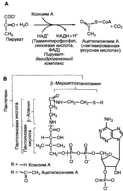
The acetyl moiety in acetyl-CoA represents "activated acetic acid," which can not only undergo catabolic processing in the citrate cycle (Fig. 6.94) but also serves as a fundamental building block for numerous synthetic reactions. Not only sugars, but also Fatty Acids and various Amino acids are broken down into acetyl-CoA, giving it a central role in metabolism.
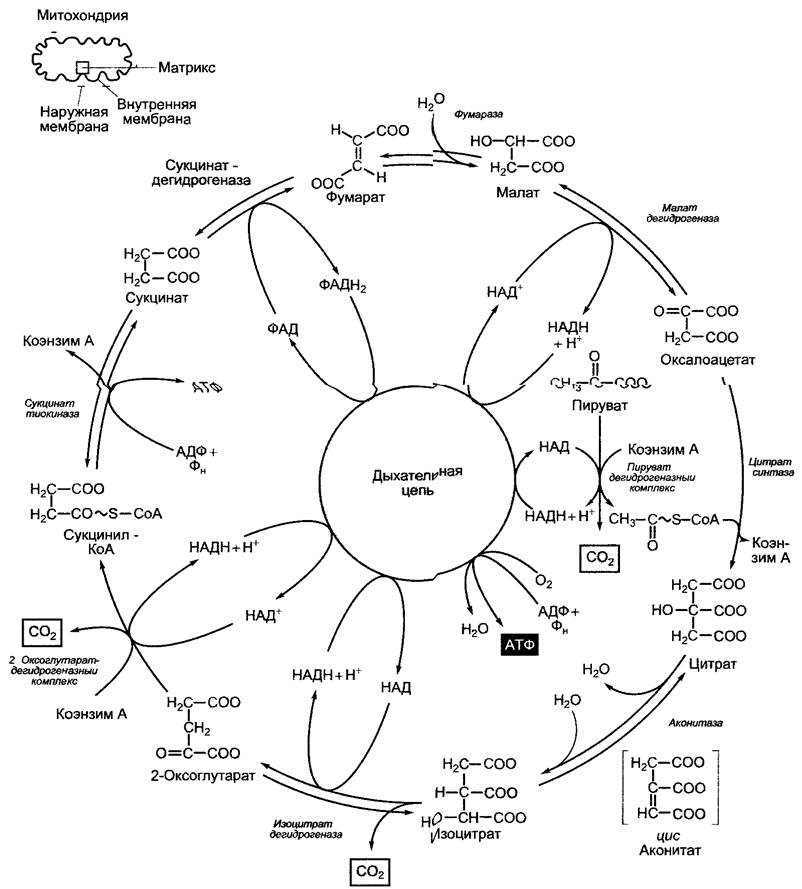
Fig. 6.94. Reaction sequence of the pyruvate dehydrogenase and citrate (Krebs) cycles. The citrate cycle results in the incorporation of one C2 unit (acetate) and the release of 2 molecules of СО2, although these are not the exact C atoms that are released in the same cycle turn (cf. grey atom labeling)
6.10.3.2. The citrate cycle (Krebs cycle)
In the citrate cycle, the acetyl residue of acetyl-CoA is oxidized to two molecules of СO2; the resulting 8 electrons are used to reduce 3 NAD+ to NADH+ + H+ and 1 FAD to FADH2 (Fig. 6.94).
The citrate cycle is also known as the Krebs-Martius cycle (named after its primary discoverers) and does not require oxygen. As is typical for cyclic processes, the citrate cycle concludes with the regeneration reactions of oxaloacetate, the acceptor molecule for the acetyl group.
Both reactions that release СO2 are oxidative decarboxylations, each leading to the transfer of a pair of electrons to NAD+. The oxidative decarboxylation of 2-oxoglutarate, catalyzed by the 2-oxoglutarate dehydrogenase complex, proceeds analogously to the reactions in the pyruvate dehydrogenase complex. Along with coenzyme A and NAD+, this process also involves thiamine pyrophosphate, Lipoic Acid, and FAD as additional coenzymes. The energy-rich thioester bond of the reaction product, succinyl-CoA, is utilized for the synthesis of ATP (or GTP in mammals) via a thiokinase reaction (substrate-level phosphorylation).
Two other pairs of electrons are released during the oxidation of succinate and malate. While NAD+ serves as the electron acceptor for malate dehydrogenase, succinate dehydrogenase utilizes covalently bound flavin adenine dinucleotide (FAD) as its electron acceptor (Fig. 6.95).
6.10.3.3. The mitochondrial respiratory chain
Thus, the 10 electrons derived from the Oxidation of Pyruvate to 3 molecules of СО2 are transferred during the pyruvate dehydrogenase reaction and the citrate cycle to four NAD+ and one FAD. The reduced coenzymes, four NADH + H+ and one FADH2, pass their electrons to the respiratory chain located in the inner mitochondrial membrane, where they are ultimately transferred to oxygen to form Н2О. The energy released by this exergonic transfer is used to establish a transmembrane proton gradient across the inner mitochondrial membrane. The resulting proton-motive force drives the synthesis of ATP (Oxidative Phosphorylation, respiratory chain phosphorylation).
There is a distinct similarity between the mitochondrial Respiratory Chain and the photosynthetic Electron Transport Chain. In cyanobacteria, both chains are located on the same membrane and share a cytochrome complex as a common module, utilizing plastoquinone as a donor and cytochrome c as an
electron acceptor for the cytochrome b6/f complex. The complexes specific exclusively to the respiratory chain are the NADH dehydrogenase complex and the cytochrome a/a3 complex. Electrons are transferred from the NADH generated in the citrate cycle via NADH dehydrogenase, plastoquinone, the cytochrome b6/f complex, and cytochrome c to the cytochrome a/a3 complex, from which they pass to molecular oxygen to form water. This situation is considered "relict" in Chloroplasts—specifically in the thylakoid membranes—where the NADH dehydrogenase complex forms part of the respiratory chain, though its precise function remains unknown.
Fig. 6.95. Redox systems of the respiratory chain acting as carriers via a 2-electron/2-proton scheme. FAD is an integral component of succinate dehydrogenase (covalently bound), FMN is a component of NADH dehydrogenase (Complex I), and ubiquinone is an electron carrier diffusing between Complexes I and III (cf. Fig. 6.96). Like plastoquinone (cf. Fig. 6.56), ubiquinone bears a prenyl residue, which typically consists of 6 isoprene units in microorganisms and 10 in higher plants (cf. 6.12.6). The lipophilic prenyl residue anchors the molecule within the mitochondrial membrane
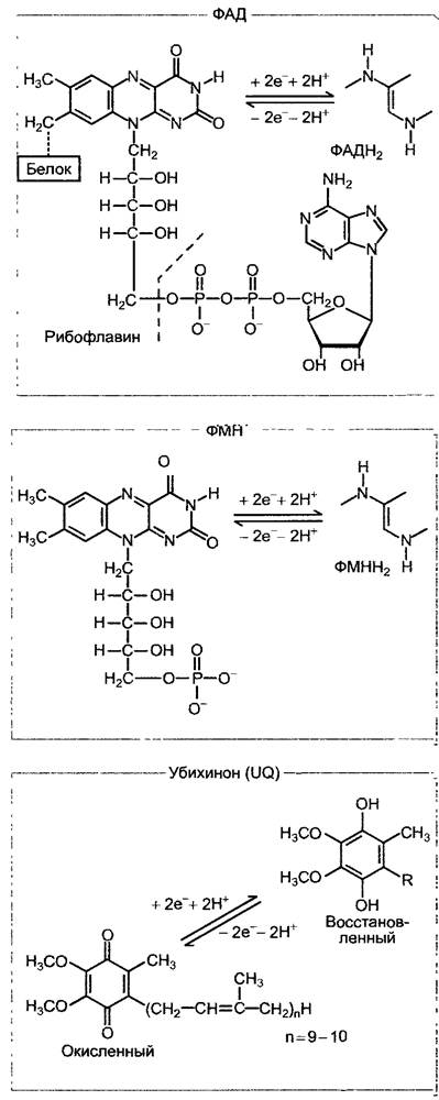
In the mitochondrial respiratory chain, instead of the components used by cyanobacteria alongside the photosynthetic electron transport chain (namely plastoquinone, the cytochrome-b6/f complex, and cytochrome c), a sequence of ubiquinone (= coenzyme Q) -> cytochrome-b/c1 complex -> cytochrome c operates. Plastoquinone and ubiquinone (see Fig. 6.56; 6.95) are functionally equivalent regarding their active groups, whereas the cytochrome b/c1 complex is structurally and functionally homologous to the cytochrome b6/f complex (cytochrome f belongs to c-type Cytochromes, see 6.4.6). The overall electron transport chain from NADH dehydrogenase to the cytochrome-a/a3 complex (also referred to as cytochrome c oxidase or terminal oxidase) likewise forms a structural unit within the inner mitochondrial membrane, maintaining a specific molar ratio of individual components and occupying a surface area of about 400 — 500 nm2. A single mitochondrion may contain up to 20,000 such Electron Transport Chains.
The Structural and functional operating principle of the respiratory chain resembles that of the photosynthetic electron transport chain. The Components of the respiratory chain are oxidoreductases, whose sequential arrangement within the chain is determined by their redox potentials (Table 6.22; Fig. 6.96). Electrons pass from a redox system with a negative standard potential (NADH + H+/NAD+, E0′ = -0.32 V) to a system with a positive standard potential (1/2O2/H2O E0' = +0.82 V); accordingly, the reaction is strongly exergonic (ΔG0' = -221 kJ • mol-1) and therefore proceeds spontaneously.
Table 6.22. Standard redox potentials of the redox systems in the respiratory chain1
Redox pair |
E0' (V) |
NAD+ + 2H+ + 2e- ⇄ NADH + H+ |
-0.32 |
FMN + 2H+ + 2e- ⇄ FMNH2 |
-0.22 |
FAD + 2H+ + 2e- ⇄ FADH2 |
-0.22 |
UQ + 2H+ + e- ⇄ UQH2 |
+0.03 |
Cytochrome b (Fe3+) + e- ⇄ cytochrome b (Fe2+) |
+0.05 |
UQH• + H+ + e- ⇄ UQH2 |
+0.19 |
Cytochrome c1 (Fe3+) + e- ⇄ cytochrome c1 (Fe2+) |
+0.23 |
Cytochrome c (Fe3+) + e- ⇄ cytochrome c (Fe2+) |
+0.24 |
Cytochrome a (Fe3+) + e- ⇄ cytochrome a (Fe2+) |
+0.28 |
Cytochrome a3 (Fe3+) + e- ⇄ cytochrome a3 (Fe2+) |
+0.35 |
O2 + 4H+ + 4e- ⇄ 2H2O |
+0.82 |
1 The list of respiratory chain components given in the table is not exhaustive. The Respiratory Electron Transport chain also includes the Rieske FeS center and a low-potential form of cytochrome b. — Ed. note
The arrangement of the respiratory chain components on the inner mitochondrial membrane is schematically shown in Fig. 6.96. Three transmembrane complexes—NADH dehydrogenase (complex I), cytochrome b/c1 complex (complex III), and cytochrome a/a3 complex (complex IV)—comprise numerous Polypeptides along with associated redox systems: flavins, iron-sulfur centers, and cytochromes, whose basic structure is already familiar from photosynthesis (see Fig. 6.95). The numbering of the complexes reflects the traditional nomenclature of isolated complexes whose structures were initially unknown, adhering to the terminology established at the time. A characteristic feature of complex IV is the presence of a copper-sulfur center and a copper-cytochrome a3 center. The latter binds molecular oxygen (O2) and presumably transfers 4 electrons to it sequentially, resulting in the formation of 2 water molecules. Instead of oxygen, the copper-cytochrome a3 center can also bind carbon monoxide (CO), azide (N-3), or cyanide (CN-)—binding so tightly that these substances act as respiratory inhibitors and are therefore highly toxic.
Soluble components serve as redox electron carriers between the transmembrane complexes. Specifically, these are ubiquinone molecules (UQ, English ubiquinone) located within the inner mitochondrial membrane between complexes I and III. Like plastoquinone in the light reactions, ubiquinone acts as a carrier for two electrons and two hydrogen ions. Cytochrome c, a soluble single-electron carrier, diffuses through the intermembrane space between complexes III and IV, thereby completing The electron transport chain.
Many details remain incompletely understood, particularly the translocation of hydrogen ions from the matrix to the intermembrane space—driven by electron transport—which takes place across the three transmembrane complexes. The stoichiometric values shown in Fig. 6.96 represent the actual state of the process, though precise data in this area are still lacking. Based on the highly probable Q-cycle in the cytochrome b/c1 complex (see the corresponding reaction in the cytochrome b6/f complex; see Fig. 6.60), it is currently believed that for every NADH + H+—and consequently for every 2 electrons transferred to oxygen—10 hydrogen ions are translocated: 4 via the NADH dehydrogenase complex, 4 via the cytochrome b/c1 complex, and 2 via the cytochrome a/a3 complex.
Fig. 6.96. Scheme of the mitochondrial respiratory chain, ATP synthesis, and export. ATP synthase consists of a transmembrane F0 stalk (inhibited by oligomycin) and factor 1 (the F1 HEAD), where ATP synthesis takes place. The structure and MECHANISM OF ACTION of the mitochondrial Fo/F1-ATP synthase are nearly identical to those of the chloroplast CFo/CF1-ATP synthase (see Fig. 6.63). Cyt. — cytochrome; ∆EM — Membrane Potential; F0 — oligomycin-sensitivity-conferring protein; (FeS)n — multiple iron-sulfur centers; Q — Q-cycle (see Fig. 6.60); UQ — ubiquinone; UQH2 — ubiquinol

The nitrate cycle enzyme succinate dehydrogenase is a peripheral membrane protein localized on the matrix side of the inner mitochondrial membrane. It transfers 2 electrons derived from succinate directly to ubiquinone, utilizing FAD as a coenzyme. Succinate dehydrogenase is also referred to as complex II of the respiratory chain. Because complex I is not involved in this reaction, only 6 hydrogen ions are transported across the inner mitochondrial membrane per pair of electrons during the oxidation of succinate. It has been suggested that not only succinate dehydrogenase, but also other Enzymes of the citrate cycle, may remain bound to the inner mitochondrial membrane in intact Organelles, forming a functional unit (a "metabolon") that likely facilitates substrate channeling between components. During organelle isolation, this interaction is disrupted; therefore, following centrifugation of the homogenate to isolate succinate dehydrogenase, all other citrate cycle enzymes remain in solution as separate components, whereas succinate dehydrogenase sediments together with the mitochondrial membrane. It is quite conceivable that a much larger number of Metabolic Pathways in living cells are organized into metabolons.
Mitochondrial ATP synthesis is carried out by ATP synthase, located in the inner mitochondrial membrane, which closely resembles the chloroplast CF0/CF1-ATP synthase in both Structure and function (see Fig. 6.63). The driving force for the synthesis of ATP from ADP + Pi is the proton-motive force (see equation
6.19). The proton-motive force can be viewed as the result of the interplay between the H+ ion concentration gradient, generated by the electron transport chain, and the electrical potential arising from restricted anion Transport Across the membrane. Consequently, the concentration of H+ decreases and a negative charge develops on the mitochondrial matrix side (or chloroplast stroma side), whereas a high concentration of H+ and a positive charge accumulate in the intermembrane space (or the thylakoid lumen of chloroplasts). The contributions of the concentration gradient and the electrical potential difference to the proton-motive force across the chloroplast and mitochondrial membranes are unequal. For instance, the movement of charges across the thylakoid membrane results in a massive hydrogen ion concentration gradient across both sides of the membrane, yet no significant electrical potential is observed. Thus, ATP synthesis in chloroplasts is "driven" primarily by the hydrogen ion concentration gradient, which represents the major contribution to the proton-motive force in chloroplasts. In contrast, blocking proton gradient formation in mitochondria rapidly leads to an electrical potential difference across the inner mitochondrial membrane (∆EM = -200 mV, with a negative charge on the matrix side), while the hydrogen ion concentration difference remains small (the pH in the intermembrane space is only 0.2 units lower than in the matrix). Therefore, ATP synthesis in mitochondria is "driven" predominantly by THE CONTRIBUTION OF the electrical potential to the proton-motive force (see equation 6.19).
Unlike chloroplasts, which utilize the ATP synthesized in the light for their own needs1 (primarily for CO2 fixation), mitochondria export ATP to the cytoplasm. This process is mediated by a translocator located in the inner membrane, which exports ATP to the cytoplasm in strict exchange for ADP (the ADP/ATP carrier). Inorganic phosphate, consumed in stoichiometric amounts, is supplied via a phosphate/OH- antiporter. The outer mitochondrial membrane, much like the plastid envelope, features porins—transmembrane proteins that form relatively large pores through which low-molecular-weight compounds and even small protein molecules can diffuse freely (the size exclusion limit is approximately 10 kDa). Consequently, the outer membrane of this organelle does not act as a barrier to metabolite exchange.
1 Despite the absence of direct ATP transport from chloroplasts to the cytoplasm, shuttle mechanisms exist that transfer energy to drive ATP synthesis in the cytoplasm. — Editor's note.
Fig. 6.97. Alternative pathway for the oxidation of ubiquihydroquinone (UQH2) by alternative oxidase. This pathway is utilized mainly when the concentration of NADH + H+ in the cytoplasm or mitochondrial matrix becomes extremely high. Energy is released as heat, and no ATP is synthesized. Under these conditions, only a small fraction of the ubiquihydroquinone is likely oxidized within the cytochrome b/c1 complex (dashed arrow).

The mitochondrial respiratory chain in plants differs from that of animals (Fig. 6.97). Unlike in animals, NADH + H+ generated during plant glycolysis is oxidized at the outer surface of the inner mitochondrial membrane by external NADH dehydrogenase. Both electrons are transferred directly to ubiquinone, bypassing complex I. This reaction becomes significant when the cytoplasmic NADH concentration is very high; thus, it is less important for ATP synthesis, but it supplies oxidized NAD+ for cytoplasmic metabolic reactions. A similar function is presumably performed by alternative NADH dehydrogenase, located on the matrix side of the inner mitochondrial membrane; it oxidizes NADH + H+ and transfers electrons to ubiquinone without translocating hydrogen ions. Under such conditions (elevated NADH levels and, consequently, an extremely high NADH/NAD ratio), ubiquihydroquinone (UQH2) is oxidized by alternative oxidase, which transfers electrons and H+ ions from UQH2 to oxygen, yielding water. Energy is released as heat, and no ATP is synthesized. This enzyme is initially activated by a high concentration of pyruvate in the matrix (an indicator of a shortage of oxidized NAD+, see Fig. 6.9). Alternative oxidase is not inhibited by cyanide, azide, or CO (inhibitors include, for example, salicylhydroxamic acid). This cyanide-resistant respiration converts the energy of NADH + H+ into heat without ATP production. In Arum maculatum and other arums, thermogenesis (heat production) mediated by alternative oxidase facilitates the volatilization of pollinator-attracting scents; in Symplocarpus foetidus, thermogenesis protects flowers against the cold; and in ripening fruits, it promotes the accelerated breakdown of organic acids and carbohydrates (respiratory Climacteric, see 7.6.5.2).
Energy yield from glucose oxidation during respiration:
C6H12O6 + 6 O2 + 6 H2O —> 6 CO2 + 12 H2O (∆G0' = -2,877 kJ • mol-1).
The energy conserved in the form of ATP accounts for 31.8% of the standard Gibbs free energy change for glucose oxidation at pH 7 (∆G0'). The fraction of conserved energy is determined as follows.
Glycolysis:
✴ net yield (substrate-level phosphorylation) —> 2 ATP;
✴ oxidation of 2 NADH + H+ by external NADH dehydrogenase —> 12 H+ —> 3 ATP1;
1 The ATP yield here is overestimated because external NADH dehydrogenase does not contribute to the proton gradient; its operation conserves 2 rather than 3 ATP molecules. — Editor's note.
✴ 2 molecules of pyruvate entering respiration.
Respiration:
✴ oxidation of 2 pyruvate molecules to CO2 via The Tricarboxylic Acid Cycle (Krebs cycle) yields
8 NADH + H+ —> 80 H+ —> 20 ATP,
2 FADH2 —> 12 H+ —> 3 ATP, and via the succinate thiokinase reaction (oxidative substrate-level phosphorylation) —> 2 ATP.
Overall, under favorable conditions where all hydrogen ions are fully utilized for ATP synthesis (which is virtually impossible in a living cell due to H+ leakage across the outer membrane into the cytoplasm)2, up to 30 ATP molecules are synthesized per glucose molecule. This corresponds to 30 ∆G0' (ADP + Pi → ATP, ∆G0' = +30.5 kJ • mol-1), totaling 915 kJ • mol-1 of free energy conserved in the form of ATP, which constitutes 31.8% of the 2,877 kJ • mol-1 of free energy released by glucose (the remainder is lost as heat). Taking into account the actual intracellular conditions that deviate from Standard States, the true energy yield is likely even higher (∆G0 for ATP synthesis within the cell is estimated to be around 50 kJ • mol-1).
2 Furthermore, the activity of alternative dehydrogenases and cyanide-resistant respiration reduces the net ATP yield. — Editor's note.
Certain methane-producing archaebacteria are capable of synthesizing methane from an energy-poor substrate, CO2, via a weakly exergonic reduction reaction:
CO2 + 4 H2 —> 2 H2O + CH4.
This conversion involves 7 enzymes and 3 coenzymes, including coenzyme F430, which contains a nickel-bearing tetrapyrrole system, and coenzyme M (HS-CH2-CH2-SO3), whose thiol group accepts the methyl group (methyl-CoM) before it is released as methane (CH4, methyl-coenzyme M reductase reaction) through the action of coenzyme F430. The hydrogen ion gradient generated during this conversion process is utilized to synthesize ATP. Although this yields less than one ATP molecule per CO2 molecule, it is nevertheless sufficient to enable these bacteria to survive in energy-depleted environments.
6.10.3.4. Connection of the Krebs Cycle with Other Metabolic Pathways
In the Krebs cycle, acetate is oxidized to CO2 coupled with the transfer of electrons to NAD+ or FAD. In addition, the tricarboxylic acid cycle (Krebs cycle) supplies intermediates for The Biosynthesis of other metabolites. The efflux of these metabolites would rapidly halt the cycle unless replenishing reactions (anaplerotic reactions) compensate for these losses. Finally, the citrate cycle links anabolic (synthetic) and catabolic (degradative) pathways of metabolism, making it amphibolic.
Some important interactions of the nitrate cycle with other Metabolic pathways are shown in Fig. 6.98.
Fig. 6.98. Some key connections of the citrate cycle (Krebs cycle) with other metabolic pathways: (1) phosphoenolpyruvate carboxylase; (2) malate dehydrogenase; (3) NAD-malic enzyme; (4) Glutamate dehydrogenase; (5) PEP carboxykinase; (6) glutamate synthase; (7) Glutamine Synthetase

Alongside plastid glutamate synthesis (see 6.6, 6.13), cytoplasmic synthesis of this amino acid also occurs. Carbon is partly supplied by 2-oxoglutarate from the nitrate cycle, while the predominant portion comes from citrate, which is converted into 2-oxoglutarate by the action of the cytoplasmic aconitase isoenzyme and NAD-isocitrate dehydrogenase. Glutamate serves as a precursor for glutamine, Arginine, and Proline (see 6.13.1), and unlike animal glutamate, it also acts as a starting point for tetrapyrrole biosynthesis (plastid-based in plants — see 6.15). To replenish the Krebs cycle with carbon compounds, mitochondria import oxaloacetate, which is produced either by the cytoplasmic phosphoenolpyruvate carboxylase reaction or from malate (via malate dehydrogenase). In addition, mitochondria are equipped with a malate translocator and take up malate from the cytoplasm. This malate can be used to raise its level in the citrate
cycle or be converted into pyruvate with the formation of NADH through a decarboxylation reaction catalyzed by matrix-localized malic enzyme. These reactions (Fig. 6.98) represent a mechanism for distributing reducing equivalents (NADH + H+) between the cytoplasm and mitochondria. Alongside pyruvate and malate, glutamate is the third major substrate for mitochondrial respiration. It represents the primary product of nitrate assimilation in chloroplasts and is present in sufficient quantities in photosynthetic cells. Following import into mitochondria, a portion of glutamate is broken down by the enzyme glutamate dehydrogenase into 2-oxoglutarate (which enters the nitrate cycle) and NH+4, generating NADH + H+ in the process (see Fig. 6.98).
The citrate cycle plays a crucial role in the conversion of fats into carbohydrates. This occurs during the germination of fat-storing seeds (see 6.12) as well as during senescence processes (for example, autumn leaf senescence), when water-insoluble Membrane Lipids (particularly during chloroplast degradation) are converted into transport carbohydrates that accumulate in storage tissues. In this sequence of biochemical reactions, which is discussed in more detail below, Fatty acids are broken down into acetate, followed by the synthesis of succinate. These reactions take place in glyoxysomes (see 6.12). Succinate diffuses into the mitochondria and is converted within the citrate cycle into oxaloacetate. The latter is transported into the cytoplasm via a translocator and converted into phosphoenolpyruvate by the action of phosphoenolpyruvate carboxylase (this reaction was introduced in section 6.5.8, Fig. 6.79). Starting from phosphoenolpyruvate, the reversible reactions of glycolysis (see Fig. 6.92) proceed all the way to the Formation of fructose-1,6-bisphosphate (Gluconeogenesis). In the irreversible fructose-
1,6-bisphosphate phosphatase reaction, it is converted into fructose-6-phosphate (ΔG0' = -17 kJ • mol-1). From this metabolite onward, the synthesis of structural and storage carbohydrates (see 6.17.1) becomes possible, as does the Synthesis of Other sugar-containing compounds (Glycolipids, Glycoproteins). Glucose-6-phosphate exists in equilibrium with fructose-6-phosphate (via the hexose isomerase reaction — see Fig. 6.72), serving as the starting metabolite for the oxidative Pentose Phosphate Pathway (see
6.10.3.5), which, alongside pentose phosphates, also provides NADPH + H+ for other cytoplasmic metabolic pathways.
Complete conversion of various respiratory substrates into CO2 requires varying amounts of oxygen, depending on their molecular structure. The volumetric ratio of produced CO2 to consumed O2 is termed the respiratory quotient (RQ = \/CO2: VO2).
Since, according to Avogadro's law, one mole occupies a volume of 22.4 L under standard conditions, the RQ value for the breakdown of a uniform substrate can be easily calculated theoretically; on the other hand, Conclusions regarding the respiratory substrate based on the RQ value must be drawn with a certain degree of caution. According to the general equation for glucose respiration (see 6.10.3.3), the RQ value for carbohydrate utilization is 1. For the breakdown of hydrogen-rich molecules such as fats and proteins, the RQ is less than 1 (about 0.7 for fats; about 0.8 for proteins):
Respiration utilizing palmitic acid;
C16H32O2 + 23 O2 -> 16 CO2 + 16 H2O,
RQ = 16/23 = 0.7.
Seedlings utilizing fats for respiration correspondingly have an RQ = 0.7. When fats are converted into carbohydrates—for example, during a specific phase of germination in fat-storing seeds or in the spring in fat-storing tree trunks—the RQ is less than 1 because a large amount of oxygen is consumed while little CO2 is produced. Conversely, reactions converting carbohydrates into fats are characterized by an RQ greater than 1 (for example, in fattening geese, RQ = 1.38).
6.10.3.5. The Oxidative Pentose Phosphate Pathway
The oxidative pentose phosphate pathway takes place in the cytoplasm and chloroplasts. In chloroplasts, a series of reactions reverse to those of the Calvin cycle occur; therefore, the Calvin cycle is sometimes referred to as the reductive pentose phosphate cycle (see 6.5.3). The oxidative pentose phosphate pathway can likewise be described as a cycle. One glucose molecule is broken down into 6 CO2 molecules upon completing six turns of this cycle. Typically, however, the purpose of this biochemical sequence is not the breakdown of glucose, but the generation of NADPH + H+ for anabolic reactions (in chloroplasts, for example, for dark fat synthesis, see 6.11.1), as well as the Synthesis of specific sugar phosphates for other synthetic pathways (such as ribose-5-phosphate for nucleic acid synthesis). Within The pentose phosphate pathway, C3-, C4-, C5-, C6-, and C7-sugars are maintained at equilibrium concentrations, as illustrated in Fig. 6.99.
Fig. 6.99. The oxidative pentose phosphate pathway. Structural formulas not shown here can be found in Fig. 6.70. Highlighted in gray are three reactions characteristic of the oxidative pentose phosphate pathway in chloroplasts; all other reactions represent reversible Reactions of the Calvin cycle (reductive pentose phosphate cycle): (1) ribulose phosphate isomerase; (2) ribulose phosphate epimerase; (3) transketolase; (4) triose phosphate isomerase; (5) aldolase; (6) fructose-1,6-bisphosphate phosphatase; (7) hexose phosphate isomerase

The characteristic enzymes of the oxidative pentose phosphate pathway that are absent in the reductive pentose phosphate cycle are glucose-6-phosphate dehydrogenase and 6-phosphogluconate dehydrogenase, which catalyze irreversible reactions, and transaldolase, which transfers a three-carbon Skeleton (from the C1 to the C3 atom of a heptose) from sedoheptulose-7-phosphate to glyceraldehyde-3-phosphate, yielding fructose-6-phosphate. The second product of the reaction is erythrose-4-phosphate.
The oxidative and reductive Pentose Phosphate Pathways in chloroplasts do not overlap temporally (a highly significant point!). This is achieved, firstly, through the light-mediated activation of certain key Calvin cycle enzymes (see 6.5.5) and, secondly, because glucose-6-phosphate dehydrogenase is inactivated in the light and activated in the dark. Fructose-1,6-bisphosphate phosphatase is also active in the light, which prevents the cyclic operation of the oxidative
pentose phosphate cycle in chloroplasts (see Fig. 6.99). The reversibility of transketolase and transaldolase reactions allows the supply of ribose-5-phosphate for nucleic acid synthesis in the light without the generation of NADPH + H+.
6.10.3.6. Dependence of Respiration on Environmental Factors
Respiration intensity depends largely on the plant species and, within a species, on the organ, developmental stage, and physiological status of the plant (Table 6.23). External factors also influence respiration rate. Temperature acts as a critically important external factor. As an enzymatic process, the dependence of respiration intensity on temperature follows an optimum curve (Fig. 6.100). The positions of characteristic points on this curve (minimum, optimum, maximum) depend on the plant species and, within a species, on the environmental conditions experienced by each individual plant (hardening, etiolation). The lower temperature limit at which respiration can still be detected generally lies around -10 °C. Frost-hardy tissues (e.g., cold-adapted conifer needles) respire at temperatures below -20 °C, whereas respiration in cold-sensitive tropical plants may cease almost entirely at 0 to 5 °C.
Fig. 6.100. Temperature dependence of respiration and photosynthesis. The temperature optimum  and maximum
  of respiration are generally higher than the optimum and maximum of overall photosynthesis (net photosynthetic productivity is shown in gray). The temperature minima for photosynthesis and respiration practically coincide
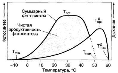
Along the rising portion of the temperature curve (for example, between 15 and 25 °С), the Q10 value (Equation 6.48, see 6.5.11,3) is generally around 2.
The upper temperature limit for respiration is usually higher than that for photosynthesis. Naturally, ATP synthesis lags behind high rates of respiration at elevated temperatures. This situation may lead to a cessation of Electron Transport and oxidative phosphorylation, or alternatively, an increase in cyanide-resistant respiration (see 6.10.3.3).
Table 6.23. Dark respiration of mature leaves in summer at 20°С relative to dry mass (mcуx.в-ва)
Plant group |
CO2 emission, mg • g-1 • mсух.в-ва • h-1 |
Plant group |
CO2 emission, mg • g' • mсух.в-ва • h-1 |
Herbaceous cultivated plants |
3-8 |
Evergreen broad-leaved |
|
trees: |
|||
Herbaceous wild plants: |
sun leaves |
- 0,7 |
|
sun-loving herbs |
5-8 |
shade leaves |
- 0,3 |
shade-tolerant herbs |
2-5 |
Evergreen conifers: |
|
Evergreen broad-leaved trees: |
|||
sun leaves |
3-4 |
sun-adapted needles |
-1 |
shade leaves |
1-2 |
shade needles |
-0,2 |
There is considerable evidence that plant adaptation to changing temperature conditions occurs by increasing the concentrations of isoenzymes suited to those conditions; in other words, the cell possesses a set of enzymes adapted to various thermal regimes.
The heat generated by plants during respiration can usually be measured only under specific experimental conditions (for example, in germinating seeds within a thermos flask). Since homoiothermic plants (i.e., plants that maintain a constant body temperature) do not exist, they lack specialized mechanisms for thermoregulation. Only in exceptional cases can heating of individual plant parts be observed due to respiration (the spadix of Arum italicum by +17 °С, flowers of Victoria regia by +10 °С, and flowers of Cucurbita by +5 °С above ambient temperature). The Biological Significance of heat production in arum inflorescences is to attract pollinators. Through the rapid breakdown of previously stored starch in the spadix—triggered by oxidative phosphorylation—heat is generated, which facilitates the enhanced dispersal of volatile Aromatic Compounds. Salicylic acid acts as the regulator (or "calorigen") in the spadix of the arum relative Sauromatum guttatum. (The spadices of Arum contain 1 – 6 µg • g-1 fresh weight of salicylic acid, whereas the heat-producing male cones of Dioon edule contain 100 µg • g-1.) The responsiveness of the spadix to salicylic acid increases as it matures and is photoperiodically controlled (see 7.7.2.2). Within damp plant debris stored in a compact state (such as in a haystack), the temperature can rise above 70 °С As a result of the respiratory activity of specific thermophilic bacteria and Fungi; the exothermic transformations triggered in this way can lead to self-ignition. The leaf temperature of plants infected with ROOT rot fungi is on average 3 – 5 °С higher than that of healthy plants. This phenomenon can be utilized for remote Diagnostics ("remote sensing") of diseased vegetation.
Water availability significantly influences The rate of respiration in plants. In submerged or waterlogged plants, respiration is limited by a shortage of oxygen due to its low solubility in water (1 L of aerated seawater contains only 7.8 mg of oxygen, whereas aquatic plants release oxygen bubbles during photosynthesis). Oxygen deficiency in the environment can be compensated for, for example, by Oxygen transport through the intercellular air space system from PARTS OF THE plant (see 3.2.1, Fig. 3.7; Vol. 1) that are exposed to the atmosphere and actively release oxygen via photosynthesis (as seen in many wetland plants). Certain specialized structures may develop to supply the plant with O2 (such as pneumatophores and respiratory roots; see Box 4.5). The highly developed intercellular system in aquatic and wetland plants (see Fig. 3.8; Vol. 1) firstly facilitates oxygen delivery and secondly stores photosynthetic oxygen for subsequent use in respiration.
Methane originating from wetland sediments can escape into the atmosphere through the intercellular spaces of rhizomes, petiole tissues, leaf surfaces, and ultimately via Stomata (for instance, in Nuphar luteum). Overall, the intercellular system undoubtedly facilitates gas transport. Approximately 25% of global methane emissions into the troposphere originate from rice paddies (3 – 5 • 1014 g of methane per year). Meristematic tissues with high metabolic activity and a poorly developed intercellular network may undergo partial fermentation processes, the products of which could play a role in the formation and subsequent Development of the intercellular system.
Certain plant Organs (most notably rhizomes) can survive under oxygen-free conditions (anoxia) for extended periods. For example, the rhizomes of Schoenoplectus lacustris can persist without oxygen for over 90 days, during which time they even develop new shoots. Their energy demands during this period are met via fermentation pathways. At the end of the anoxic period, these organs face potential danger when the influx of fresh oxygen generates reactive oxygen species. These toxic radicals can be neutralized using antioxidants such as ascorbic acid or Glutathione.
Under drought conditions, a water potential falling below a critical threshold sharply reduces respiration. Poikilohydric species (see 13.5.2) or specific developmental stages (such as seeds and spores)—which suffer no damage from depressed respiration—respire extremely slowly in an air-dry state (with a water content of about 10% of fresh weight) and consume a minimal amount of substrates. This physiological state is a prerequisite for surviving dormancy periods in seeds, spores, pollen, and entire desiccated plants (such as Lichens, certain algae, mosses, and ferns).
High concentrations of carbon dioxide also restrict respiration. On one hand, elevated CO2 levels occur within the woody tissues of tree trunks; on the other hand, they are found inside seeds with poorly CO2-permeable seed coats.
Light affects respiration in various ways. As demonstrated by Photorespiration (see 6.5.6)—which is not true respiration—prior illumination of photosynthetically active plants stimulates subsequent dark respiration through an enhanced synthesis of respiratory substrates. Although less understood, a competition between respiration and photosynthesis for various coenzymes is also plausible. For instance, mitochondrial respiration in photosynthetically active cells is suppressed in the light (the Kok effect). Furthermore, the short-wavelength (blue) region of the spectrum exerts a specific activating effect on respiration. Finally, light can modulate respiration rates via the Phytochrome system (see 7.7.2.4) by influencing Plant GROWTH AND DEVELOPMENT.
Last update: 07/08/2026
Editorial and Educational Adaptation: This material has been compiled based on the primary/original source text. The project team performed an editorial review, corrected technical inaccuracies, structured sections, and adapted the content for an educational format.
What was processed:
- elimination of formatting defects (OCR errors, structural breaks, corrupted characters);
- editorial organization of content;
- standardization of terminology in accordance with academic sources;
- verification of factual statements against the original source text.
All mentions of the author, publication year, and origin of the primary text have been preserved in accordance with the source.