LEHNINGER PRINCIPLES OF BIOCHEMISTRY - VOL. 1. THE FOUNDATIONS OF BIOCHEMISTRY: STRUCTURE AND CATALYSIS - 2011
PART I. STRUCTURE AND CATALYSIS
12. BIOSIGNALING
12.2. G-Protein-Coupled Receptors and Second Messengers
Signal Transduction via G-protein-coupled receptors (GPCRs) relies on three Key Components of this signaling system: a Cell/33.html">Plasma Membrane receptor with seven transmembrane helical segments, a plasma membrane enzyme that generates an intracellular second messenger, and a guanosine nucleotide-binding protein (G protein) that activates the enzyme. Stimulated by the activated receptor, the G protein exchanges its bound GDP for GTP; the GTP-protein complex then dissociates from the receptor and binds to a nearby enzyme, altering its activity. The Human Genome encodes approximately 350 GPCRs that recognize Hormones, growth factors, and other endogenous ligands, as well as an estimated 500 GPCRs dedicated to Olfaction and gustation (taste). A classic prototype for this type of signaling system is the β-adrenergic receptor, which mediates the Physiological effects of epinephrine on many different Tissues.
GPCRs are implicated in a wide range of common human diseases, including allergies, depression, blindness, diabetes, and various cardiovascular disorders. Nearly half of all modern pharmaceutical drugs target one GPCR or another. For instance, beta-blockers—prescribed for conditions as diverse as Hypertension, arrhythmias, glaucoma, anxiety, and migraines—function by interacting with the β-adrenergic receptor, which mediates epinephrine signaling. Roughly 150 GPCRs encoded in the human genome are still classified as "orphan receptors," meaning their endogenous ligands have yet to be identified and, consequently, their biological Functions remain unknown. Because its biological and pharmacological functions are thoroughly characterized, the β-adrenergic receptor serves as the prototype for all GPCRs. We begin our Structure/133.html">Discussion of signal transduction systems with this receptor. ■
The β-Adrenergic Receptor System Operates via the Second Messenger cAMP
The action of epinephrine begins when the hormone binds to a specific protein receptor in The Plasma Membrane of a hormone-responsive cell. There are four MAIN TYPES OF adrenergic receptors—α1, α2, β1, and β2—which differ somewhat in their binding affinities and responses to various Agonists and Antagonists. Agonists are structural analogs that bind to a receptor and mimic the effects of its natural ligands; antagonists are analogs that bind to the receptor without triggering the normal physiological response, thereby blocking the effects of agonists (Fig. 12-3). These four types of adrenergic receptors are distributed across different target tissues and mediate distinct physiological responses to epinephrine. In this section, we focus primarily on the β-adrenergic receptors found in Muscle, Liver, and adipose tissue. These receptors mediate shifts in METABOLISM/26.html">Energy Metabolism, as detailed in Chapter 23, including The stimulation of Glycogen and fat breakdown. Because the β1 and β2 subtypes operate via the identical underlying mechanism, we will use the term "β-adrenergic" to refer to both.
Class="center">Fig. 12-3. Epinephrine and its synthetic analogs. Epinephrine is secreted by the Adrenal Glands and regulates energy metabolism in muscle, liver, and adipose tissue. It also functions as a neurotransmitter in adrenergic Neurons. Its receptor binding affinity can be quantified by the dissociation constant of the Ligand-receptor complex. Isoproterenol and propranolol are synthetic analogs of epinephrine: one acts as an agonist with a higher receptor affinity than epinephrine, whereas the other functions as an antagonist with very low affinity.

The β-adrenergic receptor is an integral membrane protein containing seven hydrophobic segments of 20 to 28 amino acid residues each, which meander back and forth through the membrane seven times (hence these Proteins are also called serpentine receptors or seven-transmembrane receptors). The binding of epinephrine to a site deep within the membrane core (Fig. 12-4a, step (1)) induces a conformational change in the intracellular domain of the receptor. This change alters its interaction with the second component of the signaling pathway: a heterotrimeric GTP-binding stimulatory G protein (Gs) located on the inner face of the plasma membrane. Alfred G. Gilman and Martin Rodbell discovered that upon GTP binding to Gs, the G protein stimulates The production of cAMP by plasma membrane adenylyl cyclase (see below). The Gs protein serves as the prototype for the family of G proteins involved in biosignaling (see Box 12-2);
it is a heterodimer with α, β, and γ subunits. When GTP occupies the nucleotide-binding site, Gs (specifically the α subunit) undergoes a conformational shift that enables it to activate adenylyl cyclase (AC in Fig. 12-4a). When GDP is bound to this site, Gs is inactive. The activated β-adrenergic receptor interacts with Gs, catalyzing the replacement of bound GDP with GTP and thereby converting Gs into its active form (step (2)). As this occurs, the β and γ subunits of Gs dissociate from the α subunit to form a βγ dimer, while Gsα with its bound GTP moves laterally within the plane of the membrane from the receptor to a nearby molecule of adenylyl cyclase (step (3)). Gsα is anchored to the membrane via a covalently attached palmitoyl group (see Fig. 11-14).
Fig. 12-4. Action of epinephrine: the β-adrenergic signaling pathway, a) Mechanism coupling epinephrine (A) binding to its receptor (Rec) with the activation of adenylyl cyclase (AC); the seven steps of this mechanism are discussed in the text. The same adenylyl cyclase molecule in the plasma membrane can be regulated by a stimulatory G protein (Gs), as illustrated here, or by an inhibitory G protein (Gi) (not shown). Gs and Gi are regulated by different hormones. Hormones that induce GTP binding to Gi inhibit adenylyl cyclase, leading to a decrease in the intracellular concentration of cAMP. b) The coordinated action of Enzymes catalyzing steps (4) and (7) results first in the synthesis and subsequently in the inactivation of the second messenger, cAMP.

Adenylyl cyclase is an integral plasma membrane protein with its active catalytic site exposed on the cytoplasmic face of the membrane. The binding of active Gsα to adenylyl cyclase stimulates the enzyme to catalyze the synthesis of cAMP (Fig. 12-4a, step (4); see also Fig. 12-4b), thereby elevating the cytosolic concentration of cAMP. This productive interaction between Gsα and adenylyl cyclase occurs only when Conformational changes induced by GTP binding expose specific "switch" regions On the surface of the Gsα protein.
This stimulatory effect of Gsα is self-limiting because Gsα is also a GTPase that eventually turns itself off by hydrolyzing its bound GTP to GDP (Fig. 12-5). Following this Hydrolysis, the inactive Gsα dissociates from adenylyl cyclase, rendering the enzyme inactive. Once Gsα reassociates with the β and γ subunits (Gsβγ), it becomes competent once again to interact with a hormone-occupied receptor. Numerous G proteins function as molecular switches in GPCR-mediated signaling pathways as well as in various cellular processes involving membrane fusion or fission (Box 12-2). Trimeric G proteins: molecular switches.
Fig. 12-5. Self-inactivation of Gs. The steps of this process are described in the text. The intrinsic GTPase activity of the protein—often accelerated by RGS proteins (regulators of G protein signaling)—determines how rapidly bound GTP is hydrolyzed to GDP and, consequently, how long the G protein remains active.

Epinephrine exerts its effects by driving an increase in cAMP concentration via the action of adenylyl cyclase. Cyclic AMP, in turn, activates cAMP-dependent protein kinase—also known as protein kinase A, or PKA (Fig. 12-4a, step (5))—through Allosteric Regulation. PKA catalyzes the phosphorylation of various target proteins, including phosphorylase b kinase. This enzyme is active in its phosphorylated state and acts to initiate the mobilization of stored glycogen reserves in muscle and liver tissues when needed.
The inactive form of PKA consists of two catalytic (C) subunits and two regulatory (R) subunits (Fig. 12-6a). The R2C2 tetrameric complex is catalytically inactive because an autoinhibitory domain within each R subunit physically occupies the substrate-binding cleft of each C subunit. When cAMP binds to sites on each R subunit, the R subunit undergoes a conformational change that causes the R2C2 complex to dissociate, releasing two catalytically active C subunits. This fundamental mechanism—the displacement of an autoinhibitory domain—underlies the allosteric activation of many protein Kinases by their respective second messengers (as seen, for example, in Figs. 12-14 and 12-22). The architecture of the substrate-binding pocket in PKA is remarkably similar to that found in all other known protein kinases (Fig. 12-6b), with several conserved residues shared across thousands of different protein kinase molecules.
Fig. 12-6. Activation of cAMP-dependent protein kinase (PKA), a) At low cAMP concentrations, two identical Regulatory Subunits (R, shown in red) are bound to two identical catalytic subunits (C, blue). Within this R2C2 complex, the inhibitory sequences of the R subunits occupy the substrate-binding pockets of the C subunits, blocking substrate access and preventing catalytic activity. The N-terminal sequences of the R subunits interact with one another to form an R2 dimer, which binds to an AKAP (A-kinase anchoring protein, shown in green). When a hormonal signal triggers a rise in cAMP concentration, each R subunit binds two molecules of cAMP and undergoes a major conformational rearrangement. This shift ejects the inhibitory sequences from the substrate-binding clefts of the C subunits, liberating the active catalytic subunits. b) The crystal structure of a portion of the R2C2 complex (PDB ID 1U7E), showing one C subunit (blue) and a portion of an R subunit (two shades of red). For clarity, the N-terminal sequences of the R subunits that form the dimer are omitted. The small lobe of the C subunit contains the ATP-binding site, while the large lobe forms and surrounds the cleft where protein substrates bind and undergo phosphorylation (at Ser or Thr residues). In the inactive state, the inhibitory sequence of the R subunit (bright red) blocks the substrate-binding site on the C subunit.

Box 12-2. MEDICINE. G Proteins: Dual Molecular Switches in Health and Disease
Alfred G. Gilman and Martin Rodbell (Fig. 1) established the pivotal role of guanosine nucleotide-binding proteins (G proteins) in a vast array of cellular processes, including sensory perception, Cell Division, growth and differentiation signaling, intracellular trafficking of proteins and membrane vesicles, and Protein Biosynthesis. The human genome encodes approximately 200 such proteins, which vary in size, subunit composition, subcellular localization, and cellular function. Despite their diversity, all G proteins share a defining characteristic: they act as molecular switches with "built-in timers," capable of being activated and subsequently self-inactivating after a brief interval. This protein superfamily encompasses trimeric G proteins involved in adrenergic signaling (Gs and Gi) and Vision (Transducin); small G proteins such as Ras (which mediates Insulin signaling), ARF and Rab (involved in Vesicular Transport), Ran (mediating nuclear import and export; see Fig. 27-42), and Rho (regulating the Cell Cycle); and specific proteins essential for protein biosynthesis (the initiation factor IF2 and elongation factors EF-Tu and EF-G; see Chapter 26). Many G proteins are covalently attached to Lipids, which confer membrane affinity and dictate their intracellular localization.
Fig. 1. Alfred G. Gilman (left) and Martin Rodbell (1925–1998; right). Their Nobel Prize lecture recordings and memoirs detailing the discovery and study of G Proteins can be accessed at www.nobelprize.org.

All G proteins share a common overall structure and employ the same switching mechanism between an inactive GDP-bound conformation and an active GTP-bound conformation. The small signaling protein Ras ($M_r \approx 20$ kDa; Fig. 2) serves as a prototype for all members of this superfamily.
Fig. 2. The Ras protein, a prototype for all G proteins (PDB ID 5P21). The $\text{Mg}^{2+}\text{-GTP}$ complex (ball-and-stick model) is anchored by key residues in the phosphate-binding P-loop (blue), as well as $\text{Thr}^{35}$ in switch region I (red) and $\text{Gly}^{60}$ in switch region II (green). Residue $\text{Ala}^{146}$ ensures binding Specificity for GTP rather than ATP.

Upon GTP binding, a G protein undergoes a conformational change that exposes previously hidden regions (designated switch I and switch II), making them accessible for interaction with downstream signaling proteins. This active state persists until the G protein self-inactivates by hydrolyzing the bound GTP to GDP. The conformation of the G protein is dictated by the phosphate group of GTP, which interacts with a region known as the P-loop (short for phosphate-binding loop; Fig. 3). The phosphate group of GTP is linked to a Lys residue within the P-loop; additionally, its oxygen atoms form Hydrogen Bonds with two other critical residues: $\text{Thr}^{35}$ in switch I and $\text{Gly}^{60}$ in switch II. Like a pair of springs, these hydrogen bonds hold the protein in its active conformation. When GTP is cleaved to $\text{GDP}$ with the release of $\text{P}_i$, these hydrogen bonds break, and the protein relaxes into its inactive conformation, concealing the surfaces that previously interacted with downstream effectors. A Hydrogen bond forms between the $\text{Ala}^{146}$ residue and a guanine oxygen atom, which favors the binding of GTP over ATP.
Fig. 3. Hydrolysis of bound GTP, driven by the intrinsic GTPase activity of Ras and aided by a GAP protein, disrupts the hydrogen bonds with the $\text{Thr}^{35}$ and $\text{Gly}^{60}$ residues. As a result, the protein adopts a conformation in which switch regions I and II become inaccessible for interactions with other proteins, such as Raf.

The intrinsic GTPase activity of G proteins is accelerated roughly $10^5$-fold by GTPase-activating proteins (GAPs), which are referred to as RGS proteins (regulators of G-protein signaling) in the case of heterotrimeric G proteins (Fig. 4). Thus, GAPs (and RGS proteins) dictate the duration of the "on" state. An invariant Arg residue from these proteins inserts into the Active Site of the G-protein GTPase and participates directly in catalysis. Conversely, accelerating the exchange of bound GDP for GTP—thereby driving the protein into its active conformation—requires the assistance of guanine nucleotide exchange factors (GEFs) associated with the G protein (Fig. 4).
Fig. 4. Regulatory factors governing G-protein activity (shown in green). Inactive G proteins—both small G proteins like Ras and heterotrimeric G proteins like $\text{G}_s$—interact with nucleotide exchange factors (GEFs; shown in red; often represented by activated receptors such as rhodopsin and $\beta$-adrenergic receptors, as well as Sos) and are activated upon GTP binding. Activated G proteins subsequently stimulate downstream effector enzymes (shown in blue; Examples include cGMP phosphodiesterase, adenylate cyclase, and Raf). Activating proteins (GAPs for small G proteins) and regulators of G-protein signaling (RGS proteins, shown in yellow) modulate G-protein GTPase activity, thereby determining how long the proteins remain in their active state.

Because G proteins play pivotal roles in a multitude of signaling pathways, it is hardly surprising that defects in these proteins contribute to various diseases. Approximately 25% of all human cancers (and an even higher proportion in certain Cancer types) involve Mutations in the Ras protein—typically affecting a key residue within the GTP-binding pocket or the P-loop—that virtually abolish the protein's GTPase activity. Locked in a permanently active conformation by bound GTP, the mutant Ras protein continuously stimulates inappropriate cell division. The tumor suppressor Gene NF1 encodes a GAP that enhances the GTPase activity of normal Ras. Mutations in NF1 that yield a nonfunctional GAP deprive Ras of this external assistance, leaving it to rely solely on its own sluggish GTPase activity. Consequently, GTP-bound Ras remains active and persistently signals The Cell to divide.
Defects in heterotrimeric G proteins can likewise drive disease Pathogenesis. Mutations in the gene encoding the $\text{G}_s$ subunit (which is responsible for modulating cAMP levels in response to hormonal stimuli) can lock $\text{G}\alpha$ in either a constitutively active or constitutively inactive state. "Activating" mutations typically target residues critical for GTPase activity, leading to chronically elevated cAMP levels with severe clinical consequences, including unwanted cellular proliferation. For instance, such mutations are found in roughly 40% of pituitary adenomas. Conversely, individuals with "inactivating" $\text{G}\alpha$ mutations fail to respond to hormone signals (such as thyroid-stimulating hormone) that rely on cAMP as a second messenger. Mutations in the gene for the transducin $\text{T}\alpha$ subunit, which mediates visual signaling, lead to night blindness (nyctalopia), likely by disrupting the functional interaction between the activated $\text{T}\alpha$ subunit and the phosphodiesterase of rod outer segments (see Fig. 12-38). Sequence variations in the gene encoding the $\beta$ subunit of heterotrimeric G proteins are frequently identified in individuals with hypertension and may also contribute to The Development of obesity and atherosclerosis.
Pathogenic Bacteria that cause cholera and whooping cough produce toxins that target G proteins and disrupt normal intracellular signal transduction. Cholera toxin, secreted by Vibrio cholerae in the intestines of an infected person, is a homodimeric protein. Its B subunit binds to specific gangliosides on The surface of intestinal epithelial Cells, facilitating The entry of the A subunit into the cell. Once inside, the A subunit dissociates into two fragments — fragment A1 and fragment A2. Fragment A1 then interacts with ADP-ribosylation factor (ARF6; a small host cell G protein) via amino acid residues in the switch I and switch II regions, which are accessible only when ARF6 is in its active, GTP-bound form. Binding to ARF6 activates the A1 fragment, which catalyzes The transfer of ADP-ribose from NAD+ to an Arg residue in the P-loop of the Gs subunit (Fig. 5). ADP-ribosylation inhibits the GTPase activity of Gs, locking Gs in a permanently active state. This leads to the constitutive activation of adenylyl cyclase in intestinal epithelial cells, elevating cAMP concentrations and activating PKA. PKA then phosphorylates the CFTR chloride channel (see Box 11–3) and the Na+/H+ exchanger in intestinal epithelial cells. The resulting efflux of NaCl triggers a massive outflow of Water into the intestinal lumen as the cells attempt to counteract the impending osmotic imbalance. Consequently, the primary and most dangerous manifestation of cholera is severe dehydration and electrolyte loss. Without prompt and aggressive rehydration therapy, this condition can be fatal.
Fig. 5. Bacterial toxins responsible for cholera and whooping cough are enzymes that catalyze the transfer of an ADP-ribose group from NAD+ to an Arginine residue in Gs (in cholera; illustrated here) or to a Cysteine residue in Gi (in whooping cough). G proteins modified in this manner are unable to process normal hormonal signals. The pathophysiological effects of both diseases stem from dysregulation of Adenylyl Cyclase and the overproduction of cAMP.

Pertussis toxin, secreted by the bacterium Bordetella pertussis, catalyzes the ADP-ribosylation of the Gi subunit, thereby preventing the exchange of GDP for GTP and blocking the inhibition of adenylyl cyclase by Gi. The bacterium colonizes the respiratory tract, where it destroys the ciliated epithelial cells normally responsible for clearing mucus. The body's desperate attempts to clear the Airways in the absence of normal ciliated activity trigger severe coughing fits; this paroxysmal, suffocating cough is the hallmark symptom of the disease, and it also serves as the vehicle for aerosol transmission of the bacteria to new hosts. The precise mechanism by which the disruption of the G-protein signaling pathway leads to the destruction of ciliated epithelial cells remains to be fully elucidated.
Given the vast number of G-protein-coupled receptors encoded in the human genome, we can confidently expect that ongoing research will uncover many more examples of how defects in G-protein signaling pathways impact human health.
As shown in Figure 12–4a (step (6)), PKA regulates several enzymes involved in signaling pathways (Table 12–2). Although the proteins regulated via cAMP-dependent phosphorylation perform diverse functions, they all share a conserved Amino Acid Sequence near the phosphorylated Ser or Thr residues. It is this specific region that enables regulation by protein kinase A. The substrate-binding pocket of PKA recognizes these sequences and phosphorylates the Ser or Thr residue. A sequence alignment of various PKA protein substrates revealed a consensus sequence — the specific neighboring residues required to target a Ser or Thr residue for phosphorylation (see Table 12–2).
Table 12–2. Selected Enzymes and Other Proteins Regulated by cAMP-Dependent Phosphorylation (Protein Kinase A, PKA)
Enzyme/Protein |
Phosphorylated Sequence* |
Regulated Metabolic Pathway/Process |
Glycogen synthase |
RASCTSSS |
Glycogen synthesis |
Phosphorylase b kinase |
||
α subunit |
VEFRRLSI |
Glycogen breakdown |
β subunit |
RTKRSGSV |
|
Pyruvate kinase (rat liver) |
GVLRRASVAZL |
|
Pyruvate dehydrogenase complex (L type) |
GYLRRASV |
Pyruvate to acetyl-CoA |
Hormone-sensitive lipase |
PMRRSV |
Triacylglycerol mobilization and Fatty acid oxidation |
Phosphofructokinase-2/fructose-2,6-bisphosphatase |
LQRRRGSSIPQ |
Glycolysis/Gluconeogenesis |
Tyrosine hydroxylase |
FIGRRQSL |
Synthesis of L-DOPA, dopamine, norepinephrine, and epinephrine |
Histone H1 |
AKRKASGPPVS |
DNA Condensation |
Histone H2B |
KKAKASRKESYSVVVYK |
DNA condensation |
Cardiac phospholamban (Heart pump regulator) |
AIRRAST |
Intracellular Ca2+ concentration |
Protein phosphatase inhibitor-1 |
IRRRRPTP |
Protein dephosphorylation |
PKA consensus sequence** |
xR[RK]x[ST]B |
Multiple |
* The phosphorylated S or T residue is highlighted in red. Amino Acids are designated by their standard single-letter codes (see Table 3–1).
**X represents any amino acid; B represents any hydrophobic amino acid.
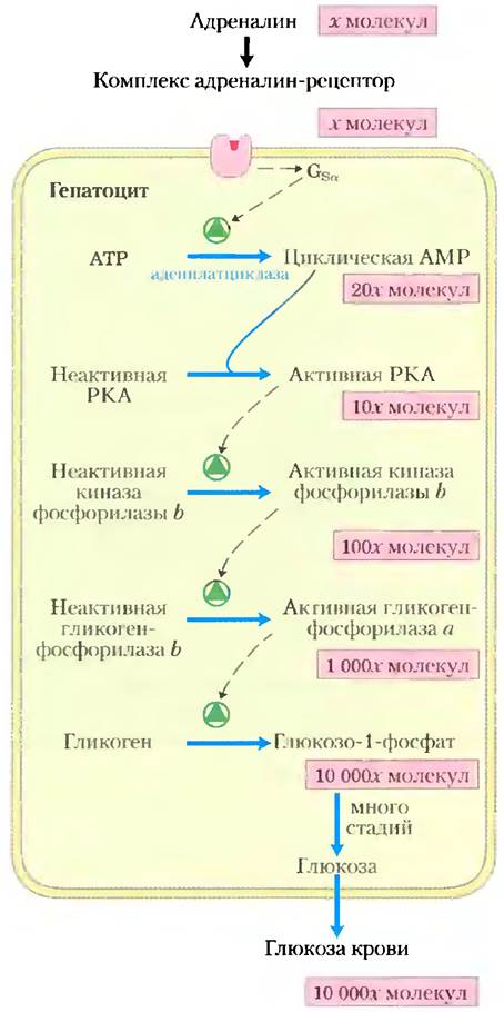
Signal transduction via adenylyl cyclase involves multiple stages that amplify the initial hormonal signal (Fig. 12–7). The binding of a single hormone molecule to a single receptor catalytically activates multiple Gs molecules. Subsequently, upon activating an adenylyl cyclase molecule, each active Gs molecule stimulates the catalytic synthesis of numerous cAMP molecules. The secondary messenger cAMP then activates PKA, and each PKA molecule in turn catalyzes the phosphorylation of many molecules of its target protein, phosphorylase b kinase (Fig. 12–7). This kinase activates phosphorylase b, which rapidly mobilizes glucose from glycogen stores. The cumulative effect of this cascade is a multi-order-of-magnitude Amplification of the hormonal signal, which explains why extremely low concentrations of epinephrine (or any other hormone) are sufficient to elicit a biological response.
Fig. 12–7. The epinephrine cascade. Epinephrine triggers a series of reactions in hepatocytes in which successive catalysts activate downstream catalysts, resulting in massive signal amplification. The binding of a small number of hormone molecules to specific β-adrenergic receptors on the cell surface activates adenylyl cyclase. The molecular counts shown are intended merely to illustrate THE PRINCIPLE OF amplification and are likely vastly underestimated. (Because 2 cAMP molecules activate 1 catalytic PKA subunit, no signal amplification occurs at this specific step.)
Several mechanisms exist for terminating the β-adrenergic response
A properly functioning signal transduction system must be able to switch off once the hormonal or other stimulus ends; consequently, all signaling systems possess signal-termination mechanisms. Furthermore, most signaling systems can adapt to prolonged stimulation by becoming less sensitive to it (desensitization). The β-adrenergic signaling system exhibits both of these properties. When the concentration of epinephrine in the Blood drops below the Kd of its receptor, the hormone dissociates from the receptor complex, and the receptor assumes an inactive conformation in which it can no longer activate Gs. A second way to halt the response to an adrenergic stimulus is the hydrolysis of GTP bound to the Gα subunit, catalyzed by the G protein's intrinsic GTPase activity. The conversion of bound GTP to GDP favors the return of Gα to a conformation in which it binds to the Gβγ subunits and can no longer interact with or stimulate adenylyl cyclase. This leads to the termination of cAMP synthesis. The rate of Gs inactivation depends on its GTPase activity, which is very low in the Gα subunit. However, GTPase-activating proteins (GAPs) greatly enhance this activity, accelerating G protein inactivation (see Box 12–2). GAPs are, in turn, regulated by other factors, ensuring a sufficiently high degree of sensitivity in the response to an adrenergic stimulus. A third mechanism for terminating the response is the removal of the secondary messenger—namely, the hydrolysis of cAMP to 5'-AMP (which cannot function as a second messenger) by the action of cyclic nucleotide phosphodiesterase (Fig. 12–4, a, step (7); 12–4, b).
Finally, at the end of the signaling pathway, the metabolic effects caused by enzyme phosphorylation are reversed by phosphoprotein Phosphatases, which hydrolyze phosphorylated Ser, Thr, or Tyr residues, releasing inorganic phosphate (Pi). Approximately 150 phosphoprotein phosphatase genes have been identified in the human genome, which is fewer than the number of protein kinase genes (500). Some of these phosphatases are known to be regulated, whereas others appear to act constitutively. When cAMP levels drop and PKA returns to its inactive state (step (7) in Fig. 12–4, a), the balance between phosphorylation and dephosphorylation is shifted by these phosphatases toward the dephosphorylated products.
Desensitization of the β-adrenergic receptor occurs via phosphorylation or arrestin binding
The signal-termination mechanisms described above operate when a stimulus ceases. The desensitization mechanism, by contrast, dampens the response even while the stimulus persists. Desensitization of the β-adrenergic receptor is mediated by a protein kinase that phosphorylates the receptor in its intracellular domain, which normally interacts with Gs (Fig. 12–8). When the receptor is bound to epinephrine, β-adrenergic receptor kinase (βARK, also known as GRK2; see below) phosphorylates Ser residues near the receptor's C-terminus. Normally localized in the Cytosol, βARK is recruited to the plasma membrane by binding to Gβγ subunits, positioning it favorably for receptor phosphorylation. Phosphorylation creates a binding site for the protein β-arrestin (βarr), also called arrestin 2, and β-arrestin binding effectively prevents interaction between the receptor and the G protein. β-Arrestin binding also facilitates receptor sequestration—the removal of receptors from the plasma membrane via endocytosis into small intracellular vesicles. Receptors within these vesicles are dephosphorylated and subsequently recycled back to the plasma membrane, completing the cycle and restoring the system's sensitivity to epinephrine. β-Adrenergic receptor kinase is a member of the G protein-coupled receptor kinase (GRK) family; all GRKs phosphorylate serpentine receptors in their C-terminal cytosolic domains and, like βARK, participate in receptor de- and resensitization. The human genome encodes at least five distinct GRKs and four arrestins; each GRK can desensitize many serpentine receptors, and each arrestin can interact with multiple phosphorylated receptors of various types.
Fig. 12–8. Desensitization of the β-adrenergic receptor during continuous exposure to epinephrine. This process is mediated by two proteins: β-adrenergic protein kinase (βARK) and β-arrestin (βarr, arrestin 2).

Cyclic AMP acts as a secondary messenger for numerous regulatory molecules
Epinephrine is just one of many hormones, growth factors, and other regulatory molecules that act by altering intracellular cAMP concentrations and, consequently, PKA activity (Table 12–3). For instance, Glucagon binds to its receptor on the plasma membrane of adipocytes, activating adenylyl cyclase (via a Gs protein). PKA, activated by the resulting rise in cAMP levels, phosphorylates and activates two proteins—perilipin and hormone-sensitive triacylglycerol lipase (see Fig. 17–3)—which are Key Enzymes in converting stored fat into Fatty acids, thereby mobilizing fatty acid reserves. Similarly, the peptide hormone ACTH (adrenocorticotropic hormone, also called corticotropin), produced by the anterior pituitary, binds to specific receptors in the adrenal cortex, activating adenylyl cyclase and thereby increasing cAMP concentrations. PKA then phosphorylates and activates several enzymes required for the synthesis of cortisol and other Steroid Hormones. In many cell types, the catalytic subunit of PKA can also enter The Nucleus, where it phosphorylates the protein CREB (cAMP response element-binding protein), which alters the expression of specific cAMP-regulated genes.
Table 12–3. Some signals that use cAMP as a secondary messenger

* Receptor subtypes are indicated in brackets. Different receptor subtypes may operate via distinct signal transduction mechanisms. For example, in some tissues, serotonin receptors belong to the 5-HT1a and 5-HT1b subtypes, which function through adenylyl cyclase and cAMP, whereas in other tissues, they belong to the 5-HT1c subtype, which acts via a phospholipase C–IP3 mechanism (Table 12–4).
Some hormones act by inhibiting adenylyl cyclase, lowering cAMP levels, and suppressing protein phosphorylation. For example, the binding of Somatostatin to its receptor leads to the activation of the inhibitory G protein Gi, which is homologous to Gs; Gi inhibits adenylyl cyclase and decreases cAMP concentration. The Effect of somatostatin thus counterbalances the action of glucagon. In adipose tissue, prostaglandin E1 (PGE1; see Fig. 10–18) inhibits adenylyl cyclase, lowering cAMP levels and slowing the mobilization of lipid reserves driven by epinephrine and glucagon. In certain other tissues, PGE1 stimulates cAMP synthesis because its receptors are coupled to adenylyl cyclase through the stimulatory G protein Gs. In tissues possessing α2-adrenergic receptors, epinephrine lowers cAMP concentration because α2-receptors are coupled to adenylyl cyclase via the inhibitory G protein Gi. In short, an extracellular signaling molecule such as epinephrine or PGE1 can produce entirely different effects in different tissues or cell types depending on three factors: the type of receptors present in the tissue, the type of G protein (Gs or Gi) to which the receptor is coupled, and the set of enzymes targeted by PKA within the cell. By utilizing all factors that lead to increases and decreases in cAMP concentration, the cell achieves the signal integration that we defined as a fundamental feature of signal transduction mechanisms (Fig. 12–1, d).
A fourth factor explaining how a single secondary messenger (cAMP) can mediate so many different signals is the restriction of signaling to specific subcellular regions by adapter proteins (noncatalytic proteins that hold together other protein molecules working in concert; see below). A-kinase anchoring proteins (AKAPs) are "bivalent"; one region binds to the regulatory (R) subunit of PKA (Fig. 12–6, a), while another binds to a specific cellular structure, anchoring PKA in close proximity to it. For instance, specific AKAPs tether PKA to microtubules, Actin filaments, Ca2+ channels, Mitochondria, and the nucleus. Different cell types contain distinct AKAPs, allowing cAMP to stimulate the phosphorylation of mitochondrial proteins in one cell and actin filaments in another. In some cases, an AKAP binds PKA alongside an enzyme that either activates PKA (adenylyl cyclase) or terminates its action (cAMP phosphodiesterase or phosphoprotein phosphatase) (Fig. 12–9). This close spatial arrangement of activating and inhibiting enzymes is presumably essential for achieving a rapid, localized response. As we will see later, certain signaling Membrane Proteins (including adenylyl cyclase) are localized to specific membrane domains within rafts or caveolae (see Section 12–5).
Fig. 12–9. Nucleation of supramolecular complexes by A-kinase anchoring proteins (AKAPs). Several types of AKAPs (green) act as multivalent scaffolds, holding the catalytic subunits of PKA (blue) near specific Organelles or cellular sites through interactions with PKA regulatory subunits (red). AKAP79, located on the cytoplasmic face of the plasma membrane, binds both PKA and adenylyl cyclase (AC). The cAMP produced by adenylyl cyclase rapidly and virtually without loss reaches nearby PKA molecules. AKAP79 can also bind PKA itself (not shown), its target protein (an ion channel), and the phosphoprotein phosphatase that dephosphorylates the target protein. AKAP250 (gravin) anchors PKA to the plasma membrane and binds cAMP phosphodiesterase (PDE), which quenches the PKA signal by converting cAMP to AMP. In both cases, AKAPs create high local concentrations of enzymes and secondary messengers, thereby achieving strict compartmentalization of the signaling pathway.

To perform a comprehensive analysis of signal transduction mechanisms, researchers must employ precise tools capable of studying the spatiotemporal aspects of signal transduction at the subcellular level and in real time. In analyzing the Intracellular Localization of biochemical processes, biochemistry intersects with cell biology, and Methods at this interface play an invaluable role in studying signaling pathways. For example, fluorescent probes are widely used in such studies. Attaching functional proteins to tags such as green fluorescent protein (GFP) allows their cellular localization to be determined (see Fig. 19–15, a). Changes in the interaction between two proteins (e.g., the R and C subunits of PKA) can be observed by measuring direct energy transfer between fluorescent labels attached to each protein—a technique known as fluorescence Resonance energy transfer (FRET; see Box 12–3).
Diacylglycerol, Inositol trisphosphate, and Ca2+ play analogous roles as secondary messengers
Another major class of GPCRs consists of receptors coupled via G proteins to plasma membrane phospholipase C (PLC), which is specific for the plasma membrane lipid phosphatidylinositol 4,5-bisphosphate (see Fig. 10–16). When a hormone of this class (Table 12–4) binds to its specific receptor on the plasma membrane (Fig. 12–10, step (1)), the hormone-receptor complex catalyzes the exchange of GTP for GDP on the associated G protein Gq (step (2)), activating it in much the same way that the β-adrenergic receptor activates Gs (Fig. 12–4). Activated Gq, in turn, activates PIP2-specific PLC (Fig. 12–10, step (3)), which catalyzes (step (4)) the generation of two potent secondary messengers: diacylglycerol and inositol 1,4,5-trisphosphate, or IP3 (not to be confused with PIP3, p. 622).
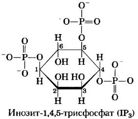
Table 12–4. Some signaling molecules that act via phospholipase C and IP3
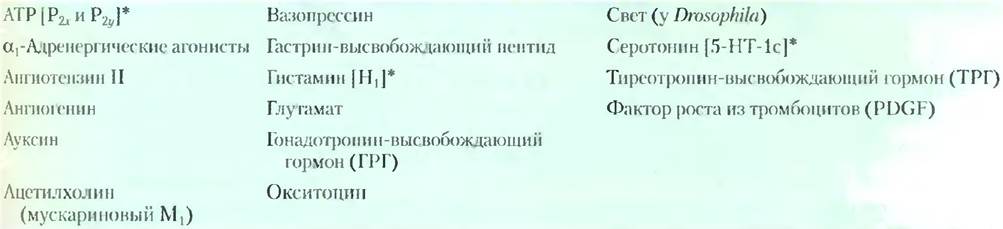
* Receptor subtypes are shown in brackets; see note to Table 12–3.
Fig. 12–10. Hormone-activated phospholipase C and IP3. Two intracellular secondary messengers are produced in the hormone-sensitive phosphatidylinositol system: inositol 1,4,5-trisphosphate (IP3) and diacylglycerol. Both substances contribute to the activation of protein kinase C. By increasing cytosolic Ca2+ concentrations, IP3 also activates other Ca2+-dependent enzymes; thus, Ca2+ likewise functions as a secondary messenger.

Water-soluble inositol trisphosphate diffuses from the plasma membrane into The Endoplasmic reticulum, where it binds to specific IP3 receptors, triggering the opening of Ca2+ channels located in the ER. As a result, Ca2+ is released into the cytosol (step (5)), and the cytoplasmic Ca2+ concentration sharply spikes to ~10-6 M. One of the effects of this elevated Ca2+ concentration is the activation of protein kinase C (PKC). Upon activation, diacylglycerol cooperates with Ca2+, also acting as a secondary messenger (step (6)). PKC phosphorylates specific Ser or Thr residues on target proteins, thereby altering their catalytic activity (step (7)). There are numerous PKC isozymes, each characterized by a distinct tissue distribution, target protein specificity, and cellular function. Among these targets are cytoskeletal proteins, enzymes, and Nuclear Proteins that regulate Gene Expression. This entire family of enzymes exhibits a broad spectrum of activities; for instance, they influence processes in the nervous and immune systems and regulate cell division.
The action of a group of compounds known as tumor promoters is driven by their effect on PKC. The most thoroughly studied of these are phorbol esters—synthetic compounds that act as potent PKC activators. They apparently mimic the action of cellular diacylglycerol as a secondary messenger, but unlike natural diacylglycerols, they resist rapid degradation. By continuously activating PKC, these synthetic tumor promoters disrupt normal Cell Growth and Division regulation (see discussion in Section 12.12), thereby provoking tumor development. ■
Calcium serves as a secondary messenger in numerous signaling pathways
Calcium ion signaling exhibits remarkable versatility. In many cells responding to extracellular signals, Ca2+ functions as a secondary messenger that triggers intracellular responses, such as exocytosis in neurons and endocrine cells, Muscle contraction, and cytoskeletal rearrangement during amoeboid movement. Under normal conditions, cytosolic Ca2+ concentration is maintained at very low levels (<10-7 M) through the action of Ca2+ pumps in the endoplasmic reticulum (ER), mitochondria, and plasma membrane. Hormonal signals, nerve impulses, or other stimuli induce either the influx of Ca2+ into the cell via specific plasma membrane Ca2+ channels or the release of calcium from the ER or mitochondria. In each case, this leads to an elevation of cytosolic Ca2+ and the initiation of a cellular response.
Changes in Ca2+ concentration are sensed by Ca2+-binding proteins that regulate a multitude of Ca2+-dependent enzymes. Calmodulin (CaM; Mr = 17,000) is an acidic protein featuring four high-affinity Ca2+-binding sites. When cellular Ca2+ levels rise to ~10-6 M (1 µM), the binding of Ca2+ to calmodulin induces a conformational change within the protein (Fig. 12-11a). Calmodulin interacts with numerous proteins, modulating their activity upon Ca2+ binding (Fig. 12-11b). Calmodulin belongs to the family of Ca2+-binding proteins, which also includes troponin (p. 247), an initiator of Skeletal Muscle contraction in response to elevated calcium ion concentrations. This protein family is characterized by a conserved Ca2+-binding structural motif known as the "EF hand" (Fig. 12-21c).
Box 12–3. METHODS. FRET: Biochemistry That Can Be Visualized in a Living Cell
Fluorescent probes are widely used to detect rapid biochemical changes within individual living cells. They enable the near-instantaneous (nanosecond-scale) tracking of fluctuations in intracellular secondary messenger concentrations or protein kinase activity. Furthermore, Fluorescence Microscopy offers sufficient resolution to pinpoint precisely where within the cell these changes occur. One widely adopted technique utilizes fluorescent probes derived from a naturally occurring protein: the green fluorescent protein (GFP) from the jellyfish Aequorea victoria (Fig. 1).
Fig. 1. The jellyfish Aequorea victoria, commonly found in Puget Sound, Washington, USA.

Upon absorbing a light quantum, GFP subsequently emits a photon (i.e., fluoresces) in the green region of the spectrum. The GFP protein consists of 11 strands folded into a β barrel, at the center of which lies the chromophore (the photon-absorbing and emitting unit)—the tripeptide Ser65-Tyr66-Gly67 (Fig. 2). Oxidation of this tripeptide is catalyzed autocatalytically by GFP itself (Fig. 3), meaning the reaction requires no additional proteins or Cofactors (other than molecular oxygen). Consequently, GFP can be expressed via cloning in virtually any cell type, where it serves as a fluorescent tag—for instance, as a fusion protein linked to another protein of interest (see Fig. 9-15a).
Fig. 2. Green fluorescent protein (GFP) and a ball-and-stick model of its fluorophore (from PDB ID 1GFL).

Fig. 3. The chromophore of GFP is formed by The amino acid sequence -Ser65-Tyr66-Gly67-. Chromophore maturation involves an internal rearrangement coupled with a multi-step oxidative reaction. A simplified mechanism of this process is shown here.

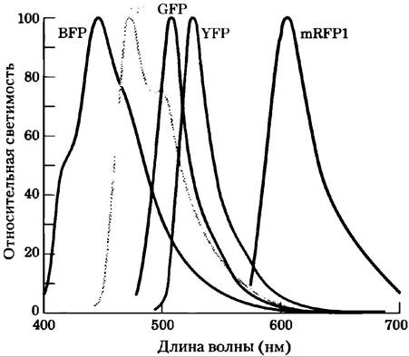
Introduction/32.html">Genetic Engineering OF the GFP gene has yielded variants with diverse fluorescence spectra. For instance, in yellow fluorescent protein (YFP), substitution of Ala206 with Lys alters both the Light absorption and emission wavelengths. Other GFP variants emit blue (BFP) or cyan (CFP) light, whereas a related protein (mRFP1) fluoresces in the red region (Fig. 4). GFP and its variants form compact structures that retain The ability to fold into their native β-barrel conformation even when fused to another protein. Researchers employ these chimeric fluorescent proteins as "spectral rulers" to determine distances between interacting cellular components and (indirectly) to measure the concentration of substances that alter the distance between two tagged proteins.
Fig. 4. Emission spectra for various GFP derivatives.
An excited fluorescent molecule, such as GFP or YFP, can release the energy of an absorbed photon via one of two pathways: (1) through fluorescence, wherein a photon of slightly longer wavelength (lower energy) than the exciting light is emitted, or (2) through fluorescence resonance energy transfer (FRET), a nonradiative process in which the excitation energy of a donor molecule is transferred directly to a neighboring acceptor molecule—without photon emission—thereby driving the acceptor into an excited state (Fig. 5). The acceptor can subsequently return to its ground state via fluorescence; the emitted photon has a longer wavelength (lower energy) than either the initial excitation light or the donor's fluorescence. This second quenching pathway (FRET) is strictly dependent on close proximity between the donor and acceptor (typically 1 to 50 Å); the efficiency of FRET is inversely proportional to the sixth power of the distance between them. Consequently, minute shifts in the distance between donor and acceptor manifest as dramatic changes in FRET efficiency, measured as an alteration in acceptor fluorescence upon donor excitation. With sufficiently sensitive light detectors, this fluorescent signal can be measured in localized regions of a single living cell.
Fig. 5. When a donor protein (CFP) is excited with monochromatic light at 433 nm, it fluoresces at 476 nm (left). When a protein fused to CFP (red) interacts with a protein fused to YFP (purple), this interaction brings CFP and YFP close enough to enable fluorescence resonance energy transfer (FRET) between them. Upon absorbing light at 433 nm, CFP now transfers its energy directly to YFP rather than fluorescing at 476 nm; YFP then emits light at its characteristic wavelength of 527 nm. The ratio of emission intensities at 527 nm to that at 476 nm serves as a quantitative measure of the interaction between the red and purple proteins.

The FRET technique has been successfully utilized to measure cAMP concentrations in living cells. The GFP gene was fused to the gene encoding the regulatory (R) subunit of cAMP-dependent protein kinase, while the BFP gene was fused to the catalytic (C) subunit gene (Fig. 6). When these two hybrid proteins are coexpressed within a cell, BFP (donor; excitation at 380 nm, emission at 460 nm) and GFP (acceptor; excitation at 475 nm, emission at 545 nm) within the inactive PKA holoenzyme (R2C2 tetramer) reside close enough to undergo FRET. Wherever cellular cAMP levels rise, the R2C2 complex dissociates into R2 and 2C, causing the FRET signal to vanish because the donor and acceptor are now too far apart. Under fluorescence microscopy, regions of higher cAMP concentration exhibit minimal GFP signal alongside an enhanced BFP signal. Calculating the ratio of emissions at 460 nm to 545 nm provides a precise measurement of changes in cAMP concentration. By mapping this ratio across all cellular regions, researchers can generate false-color ratiometric images of the cell, where relative cAMP concentrations are represented by color intensity. Time-lapse imaging captures dynamic fluctuations in cAMP content over time.
A variation of this technology was employed to measure PKA activity within living cells (Fig. 7). Researchers engineered a PKA biosensor by synthesizing a chimeric protein comprising four distinct modules: YFP (acceptor); a short peptide containing a Ser residue embedded within a PKA consensus phosphorylation sequence; a  Ser-binding domain (known as 14-3-3); and CFP (donor). When the Ser residue is unphosphorylated, the 14-3-3 domain lacks affinity for it, causing the fusion protein to adopt an extended conformation that keeps the donor and acceptor too far apart to generate a FRET signal. Wherever active PKA is present in the cell, it phosphorylates the Ser residue within the biosensor, prompting 14-3-3 to bind the resulting
  phosphoserine. This binding event brings YFP and CFP into close proximity, generating a FRET signal detectable by fluorescence microscopy that signals the presence of active PKA.
Fig. 7. Measuring PKA activity via FRET. An engineered protein links YFP and CFP through a peptide containing a Ser residue, flanked by a PKA consensus phosphorylation sequence and a phosphoserine-binding 14-3-3 domain. Active PKA phosphorylates the Ser residue, which then docks with the 14-3-3 domain; this brings the fluorescent proteins close enough for FRET to occur, thereby indicating active PKA activity.

Calmodulin also serves as an integral subunit of a family of enzymes known as Ca2+/calmodulin-dependent protein kinases (CaM kinases I–IV). When the intracellular Ca2+ concentration increases in response to a stimulus, calmodulin binds Ca2+, undergoes a conformational change, and activates CaM kinase. This kinase then phosphorylates A number of target enzymes, thereby regulating their activity. Calmodulin also functions as a regulatory subunit of muscle phosphorylase b kinase, which is activated by Ca2+ ions. Thus, Ca2+ triggers ATP-dependent muscle contraction while simultaneously activating glycogen breakdown to provide the "fuel" required for ATP synthesis. Numerous Other Enzymes are likewise known to be regulated by Ca2+ ions with the assistance of calmodulin (Table 12-5). The activity of Ca2+ as a second messenger, much like that of cAMP, can be spatially restricted; after its release has triggered a local response, Ca2+ is typically cleared before it can diffuse to distant PARTS OF THE cell.
Figure 12-11. Calmodulin is a protein mediator for many enzymatic reactions stimulated by Ca2+ ions. Calmodulin contains four high-affinity Ca2+-binding sites (Kd ≈ 0.1 – 1 µM). (a) Ribbon model of the crystal structure of calmodulin (PDB ID 1GLL). The four Ca2+-binding sites are occupied by Calcium Ions (purple). Left, N-terminal domain; right, C-terminal domain. (b) Calmodulin bound to the helical domain (red) of one of the many proteins it regulates—calmodulin-dependent protein kinase II (PDB ID 1GDl). Note that the long central α-Helix visible in (a) bends back upon binding to the substrate's helical domain. The central helix is, of course, more flexible in solution than in the crystal. (c) Each of the four Ca2+-binding sites resides in a helix-loop-helix motif known as an "EF hand," which is also present in many other Ca2+-binding proteins.

Table 12-5. Some proteins regulated by Ca2+ and calmodulin
Plasma membrane Ca2+-ATPase (Ca2+ pump) |
Sarcoplasmic reticulum Ca2+-release channel |
Ca2+-dependent Na+ channel (in Paramecium) |
Ca2+/calmodulin-dependent protein kinases (CaM kinases I through IV) |
cAMP-regulated olfactory channel |
cAMP phosphodiesterase |
cGMP-regulated Na+, Ca2+ channels (rod and cone cells) |
NAD+ kinase |
NO synthase (nitric oxide synthase) |
Adenylyl cyclase (in the Brain) |
Glutamate decarboxylase |
Calcineurin (phosphoprotein phosphatase 2B) |
Myosin light-chain kinases |
RNA helicase (p68) |
Phosphoinositide 3-kinase |
Very often, the concentration of Ca2+ does not merely rise and fall, but rather oscillates with a period of several seconds (Figure 12-12), even when the extracellular hormone concentration remains constant. The mechanism underlying these calcium oscillations appears to involve feedback regulation of either the phospholipase that generates IP3, the ion channel responsible for releasing Ca2+ from the ER, or both. Whatever the mechanism, the effect is to translate one type of signal (e.g., hormone concentration) into another (the frequency and amplitude of oscillations in intracellular Ca2+ content). Another possible manifestation is local calcium "spots," "puffs," or "waves"—transient increases in Ca2+ concentration restricted to a specific region of the cell (Figure 12-13). The Ca2+ signal subsides as calcium ions diffuse away from the source (the calcium channel), are sequestered back into the ER, or are pumped out of the cell.
Figure 12-12. Initiation of intracellular Ca2+ oscillations by extracellular signals. (a) A dye (fura) whose fluorescence changes upon binding to Ca2+ was allowed to diffuse into cells, and the fluorescence intensity was subsequently measured by fluorescence microscopy. Fluorescence intensity is shown in pseudocolor; the Ca2+ concentration can be estimated from the color scale. In this case, thymocytes (Thymus cells) were stimulated with exogenous ATP, which raises their internal Ca2+ concentration. The cellular response is heterogeneous; some cells exhibit a high intracellular Ca2+ concentration (red), while others show a much lower concentration (blue). (b) When such a probe is used to measure Ca2+ levels in a single hepatocyte, we observe that the agonist norepinephrine (addition time indicated by the arrow) triggers Ca2+ oscillations ranging from 200 to 500 nM. Similar oscillations are induced in other cell types by different extracellular signals.

Figure 12-13. Transient and strictly localized increases in Ca2+ concentration. (a) IP3-gated calcium channels of the endoplasmic reticulum are organized into clusters, but each channel can respond independently to an IP3 signal. A relatively weak stimulus causing a minor increase in IP3 concentration may trigger the rapid opening of a single channel, resulting in the transient, localized appearance of a calcium "spot" (or spark). (b) A somewhat stronger stimulus, eliciting a greater increase in IP3 concentration, can cause all calcium channels within a cluster to open, generating a Ca2+ "puff," with a greater concentration increase, duration, and spatial spread than in the first case. (c) A sufficiently large cloud of elevated calcium concentration can recruit neighboring clusters of calcium channels. The opening of channels in adjacent clusters amplifies the effect, resulting in a wave of elevated calcium ion concentration propagating along the ER.

The signaling systems involving Ca2+ and cAMP are interconnected. In some tissues, calcium ions stimulate both adenylyl cyclase (which produces cAMP) and phosphodiesterase (which degrades cAMP). Consequently, transient and local changes in calcium ion concentration can lead to transient and local changes in cAMP concentration. As we mentioned earlier, the cAMP-dependent enzyme PKA is frequently part of a tightly localized supramolecular complex assembled around scaffold proteins such as AKAPs. The intracellular localization of target enzymes, combined with the temporal and spatial gradients of Ca2+ and cAMP, enables the cell to respond to one or multiple signals with precise metabolic adjustments localized in both time and space.
Section 12.2 Summary: G Protein-Coupled Receptors and Second Messengers
■ G protein-coupled receptors (GPCRs) act via heterotrimeric G proteins. Upon ligand binding, these receptors catalyze the exchange of GTP for GDP bound to the associated G protein, triggering the dissociation of the G protein α subunit. This subunit stimulates or inhibits a target enzyme, thereby altering the concentration of its product—a second messenger.
■ The β-adrenergic receptor activates the stimulatory G protein Gs, thereby activating adenylyl cyclase and increasing the concentration of the second messenger cAMP. Cyclic AMP stimulates cAMP-dependent protein kinase, which in turn phosphorylates key proteins, altering their activity.
■ Enzymatic cascades, in which one hormone molecule activates a catalyst that in turn activates another catalyst, and so forth, result in massive signal amplification—a hallmark of most hormone-activated systems.
■ Cyclic AMP is ultimately degraded by cAMP phosphodiesterase, while Gs turns itself off by hydrolyzing its bound GTP to GDP, acting as a self-limiting binary switch.
■ When the adrenaline signal is prolonged, a β-adrenergic receptor-specific protein kinase and β-arrestin transiently desensitizing the receptor and target it for internalization into intracellular vesicles.
■ Some receptors stimulate adenylyl cyclase via Gs; others inhibit it via Gi. Thus, the cellular concentration of cAMP represents the net integrated response to two (or more) signals.
■ Noncatalytic adapter proteins, such as AKAPs, tether proteins involved in the same signaling pathway in close proximity, enhancing the efficiency of their interaction and, in some cases, determining the subcellular Location OF THE process.
■ Some GPCRs operate via plasma membrane phospholipase C, which cleaves PIP2 to yield diacylglycerol and IP3. By opening Ca2+ channels in the endoplasmic reticulum, IP3 increases the cytosolic Ca2+ concentration. Diacylglycerol and Ca2+ act synergistically to activate protein kinase C, which
phosphorylates specific cellular proteins and alters their activity. The intracellular Ca2+ concentration also regulates (often via calmodulin) the activity of numerous other enzymes and proteins involved in secretion, cytoskeletal rearrangement, or contraction.
Last update: 06/08/2026
Editorial and Educational Adaptation: This material has been compiled based on the primary/original source text. The project team performed an editorial review, corrected technical inaccuracies, structured sections, and adapted the content for an educational format.
What was processed:
- elimination of formatting defects (OCR errors, structural breaks, corrupted characters);
- editorial organization of content;
- standardization of terminology in accordance with academic sources;
- verification of factual statements against the original source text.
All mentions of the author, publication year, and origin of the primary text have been preserved in accordance with the source.