LEHNINGER PRINCIPLES OF BIOCHEMISTRY - VOLUME 1. THE FOUNDATIONS OF BIOCHEMISTRY: STRUCTURE AND CATALYSIS - 2011
PART I. STRUCTURE AND CATALYSIS
12. BIOSIGNALING
12.3. Receptor Enzymes
Receptor Tyrosine Kinases (RTKs) constitute a large family of Plasma Membrane Receptors that possess intrinsic protein kinase activity. The Mechanism of extracellular signal Transduction via these receptors differs fundamentally from that mediated by GPCRs. The Ligand-binding domain of an RTK, located on the outer surface of Cell/30.html">The Plasma Membrane, and the enzyme's Active Site, located on the cytoplasmic surface, are connected by a single transmembrane segment. The cytoplasmic domain is a protein kinase (tyrosine kinase) that phosphorylates Tyr residues in specific target Proteins. Prototypical Examples of this protein group are the Insulin receptor and the epidermal growth factor receptor.
Insulin receptor stimulation triggers a cascade of protein phosphorylation reactions
Insulin regulates both METABOLISM and Gene Expression: the insulin signal travels from the plasma membrane receptor to insulin-sensitive metabolic Enzymes and to The Nucleus, where it stimulates the Transcription of specific genes. The active insulin receptor (INS-R) consists of two identical α chains protruding from the extracellular side of the plasma membrane and two transmembrane β subunits whose C-termini extend into the Cytosol (Fig. 12-14). The α chains contain the insulin-binding domain, whereas the intracellular domains of the β subunits, which possess protein kinase activity, transfer a phosphoryl group from ATP to the hydroxyl group of Tyr residues in specific target proteins. Signal transduction through the insulin receptor begins when the binding of insulin to the α chains activates the tyrosine kinase activity of the β subunits, and each αβ monomer phosphorylates three key Tyr residues near the C-terminus of its partner's β chain within the dimer. Autophosphorylation opens the active site so that the enzyme can phosphorylate Tyr residues on other target proteins. Insulin receptor stimulation initiates a protein phosphorylation cascade. The mechanism of activation of the insulin receptor's protein kinase activity is analogous to that of PKA and PKC: a segment of the cytoplasmic domain (the inhibitory sequence) that normally blocks the active site moves away from the active site following phosphorylation, making it accessible for binding target proteins (Fig. 12-14).
One such target protein (Fig. 12-15, step (1)) is insulin receptor substrate 1 (IRS-1; step (2)). Once phosphorylated at its Tyr residues, IRS-1 serves as a docking center for a complex of proteins (step (3)) that relays information from the insulin receptor to downstream targets in the cytosol and nucleus through a series of intermediate proteins. First, the  Tyr residue in IRS-1 binds to the SH2 domain of the Grb2 protein. (SH2 stands for Src Homology 2; the Amino acid sequences of SH2 domains are similar to the homologous domain found in another protein tyrosine kinase, Src, pronounced "sark"). Several signaling proteins contain SH2 domains, all of which bind
  -Tyr residues of a partner protein. Grb2 also contains a second protein-binding domain, SH3, which binds to Proline-rich regions. Grb2 associates with the proline-rich region of Sos, recruiting Sos into the expanding receptor complex. Bound to Grb2, Sos acts as a nucleotide exchange factor (GEF), catalyzing the replacement of bound GDP with GTP on the Ras protein (a G protein).
Ras is a member of the small G protein family, which mediates the transmission of A wide variety of diverse signals (Box 12-2). Like the trimeric G protein involved in the β-adrenergic signaling system (Fig. 12-5), the Ras protein can exist in an active conformation (complexed with GTP) and an inactive conformation (complexed with GDP), but Ras is a monomer (~20 kDa). When GTP is bound, Ras can activate the Raf-1 protein kinase (Fig. 12-15, step (4)), the first of three protein kinases—Raf-1, MEK, and ERK—that form a cascade in which each kinase activates the next by phosphorylation (step (5)). The protein kinases MEK and ERK are activated by the phosphorylation of Thr and Tyr residues. In their activated state, they mediate some of the biological effects
of insulin by entering the nucleus and phosphorylating proteins such as Elk-1 (step (6)), which modulates the transcription of nearly 100 insulin-regulated genes (step (7)).
Class="center">Fig. 12-14. Activation of insulin receptor tyrosine kinase by autophosphorylation. (a) The insulin-binding domain (pink; shown as a molecular surface contour of the crystal Structure; adapted from PDB ID 2DTG) of the insulin receptor is extracellular and consists of (b) two α subunits and the extracellular portions of two β subunits. (The structure of the transmembrane domain has not been resolved by X-ray crystallography.) Information regarding insulin binding (red; PDB ID 2CEU) is transmitted via the single transmembrane helix of each β subunit to the respective intracellular tyrosine kinase domains, causing them to activate and phosphorylate each other on three tyrosine residues, (c) In the inactive form of the tyrosine kinase domain (PDB ID 1IPK), the activation loop (blue) blocks the active site, and none of the key Tyr residues (black-and-red Ball-and-stick models) are phosphorylated. This conformation is stabilized by a Hydrogen bond between Tyr1162 and Asp1132. (d) Tyrosine kinase activation enables each β subunit of the dimer to phosphorylate three Tyr residues (Tyr1158, Tyr1162, and Tyr1163) on the opposing subunit (PDB ID 1IR3). (Phosphoryl groups are shown as ball-and-stick models, with phosphorus atoms in orange and oxygen atoms in red.) The Effect of the three charged (P)-Tyr residues is to displace the activation loop by 30 Å away from the substrate-binding center, allowing it to bind and phosphorylate the target protein, indicated by the red arrow.

Fig. 12-15. Introduction/30.html">Regulation of Gene Expression by insulin. The insulin receptor (INS-R) consists of two α chains located on the extracellular side of the plasma membrane and two β subunits that span the membrane and protrude into the cytosol. The binding of insulin to the α chains triggers a conformational change that enables the autophosphorylation of Tyr residues in the C-terminal domain of the β subunits. Autophosphorylation activates the tyrosine kinase domain, which then catalyzes the phosphorylation of other target proteins. The signaling pathway by which insulin regulates the expression of specific genes consists of a cascade of protein kinases, each activating the subsequent one. The insulin receptor is a tyrosine-specific kinase; other kinases (all shown in blue) phosphorylate Ser or Thr residues. MEK is a dual-Specificity kinase that phosphorylates both Thr and Tyr residues in ERK (extracellular signal-regulated kinase); MEK is a mitogen-activated protein kinase kinase that activates ERK; SRF is the serum response factor.

The Raf-1, MEK, and ERK proteins are members of three large families classified in several ways. ERK is a member of the MAPK family (mitogen-activated protein kinases; mitogens are extracellular signaling molecules that induce mitosis and Cell Division). Soon after the Discovery of the first MAPK, it was found that this enzyme is activated by another protein kinase, which came to be known as MAP kinase kinase (to which MEK belongs); when a third kinase was discovered that activates MAP kinase kinase, it was given the somewhat cumbersome name "MAP kinase kinase kinase" (Raf-1 is a member of this family; Fig. 12-15, step (4)). The acronyms for these three families are much less cumbersome: MAPK, MAPKK, and MAPKKK. Kinases of the MAPK and MAPKKK families are specific for Ser or Thr residues, whereas MAPKK kinases (such as MEK here) phosphorylate both Ser and Tyr residues in their substrates (MAPK, such as ERK here).
Biochemists now understand that the insulin pathway is not the sole example of the universal mechanism by which hormonal signals lead to the phosphorylation of target enzymes by protein kinases in ways similar to those shown in Fig. 12-15. The target of phosphorylation is often another protein kinase, which in turn phosphorylates a third protein kinase, and so on. As a result, an enzymatic cascade amplifies the initial signal by many orders of magnitude (see Fig. 12-1, b). MAPK cascades (Fig. 12-15) mediate signal transduction triggered by various growth factors, such as platelet-derived growth factor (PDGF) and epidermal growth factor (EGF). Another general paradigm, illustrated here using the insulin receptor, is The Use of adapter proteins that link the components of a branched signaling pathway.
The membrane phospholipid PIP3 Functions in one branch of insulin signaling
The insulin-initiated signaling pathway branches at the level of IRS-1 (Fig. 12-15, step (2)). The Grb2 protein is not the only protein that binds to phosphorylated IRS-1. The enzyme phosphoinositide 3-kinase (PI3K) associates with IRS-1 via its SH2 domain (Fig. 12-16). Once activated in this manner, PI3K converts the membrane lipid phosphatidylinositol 4,5-bisphosphate (see Fig. 10-16), also known as PIP2, into phosphatidylinositol 3,4,5-trisphosphate (PIP3). The multiply charged HEAD group of PIP3, protruding from the cytoplasmic surface of the plasma membrane, serves as the docking site for a second branch of the signaling pathway involving another protein kinase cascade. When protein kinase B (PKB) binds to PIP3, it is phosphorylated and activated by another protein kinase, PDK1. Activated PKB then phosphorylates Ser or Thr residues on its target proteins, one of which is Glycogen synthase kinase 3 (GSK3). In its active, unphosphorylated form, GSK3 phosphorylates glycogen synthase, inactivating it and thereby slowing down glycogen synthesis. (This mechanism represents only part of the process describing the effect of insulin on Glycogen Metabolism.) When phosphorylated by PKB, GSK3 is inactivated. By preventing the inactivation of glycogen synthase in Liver and Muscle, the protein phosphorylation cascade initiated by insulin stimulates glycogen synthesis (Fig. 12-16). In muscle, PKB triggers the translocation of glucose transporters (GLUT4) from intracellular vesicles to the plasma membrane, stimulating glucose uptake from the Blood (Fig. 12-16, step (5); see also Box 11-2).
Fig. 12-16. Activation of glycogen synthase by insulin. Signal transduction is mediated by PI 3-kinase (PI3K) and protein kinase B (PKB).

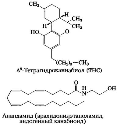
Protein kinase B is also involved in other signaling pathways, including one triggered by Δ9-tetrahydrocannabinol (THC), the active ingredient in marijuana and hashish. THC activates the CB1 receptor in the plasma membrane of Brain Neurons, initiating a signaling cascade that includes MAP kinases. One of the consequences of CB1 activation manifests as appetite stimulation, a well-known effect of marijuana use. In the body, the endogenous ligands for CB receptors are endocannabinoids, such as anandamide, which serve to protect the brain from the Toxic effects of excessive neural activity, such as epileptic seizures. Hashish has been used for centuries to treat Epilepsy.
As in all signaling pathways, a mechanism exists to terminate signal transmission through the PI3K-PKB cascade. A PIP3-specific phosphatase (PTEN in humans) removes the 3-phosphate group from PIP3 to yield PIP2, which is no longer a ligand for PKB, thereby interrupting the signaling chain. In late-stage cancers of various Tissues, tumor Cells frequently harbor a defect in the PTEN gene and consequently exhibit abnormally high levels of PIP3 and PKB activity. This presumably results in a persistent, unremitting signal that drives cell division and tumor growth. ■
In addition to numerous receptors that function as protein tyrosine kinases, a variety of receptor-like proteins in the plasma membrane exhibit protein tyrosine phosphatase activity. Based on the structures of these proteins, we can infer that their ligands are Components of the Extracellular matrix or the surfaces of neighboring cells. Although their precise role in signaling is not yet fully understood—much like that of receptor tyrosine kinases—it is clear that they have the capacity to reverse the effects of signals stimulated by these kinases.
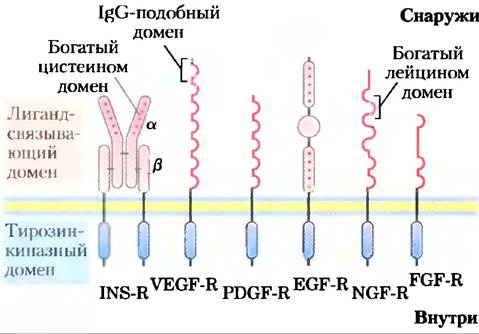
The insulin receptor is the prototype for an entire class of receptor enzymes with similar structures and receptor tyrosine kinase activity (Fig. 12-17). For instance, the epidermal growth factor receptor and the platelet-derived growth factor receptor share structural and Amino Acid Sequence similarities with the insulin receptor, and both possess protein tyrosine kinase activity that phosphorylates IRS-1. Many of these receptors dimerize upon ligand binding; the insulin receptor already exists as a dimer prior to insulin attachment. The binding of adapter proteins, such as Grb2, to (P)-Tyr residues is a common mechanism for facilitating Protein-Protein Interactions. We will return to this topic in Section 12.5.
Fig. 12-17. Receptor tyrosine kinases. Growth factor receptors that transmit signals via tyrosine kinase activity include the receptors for insulin (INS-R), vascular endothelial growth factor (VEGF-R), platelet-derived growth factor (PDGF-R), epidermal growth factor (EGF-R), nerve growth factor (NGF-R), and fibroblast growth factor (FGF-R). All these receptors contain a tyrosine kinase domain on the cytoplasmic side of the plasma membrane (shown in blue). The extracellular domain is unique to each receptor because each specifically binds only its corresponding growth factor. These extracellular domains typically consist of a combination of Structural motifs, such as Cysteine- or leucine-rich sequences and regions containing one or more immunoglobulin-like motifs (Ig-like domains; see p. 203). The Human Genome encodes many other receptors of this type, each featuring its own distinct extracellular domain.
What drove the evolution of such a complex regulatory mechanism? This system allows a single activated receptor to activate multiple IRS-1 molecules, amplifying the insulin signal and enabling the integration of signals from several receptors, each capable of phosphorylating IRS-1. Moreover, because IRS-1 can activate any of the proteins containing SH2 domains, a single receptor acting through IRS-1 can initiate two (or more) signaling pathways. For example, insulin regulates gene expression via the Grb2-Sos-Ras-MAPK cascade and glycogen metabolism through the PI3K-PKB cascade. Finally, there are several closely related IRS proteins (IRS-1, IRS-2), each with a characteristic tissue distribution and specialized functions, which further expands the repertoire of signaling pathways initiated by RTKs.
The JAK-STAT signaling system also utilizes tyrosine kinase activity
A variation on the basic theme of receptor tyrosine kinases is seen in receptors that lack intrinsic protein kinase activity but associate with a cytosolic tyrosine kinase upon ligand binding. One example is the system that regulates mammalian erythrocyte production. The cytokine (a signaling molecule involved in development) for this system is Erythropoietin (EPO), a 165-amino-acid protein produced by the Kidneys. When EPO binds to its receptor on the plasma membrane (Fig. 12-18), the receptor dimerizes and acquires The ability to bind a soluble protein kinase, JAK (Janus kinase). This binding activates JAK, which phosphorylates several Tyr residues in the cytoplasmic domain of the EPO receptor. A family of transcription factors known as STATs (signal transducers and activators of transcription) also serves as targets for JAK activity. An SH2 domain in STAT5 binds to (P)-Tyr residues on the EPO receptor, positioning them favorably for phosphorylation by the protein kinase (JAK). When STAT5 is phosphorylated in response to EPO, it forms dimers capable of translocating to the nucleus. Inside the nucleus, STAT5 induces the expression (transcription) of specific genes required for erythrocyte maturation. The JAK-STAT system operates in several signaling pathways, including that of the hormone leptin, described in detail in Chapter 23 (Fig. 23-37). Activated JAK can also recruit Grb2 to trigger the MAPK cascade (Fig. 12-18b), leading to changes in specific gene expression.
Fig. 12-18. Mechanism of JAK-STAT signaling for the erythropoietin receptor. Binding of erythropoietin (EPO) induces dimerization of the EPO receptor, allowing the soluble tyrosine kinase JAK to associate with the intracellular domain of the receptor and phosphorylate it at multiple Tyr residues. (a) In one signaling pathway, the SH2 domain of the STAT5 protein binds to  -Tyr residues on the receptor, bringing the receptor and JAK into close proximity. Phosphorylation of STAT5 by the JAK kinase enables two STAT molecules to form a dimer, with each molecule binding to different
  -Tyr residues. Dimerization of STAT5 exposes a nuclear localization sequence (NLS), which mediates The transport of STAT5 into the nucleus. In the nucleus, STAT induces the expression of EPO-controlled genes. (b) A second signaling pathway is triggered by autophosphorylation of JAK associated with the receptor-bound EPO; the adapter protein Grb2 binds to
  -Tyr on JAK and initiates the MAPK cascade, much like in the insulin system (see Fig. 12-15).

The Src protein is another soluble protein tyrosine kinase that associates with certain receptors upon ligand binding. The characteristic  -Tyr-binding domain was first discovered in the Src protein and was subsequently named the "SH2 domain".
Signaling systems are intricately interconnected
For the sake of simplicity, we have thus far treated individual signal transduction pathways as independent sequences of events leading to distinct metabolic outcomes. In reality, however, signaling systems are densely interconnected. The metabolic regulatory network is intricately layered and interwoven. Although we discussed the insulin and epinephrine signaling pathways separately, they do not operate in isolation. In many tissues, the Physiological effects of insulin oppose those of epinephrine, and activation of the insulin signaling pathway attenuates $\beta$-adrenergic signaling. For instance, the insulin receptor tyrosine kinase directly phosphorylates two Tyr residues in the cytoplasmic domain of the $\beta_{2}$-adrenergic receptor, while insulin-activated PKB (Fig. 12-19) phosphorylates two Ser residues in the same region. The phosphorylation of these four residues triggers the internalization of the $\beta_{2}$-adrenergic receptor, effectively desensitizing it and rendering The Cell less responsive to epinephrine. Another layer of cross-talk is evident when the  Tyr residues of the $\beta_{2}$-adrenergic receptor—phosphorylated by the insulin receptor—serve as docking sites for SH2 domain-containing proteins, such as Grb2 (Fig. 12-19, left). Perhaps facilitated by this interaction, insulin-induced activation of ERK in the MAPK cascade (see Fig. 12-15) proceeds 5- to 10-fold more efficiently in the presence of the $\beta_{2}$-adrenergic receptor. Signaling systems utilizing cAMP and Ca2+ are similarly interdependent, with each secondary messenger modulating the production and concentration of the other. A primary objective of systems biology is to decipher the network-level outcomes of these complex interactions across all Tissues of the Organism—a formidable challenge, to put it mildly!
Fig. 12-19. Cross-talk between the insulin receptor and the $\beta_{2}$-adrenergic receptor (or other GPCRs). Upon insulin binding, the tyrosine kinase activity of the activated INS-R directly phosphorylates the $\beta_{2}$-adrenergic receptor (right) at two tyrosine residues (Tyr350 and Tyr364) near the C-terminus. Simultaneously, activation of protein kinase B (PKB; see Fig. 12-16) promotes phosphorylation of two Ser residues in the same region. This dual phosphorylation drives receptor internalization and dampens the cellular response to epinephrine. Conversely, insulin receptor-catalyzed phosphorylation of the GPCR (whether adrenergic or another type) at C-terminal Tyr residues (left) creates docking sites that promote the MAPK cascade (see Fig. 12-15), with Grb2 acting as an adaptor protein. In this scenario, INS-R co-opts the GPCR to amplify its own downstream signaling.

Summary of Section 12.3 Receptor Enzymes
■ The insulin receptor (INS-R) serves as the prototypical receptor tyrosine kinase. Upon insulin binding, each $\alpha\beta$-dimer of the receptor phosphorylates the $\beta$-chain of its partner, thereby stimulating its intrinsic tyrosine kinase activity. This kinase subsequently catalyzes the phosphorylation of Tyr residues on downstream target proteins, such as IRS-1.
■ Phosphorylated IRS-1 residues act as docking sites for proteins containing SH2 domains. Several of these proteins, including Grb2, possess multiple protein-binding domains and function as adaptors that bridge distinct signaling components.
■ Sos binds to Grb2 and catalyzes the GDP-to-GTP exchange on the small G-protein Ras, which in turn triggers a protein kinase cascade culminating in the phosphorylation of specific targets in the cytosol or nucleus, ultimately driving changes in metabolism and gene expression.
■ PI-3K, activated through its interaction with the IRS-1 substrate, converts the membrane lipid PIP2 into PIP3, establishing a critical docking platform for the assembly of multi-Protein Complexes driving the second and third Branches of the insulin signaling pathway.
■ In the JAK-STAT signaling pathway, a soluble JAK tyrosine kinase is activated upon ligand binding to the receptor and subsequently phosphorylates a STAT transcription factor; STAT then translocates to the nucleus to regulate the expression of specific target genes. Extensive and intricate cross-talk between these signaling networks enables precise coordination and fine-tuning of diverse hormonal responses.
Last update: 06/08/2026
Editorial and Educational Adaptation: This material has been compiled based on the primary/original source text. The project team performed an editorial review, corrected technical inaccuracies, structured sections, and adapted the content for an educational format.
What was processed:
- elimination of formatting defects (OCR errors, structural breaks, corrupted characters);
- editorial organization of content;
- standardization of terminology in accordance with academic sources;
- verification of factual statements against the original source text.
All mentions of the author, publication year, and origin of the primary text have been preserved in accordance with the source.