LEHNINGER PRINCIPLES OF BIOCHEMISTRY - VOLUME 1. THE FOUNDATIONS OF BIOCHEMISTRY: STRUCTURE AND CATALYSIS - 2011
PART I. STRUCTURE AND CATALYSIS
Class="center">When I first began to study the Action of Hormones some 25 years ago, it was widely believed among biologists that it was meaningless to study hormone action outside the organized cellular Structure. However, reflecting upon The history of biochemistry, it seemed quite plausible to me that hormones might act at THE MOLECULAR LEVEL.
Earl Wilbur Sutherland, Nobel Lecture, 1971
12. BIOSIGNALING
The ability of Cells to receive and respond to signals from beyond their Cell/33.html">Plasma Membrane is fundamental to life. Bacterial cells receive a continuous influx of signals from Membrane Proteins acting as environmental sensors that monitor local pH, osmotic strength, the availability of nutrients, oxygen, and light, as well as the presence of toxic chemicals, predatory organisms, or food competitors. These signals elicit appropriate responses, such as moving toward food or away from toxic substances, or forming resting spores in nutrient-depleted environments. In Multicellular Organisms, cells with diverse Functions exchange a multitude of signals. Plant cells respond to growth hormones and sunlight intensity. Animal cells communicate regarding the concentration of ions and glucose in extracellular fluids, allowing interdependent metabolic processes to occur across different Tissues and coordinating proper cell arrangement during embryonic development. In all these cases, a signal represents information that is recognized and received by specific receptors and converted into a cellular response—a process that invariably involves chemistry. This conversion of information into Chemical Reactions is called signal Transduction and is a universal property of living cells.
12.1. General Features of Signal Transduction Systems
Signal transduction is characterized by high Specificity and exquisite sensitivity. Specificity is achieved by precise molecular complementarity between the signal molecule and receptor molecules (Fig. 12-1a), mediated by the same types of weak (noncovalent) forces that govern enzyme-substrate and antigen-antibody interactions. Multicellular organisms possess an additional layer of specificity because receptors for a given signal—or intracellular targets for a given signaling pathway—are present only in certain cell types. For example, thyrotropin-releasing hormone triggers responses in anterior pituitary cells, but not in hepatocytes (Liver cells), which lack receptors for this hormone. Epinephrine alters Glycogen METABOLISM in hepatocytes but not in erythrocytes; although both cell types have epinephrine receptors, liver cells contain glycogen and the metabolizing Enzymes stimulated by epinephrine, whereas erythrocytes do not.
Figure 12-1. Four hallmark features of signal transduction systems.
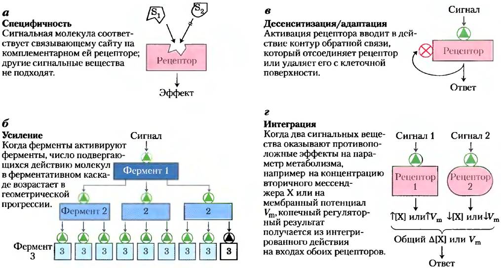
The exceptionally high sensitivity of signal transducers is ensured by three factors: high receptor affinity for signal molecules, cooperativity (frequent, though not universal) in Ligand-receptor interactions, and signal Amplification by enzymatic cascades. The affinity between the signaling
molecule (ligand) and the receptor can be expressed by the dissociation constant Kd, typically ≤10-10 M, meaning the receptor detects picomolar concentrations of the signal molecule. Ligand-receptor interactions are quantified using Scatchard analysis to determine affinity (Kd) and the number of ligand-binding sites on the receptor (Box 12-1).
Cooperativity in ligand-receptor interactions leads to a steep activation response of the receptor with small changes in ligand concentration (recall the cooperative effect in oxygen binding by Hemoglobin; Fig. 5-12). Amplification by enzymatic cascades occurs when an enzyme associated with a signal receptor is activated and in turn catalyzes the activation of many molecules of a second enzyme, each of which activates many molecules of a third enzyme, and so on (Fig. 12-1b). Such cascades can amplify the signal magnitude by orders of magnitude within milliseconds. At some point, the response to an incoming signal must cease so that the resulting reactions remain proportional to the strength of the original stimulus.
Box 12-1. Methods. Scatchard Analysis Quantifies Ligand-Receptor Interactions
Hormone action within a cell begins when the hormone (ligand, L) binds specifically and tightly to its protein receptor (R) On the surface of or inside the target cell. This binding is mediated by noncovalent interactions (hydrogen bonding, hydrophobic, and Electrostatic Interactions) between the complementary surfaces of the ligand and receptor. Receptor-ligand binding induces a conformational change that alters the biological activity of the receptor, which may be an enzyme, an enzyme regulator, an ion channel, or a regulator of Gene Expression.
Ligand-receptor binding is described by the equation
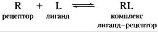
Like enzyme-substrate binding, this binding depends on the concentrations of the interacting components and can be characterized by an Equilibrium Constant:
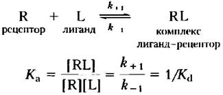
where Ka is the association constant and Kd is the dissociation constant.
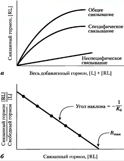
Like enzyme-substrate binding, ligand-receptor binding is saturable. As increasing amounts of ligand are added to a fixed amount of receptor, the number of receptor molecules occupied by the ligand increases (Fig. 1a). A rough estimate of ligand-receptor affinity is provided by the ligand concentration required for half-saturation of the receptor. Using ligand-receptor Scatchard analysis, we can estimate both the dissociation constant Kd and the number of binding sites in a given preparation. When binding reaches equilibrium, the total number of available binding sites Bmax equals the sum of unoccupied sites ([R]) and ligand-occupied sites ([RL]); i.e., Bmax = [R] + [RL]. The number of unoccupied sites can be expressed as the total number of all sites minus the occupied sites: [R] = Bmax — [RL]. The association reaction can now be written as
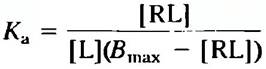
Figure 1. Scatchard analysis of ligand-receptor interaction. A radiolabeled ligand L (such as a hormone) at various concentrations is added to a fixed amount of receptor R, and the fraction of hormone is determined after separating the hormone-receptor complex RL from the free hormone. (a) A plot of [RL] versus [L] + [RL] (the sum of free and bound hormone, i.e., total added hormone) yields a hyperbola that approaches [RL] at receptor saturation. A separate binding series of experiments is necessary to control for non-saturating, nonspecific binding sites (for example, eicosanoid hormones bind nonspecifically to The Lipid Bilayer). A large excess of unlabeled hormone is added along with the dilute radiolabeled ligand solution. Unlabeled molecules compete with labeled ones for specific binding to saturable sites on the receptor, but not for nonspecific binding. To determine specific binding, nonspecific binding is subtracted from total binding. (b) A linear plot of [RL]/[L] versus [RL] yields Kd and Bmax for the receptor-hormone complex. Compare these plots with the V0 versus [S] and 1/V0 versus 1/[S] plots for the enzyme-substrate complex (Fig. 6-12, Box 6-1).
Rearranging the equation gives The ratio of bound to free (unbound) ligand:
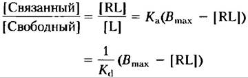
Plotting [bound ligand]/[free ligand] — [free ligand] yields a straight line with a slope of -Kа(-1/Kd) that intercepts the abscissa at a value equal to Vmах (the total number of binding sites) (Fig. 1b). When the ligands are hormones, Kd values typically range from 10-9 to 10-11 M, which indicates very tight binding.
Scatchard analysis is reliable for the simplest cases, but, much like the Lineweaver–Burk plots for enzymes, deviations from linearity occur if the receptor is an allosteric protein.
The sensitivity of receptor systems can change. When a signaling molecule acts continuously (uninterruptedly), desensitization (loss of sensitivity) of the receptor system occurs (Fig. 12-1b); when the stimulus drops below the threshold level, the system regains its sensitivity. Imagine what happens to your visual signaling system when you walk from bright sunlight into a dark room or step out from the darkness into the light.
A final distinctive feature of signal transduction systems is integration (Fig. 12-1d), the ability of the system to receive multiple signals and produce a unified response tailored to the needs of The Cell or Organism. Different signaling pathways "crosstalk" with one another at various levels, generating a complex network of interactions that maintain Homeostasis within the cell and the organism.
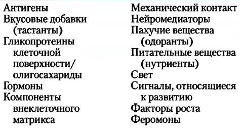
The Study of signal transduction mechanisms has revealed a striking feature: their high degree of evolutionary conservation. Although the total number of distinct biological signals (see Table 12-1) is apparently a few thousand—and the number of potential response types is equally vast—the machinery for transducing all these signals consists of only a dozen or so core protein components. In this chapter, we will examine several Examples of fundamental signal transduction mechanisms and discuss how they are deployed to carry out specific biological functions, such as Nerve Impulse propagation, responses to hormones and growth factors, Vision, Olfaction, gustation, and Cell Cycle control. Often, the end point of a signaling pathway is the phosphorylation of specific target cell proteins, leading to an alteration in their activity and, consequently, A change in cellular behavior. Throughout our Discussion, we will repeatedly emphasize the conservation of these fundamental biosignaling mechanisms and how these core pathways are adapted to a wide array of signaling networks.
Table 12-1. Some Signals to Which Cells Respond
In this chapter, we examine the molecular details of several representative signal transduction systems. While the triggers vary among different systems, the core stages of signal transduction are universal: a signaling molecule interacts with a receptor; the activated receptor engages cellular machinery to generate a secondary signal or alter cellular protein activity; the metabolic activity (broadly defined to include RNA, DNA, and Protein metabolism) of the target cell is modified; and finally, the transduction process concludes, returning the cell to its basal (pre-stimulus) state. To illustrate these common properties of signaling systems, we present six primary signaling mechanisms as examples (Fig. 12-2).
1. G protein-coupled receptors, which indirectly activate enzymes (via GTP-binding proteins, or G proteins) that generate intracellular second messengers. This is exemplified by the β-adrenergic receptor system, which responds to epinephrine (Section 12.2).
2. Receptor Tyrosine Kinases, which are Plasma Membrane Receptors that function directly as enzymes. When one of these receptors is activated by its extracellular ligand, it catalyzes The formation of an intracellular second messenger. Examples include the Insulin receptor (Section 12.3) and the epidermal growth factor receptor (EGF-R).
3. Receptor guanylyl cyclases, which are plasma membrane receptors possessing a cytoplasmic enzymatic domain. The intracellular second messenger generated by these receptors, cyclic guanosine monophosphate (cGMP), activates a cytosolic protein kinase that phosphorylates cellular proteins, thereby altering their activity (Section 12.4).
4. Gated Ion Channels of The Plasma Membrane, which open and close in response to the binding of chemical ligands or changes in transmembrane potential. These channels represent the simplest signal transducers. An example of this mechanism is the acetylcholine ion channel (Section 12.2).
Figure 12-2. Six Major Types of signal transduction mechanisms.
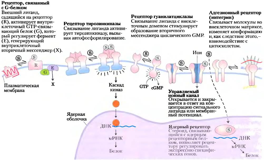
5. Adhesion receptors, which interact with macromolecular Components of the Extracellular matrix (such as Collagen) and Relay instructions to the Cytoskeleton regarding cell migration or attachment to the matrix. Integrins (discussed in Chapter 10) exemplify this general type of signal transduction mechanism.
6. Nuclear receptors (steroid receptors), which, upon binding a specific ligand (such as estrogen), alter the rate at which specific Genes are transcribed and translated into cellular proteins. Because Steroid Hormones function through a mechanism closely tied to the Introduction/30.html">Regulation of Gene Expression, we will cover them only briefly here (Section 12.8), deferring a detailed discussion of their action to Chapter 28.
We now turn to the mechanisms of biosignaling, beginning with a few remarks on the nomenclature of signaling proteins. In most cases, these proteins were discovered while investigating a specific problem and were named accordingly. Later, it became apparent that they participate in a much wider range of biological functions entirely unrelated to their original names. For example, the retinoblastoma protein, pRb, was initially identified as a protein whose mutation contributes to The Development of Cancer of the retina (retinoblastoma). However, we now know that this protein is involved in many essential Cell Division processes across all cell types, not just in retinal cells. Some genes and proteins have been given rather obscure designations: for instance, the tumor suppressor p53 is named for its apparent molecular mass of 53 kDa, yet this name conveys nothing about the protein's critically important role in regulating cell division and cancer development. As we progress through the material, we will clarify the meaning of such
names and provide the terminology commonly used by researchers in the field. Don't be discouraged if a protein's name seems confusing when you first encounter it!
Summary of Section 12.1 Selection/11.html">General features of Signal Transduction
■ All cells possess specific, highly sensitive signal transduction mechanisms that have been conserved throughout evolution.
■ A vast array of diverse stimuli act through specific protein receptors located in the plasma membrane.
■ Receptors bind the signaling molecule, amplify the signal, integrate it with inputs from other receptors, and relay it into the cell. If the signal persists, receptor desensitization dampens or terminates the response.
■ Multicellular organisms utilize six major types of signaling mechanisms: membrane proteins that operate via G proteins; receptor tyrosine kinases; receptor guanylyl cyclases that act through protein kinases; gated ion channels; enzymes; adhesion receptors that convey information between the extracellular matrix and the cytoskeleton; and Nuclear Proteins that bind Steroids and influence gene expression.
Last update: 06/08/2026
Editorial and Educational Adaptation: This material has been compiled based on the primary/original source text. The project team performed an editorial review, corrected technical inaccuracies, structured sections, and adapted the content for an educational format.
What was processed:
- elimination of formatting defects (OCR errors, structural breaks, corrupted characters);
- editorial organization of content;
- standardization of terminology in accordance with academic sources;
- verification of factual statements against the original source text.
All mentions of the author, publication year, and origin of the primary text have been preserved in accordance with the source.