LEHNINGER PRINCIPLES OF BIOCHEMISTRY - VOL. 2. BIOENERGETICS AND METABOLISM - 2014
CHAPTER II. BIOENERGETICS AND METABOLISM
15. PRINCIPLES OF METABOLIC REGULATION
15.4. Glycogen Metabolism in Animals
Continuing our Structure/133.html">Discussion of Carbohydrate METABOLISM, we now focus on the Synthesis and Breakdown of Glycogen. In this section, we examine the reactions of this metabolic pathway, and in Section 15.5 we turn to the mechanisms of its regulation.
In vertebrates and many microorganisms, excess glucose is stored as high-molecular-weight glycogen, whereas in plants it is stored as starch. In vertebrates, glycogen is deposited primarily in The Liver and Muscles, where it can account for up to 10% of the liver's mass and 1–2% of Muscle tissue mass. If an equivalent amount of glucose were dissolved in the Cytosol as free monomer, its concentration would reach 0.4 M, which would severely disrupt The Cell's osmotic balance. However, the concentration of polymeric glycogen molecules at this same glucose mass is only about 0.01 µM. Glycogen is stored in the form of large granules. A single glycogen granule (β-particle) has a diameter of about 21 nm, consists of roughly 55,000 glucose residues, and contains approximately 2,000 nonreducing ends. From 20 to 40 such particles aggregate to form α-rosettes, which are readily visible under a Microscope in tissue samples from well-fed animals (Fig. 15-24) but disappear after 24 hours of fasting.
Class="center">Fig. 15-24. Glycogen granules in a hepatocyte. Glycogen is the primary form of carbohydrate storage in Cells, particularly in hepatocytes. It forms electron-dense structures, often appearing as aggregates or rosettes. In hepatocytes, glycogen is closely associated with the tubules of the smooth Endoplasmic reticulum. Numerous Mitochondria are also visible in the micrograph. Glycogen is deposited in Regions of the smooth endoplasmic reticulum rich in vesicles and tubules.

Muscle glycogen serves as a readily available energy source for both aerobic and Anaerobic Metabolism. During intense physical exertion, these reserves can be depleted in less than an hour. Liver glycogen acts as a source of glucose for other Tissues when dietary glucose is unavailable (between meals or during fasting); this is especially crucial for Brain Neurons, which cannot use Fatty acids as an energy source. Liver glycogen stores can be depleted within 12 to 24 hours. In humans, the total amount of energy stored as glycogen is much smaller than that stored as fats (triacylglycerols; see Table 23-5); however, mammals are incapable of converting fats into glucose or breaking them down under anaerobic conditions.
Glycogen granules are complex aggregates composed of glycogen, the Enzymes that synthesize and degrade it, and a specialized apparatus for regulating these enzymes. Although the mechanisms of glycogen storage and utilization are shared between muscle and liver, the tissue-specific enzymes differ slightly to reflect the distinct physiological roles of glycogen in these two tissues. Dietary glycogen is also ingested and broken down in the intestine; this process involves a different set of hydrolytic enzymes that convert glycogen (and starch) into glucose. Dietary starch is degraded in a similar manner. We begin by examining The breakdown of glycogen to glucose-1-phosphate (Glycogenolysis), followed by the pathways of glycogen synthesis (Glycogenesis).
Glycogen breakdown is catalyzed by Glycogen phosphorylase
In muscle and liver, glucose units from the outer branches of glycogen enter Glycolysis through the sequential action of three enzymes: glycogen phosphorylase, a debranching enzyme, and phosphoglucomutase. Glycogen Phosphorylase catalyzes the attack of inorganic phosphate Pi on the (α1 → 4) glycosidic bond between two glucose residues at a nonreducing end of glycogen, releasing the terminal glucose residue as α-D-glucose-1-phosphate (Fig. 15-25). This phosphorolysis reaction differs from the hydrolytic Cleavage of glycosidic bonds by amylase during the Digestion of dietary glycogen and starch. In phosphorolysis, a portion of the glycosidic bond energy is preserved in the form of the sugar ester glucose-1-phosphate (see Section 14-2).
Fig. 15-25. Removal of the terminal glucose residue from the nonreducing end of a glycogen chain by glycogen phosphorylase. This process repeats continuously; the enzyme successively removes glucose residues until it stops four residues away from a branch point (see Fig. 15-26).

The reaction catalyzed by glycogen phosphorylase requires the cofactor Pyridoxal phosphate, whose phosphate group acts as a general acid, catalyzing the attack of Pi on the glycosidic bond. Notably, this catalytic role is unusual for this cofactor, which much more frequently Functions in Amino acid metabolism (see Fig. 18-6).
Glycogen phosphorylase acts repeatedly at the nonreducing ends of the glycogen molecule until it reaches a branch point with an α1 → 6 configuration (see Fig. 7-14); the enzyme's action halts four glucose residues away from this point. Further degradation of glycogen by glycogen phosphorylase can only proceed after the intervention of the debranching enzyme. This enzyme, which possesses both 4-α-D-glucanotransferase and amylo-1,6-glucosidase activities, sequentially catalyzes The transfer of a three-residue side-chain segment and the Cleavage of the final glucose residue (Fig. 15-26); after this, glycogen phosphorylase can resume its activity.
Fig. 15-26. Degradation of glycogen near an (α1 → 6) branch point. After the removal of outer glucose residues by glycogen phosphorylase (see Fig. 15-25), the residues at the branch point are cleaved by a specialized bifunctional enzyme. The transferase activity of this enzyme shifts a block of three glucose residues from the branch to a nearby nonreducing end, forming a new (α1 → 4) bond. The remaining single glucose residue, attached by an (α1 → 6) bond, is then hydrolyzed as free glucose by the 1,6-glucosidase activity of the enzyme. Glucose residues are depicted here as pyranose rings without explicit -H, -OH, and -CH2OH groups.

Glucose-1-phosphate can enter glycolysis or, via hepatic conversion, replenish Blood glucose levels
The end product of the glycogen phosphorylase reaction, glucose-1-phosphate, is converted to glucose-6-phosphate by phosphoglucomutase:
Glucose-1-phosphate ⇄ glucose-6-phosphate
The enzyme, which is initially phosphorylated on a Ser residue, transfers its phosphoryl group to the C-6 hydroxyl of the substrate and subsequently accepts the phosphoryl group from C-1 (Fig. 15-27).
Fig. 15-27. The reaction catalyzed by phosphoglucomutase. At THE START OF the reaction, the enzyme is phosphorylated on a Ser residue. In step (1), the enzyme transfers its phosphoryl group (highlighted in green) to glucose-1-phosphate, yielding glucose-1,6-bisphosphate. In step (2), the phosphoryl group at the C-1 position of glucose-1,6-bisphosphate (highlighted in red) is transferred back to the enzyme, regenerating the phosphoenzyme and producing glucose-6-phosphate.

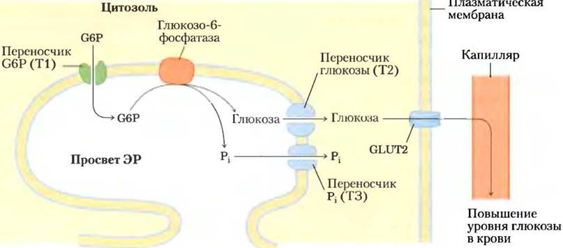
Glucose-6-phosphate derived from muscle glycogen can enter glycolysis to provide energy for Muscle contraction. In the liver, glycogen breakdown serves a different purpose: maintaining blood glucose levels, such as between meals. This process relies on glucose-6-phosphatase, an enzyme found exclusively in the liver and Kidneys. This enzyme is an integral membrane protein of The endoplasmic reticulum (ER), believed to contain nine transmembrane helices, with its Active Site facing the lumen of the ER. Glucose-6-phosphate generated in the cytosol is transported into the ER lumen by a specialized transport protein, T1 (Fig. 15-28), and is hydrolyzed by glucose-6-phosphatase on the luminal surface of the ER membrane. The resulting Pi and glucose molecules are presumably transported back into the cytosol by two other carrier Proteins (T2 and T3), after which glucose leaves the hepatocyte via another transporter, GLUT2, located in The Plasma Membrane. Note that by orienting the Active Site of glucose-6-phosphatase toward the ER lumen, the cell successfully compartmentalizes this reaction away from glycolysis, which occurs in the cytosol and could otherwise be futilely disrupted by glucose-6-phosphatase activity. Genetic Defects in either glucose-6-phosphatase or T1 lead to severe Metabolic Disorders manifested as glycogen storage disease type Ia (Box 15-4).
Fig. 15-28. Hydrolysis of glucose-6-phosphate (G6P) by glucose-6-phosphatase in the endoplasmic reticulum (ER). The catalytic site of glucose-6-phosphatase faces the ER lumen. The glucose-6-phosphate transporter T1 transports G6P from the cytosol into the ER lumen, whereas transporters T2 and T3 transport the resulting glucose and Pi back into the cytosol. Glucose leaves the cell via the specialized transporter GLUT2 located in the plasma membrane.
Because muscle and adipose tissue lack glucose-6-phosphatase, these tissues cannot convert the glucose-6-phosphate derived from glycogen breakdown into free glucose; consequently, they do not contribute to maintaining blood glucose levels.
The sugar nucleotide UDP-glucose participates in glycogen synthesis
Many Reactions Involving the transformation or polymerization of hexoses utilize sugar NUCLEOTIDES—compounds in which the anomeric carbon of the sugar is activated by attachment to a nucleotide via an ester linkage. These nucleotide sugars serve as substrates for the polymerization of Monosaccharides into Disaccharides, glycogen, starch, Cellulose, and more complex extracellular Polysaccharides. They are also important intermediates in The formation of amino hexoses and deoxy hexoses found in some of these polysaccharides, as well as in the synthesis of Vitamin C (L-ascorbic acid). The Argentine biochemist Luis Leloir was the first to recognize the involvement of sugar nucleotides in The Biosynthesis of glycogen and many other carbohydrate derivatives in 1953.
Luis Leloir, 1906–1987


Box 15–4. Carl and Gerty Cori: Pioneers in Glycogen Metabolism and Related Disorders
Much of what is written in modern biochemistry textbooks about glycogen metabolism was discovered between 1925 and 1950 by the remarkable research team of Carl F. Cori and Gerty T. Cori. Both received their medical training in Europe at the end of World War I (Gerty Cori completed her preparatory studies and medical school in just one year!). The couple left Europe in 1922 and established research laboratories in the United States: they spent their first nine years in Buffalo, New York—now the Roswell Park Memorial Institute—and then, from 1931 until the end of their days, at Washington University in St. Louis.
In their early physiological studies on the origin and transformations of glycogen in animal tissues, the Coris demonstrated that glycogen in muscle is converted into lactate, which is then transported via the blood to the liver and resynthesized into glycogen; this pathway later became known as the Cori cycle (see Fig. 23-20). In subsequent biochemical studies, they showed that glycogen degradation is initiated by a phosphorylation reaction catalyzed by an enzyme they discovered, which was named glycogen phosphorylase. They demonstrated that the reaction product, glucose-1-phosphate ("Cori ester"), can be reused for glycogen resynthesis via the reverse pathway. Although this discovery did not prove that intracellular glycogen is synthesized precisely by this route, it was nevertheless the first demonstration of macromolecular synthesis from monomeric subunits in vitro, prompting other scientists to search for enzymes with polymerase activity. Arthur Kornberg, who worked in the Cori laboratory and discovered the first DNA polymerase, remarked: "It was glycogen phosphorylase, not base pairing, that led me to the Discovery of DNA polymerase."
Table 1. Human Glycogen Storage Diseases (glycogenoses)
Glycogen storage disease type (disease name) |
Defective enzyme |
Affected organ |
Symptoms |
Type 0 |
Glycogen synthase |
Liver |
Low blood glucose, high Ketone Bodies, early death |
Type Ia (von Gierke disease) |
Glucose-6-phosphatase |
Liver |
Hepatomegaly, renal failure |
Type Ib |
Microsomal glucose-6-phosphate translocase |
Liver |
Same as Ia; susceptibility to bacterial infections |
Type Ic |
Microsomal Pi transporter |
Liver |
Same as Ia |
Type II (Pompe disease) |
Lysosomal glucosidase |
Skeletal and cardiac muscle |
Infantile form: death before age 2; juvenile form: myopathy; adult form: resembles muscular dystrophy |
Type IIIa (Forbes disease or Cori disease) |
4-α-D-glucanotransferase and/or amylo-1,6-glucosidase |
Liver, skeletal and cardiac muscle |
Hepatomegaly in infants; myopathy |
Type IIIb |
Hepatic 4-α-D-glucanotransferase and/or amylo-1,6-glucosidase (muscle enzyme normal) |
Liver |
Hepatomegaly in infants |
Type IV (Andersen disease) |
Branching enzyme (amylo-(1,4→1,6)-transglycosylase) |
Liver, Skeletal Muscle |
Hepatosplenomegaly, myoglobinuria |
Type V (McArdle disease) |
Muscle phosphorylase |
Skeletal muscle |
Cramps and pain with physical exertion; myoglobinuria |
Type VI (Hers disease) |
Liver phosphorylase |
Liver |
Hepatomegaly |
Type VII (Tarui disease) |
Muscle PFK-1 |
Muscle, erythrocytes |
Same as V; hemolytic anemia |
Type VIb, VIII, or IX |
Phosphorylase kinase |
Liver, leukocytes, muscle |
Hepatomegaly |
Type XI (Fanconi-Bickel syndrome) * |
Glucose transporter (GLUT2) |
Liver |
Growth retardation, Rickets, hepatomegaly, Renal Dysfunction |
Gerty Cori became interested in human Genetic Disorders characterized by the abnormal accumulation of glycogen in the liver. In several cases, she successfully identified the BIOCHEMICAL BASIS OF the defect and showed that diseases could be diagnosed by assaying glycogen-metabolizing enzymes in small tissue biopsy samples. Table 1 summarizes our current knowledge of 13 such disorders. ■
Carl and Gerty Cori shared the 1947 Nobel Prize in Physiology or Medicine with Bernardo Houssay of Argentina, who was awarded for his research on the hormonal REGULATION OF CARBOHYDRATE Metabolism. During the 1940s and 1950s, the Cori laboratory in St. Louis served as an international hub for biochemical research. Six scientists who trained in the Cori laboratory went on to win Nobel Prizes: Arthur Kornberg (1959) for discovering The Mechanism of DNA Synthesis, Severo Ochoa (1959) for discovering the mechanism of RNA Synthesis, Luis Leloir (1970) for elucidating The Role of sugar nucleotides in Polysaccharide Biosynthesis, Earl Sutherland (1971) for discovering cAMP and establishing its role in Metabolic Regulation, Christian de Duve (1974) for discoveries concerning the Structural and functional Organization OF THE cell, and Edwin Krebs (1991) for the discovery of phosphorylase kinase.
The Coris in Gerty Cori's laboratory, ca. 1947.

The involvement of sugar nucleotides in biosynthetic reactions is supported by several key considerations outlined below.
1. The formation of these compounds is irreversible, which helps drive the Metabolic Pathways in which they participate. The Condensation reaction of a nucleoside triphosphate with a hexose-1-phosphate has a small positive standard free-energy change, but it releases inorganic pyrophosphate (PPi), which is rapidly hydrolyzed by inorganic pyrophosphatase in an exergonic reaction
(ΔG′° = -19.2 kJ/mol; Fig. 15-29). As a result, the intracellular concentration of PPi remains low, making the overall standard free-energy change for the process favorable. As is characteristic of many biological polymerization reactions, the rapid removal of a product—in this case driven by the large negative free-energy change of PPi hydrolysis—pulls the entire synthetic pathway forward.
Fig. 15-29. Formation of nucleoside diphosphate sugars. A sugar phosphate and a nucleoside triphosphate (NTP) undergo a condensation reaction. The negatively charged oxygen of the sugar phosphate acts as a nucleophile, attacking the α-phosphate of the nucleoside triphosphate and displacing pyrophosphate. The reaction equilibrium lies far to the right because the resulting PPi is rapidly hydrolyzed by inorganic pyrophosphatase.

2. Although the nucleoside portion of a sugar nucleotide does not directly participate in the chemical reaction, various Functional groups of the nucleoside can engage in noncovalent interactions with the enzyme. The additional binding Free energy thus contributed can significantly enhance the catalytic efficiency of the enzyme (Chapter 6; see also Vol. 1, p. 425).
3. Like phosphate, nucleotides (such as UMP or AMP) are excellent leaving groups that facilitate nucleophilic attack by activating the specific carbon atom of the sugar moiety to which they are attached.
4. By "tagging" specific hexose molecules with nucleotide groups, the cell can earmark them for specialized pathways (such as glycogen synthesis), effectively separating them from the pool of hexose phosphates directed toward other metabolic fates (such as glycolysis).
Glycogen synthesis occurs in virtually all body tissues, but it is particularly intensive in the liver and skeletal muscles. The starting point for synthesis is glucose-6-phosphate. As we have already shown, this compound can be formed from free glucose through the action of Isoenzymes—hexokinase I and hexokinase II in muscles, or hexokinase IV (glucokinase) in the liver:
D-Glucose + ATP —> D-glucose-6-phosphate + ADP
However, some of the glucose entering the body with food is channeled into glycolysis via a more complex pathway. It is first taken up by erythrocytes and converted into lactate during glycolysis; the lactate then reaches the liver, where it is converted back into glucose-6-phosphate via Gluconeogenesis.
In The First stage of glycogen synthesis, glucose-6-phosphate is converted into glucose-1-phosphate by the action of phosphoglucomutase:
Glucose-6-phosphate ⇄ glucose-1-phosphate
A key moment in glycogen synthesis is The conversion of the reaction product into UDP-glucose using the enzyme UDP-glucose pyrophosphorylase:
Glucose-1-phosphate + UTP —> UDP-glucose + PPi
Note that the name of this enzyme is derived from the reverse reaction. Within the cell, the reaction proceeds in the direction of UDP-glucose formation because the released pyrophosphate is rapidly hydrolyzed by inorganic pyrophosphatase (Fig. 15-29).
UDP-glucose acts as a direct donor of glucose residues in the reaction catalyzed by glycogen synthase, which facilitates the transfer of a glucose residue from UDP-glucose to the non-reducing end of a branched glycogen molecule (Fig. 15-30). The overall equilibrium for the incorporation of glucose-6-phosphate into the growing glycogen chain is strongly shifted toward synthesis.
Fig. 15-30. Glycogen synthesis. Elongation of the glycogen chain proceeds via glycogen synthase. The enzyme transfers a glucose residue from UDP-glucose to the non-reducing end of the glycogen chain (see Fig. 7-14), forming a new (α1 —> 4) bond.

Glycogen synthase cannot form (α1 —> 6) bonds found at the branching points of glycogen molecules. These bonds are created by a special glycogen-branching enzyme, 1,4 —> 1,6-transglycosylase (glucosyl-4,6-transferase). This enzyme catalyzes the transfer of a terminal segment of 6-7 glucose residues from the non-reducing end of a glycogen chain (consisting of at least 11 residues) to the OH group of the C-6 carbon atom of a glucose residue located closer to the beginning of the same or another glycogen chain, thereby creating a new branch (Fig. 15-31). Further elongation of this fragment is carried out by glycogen synthase. The biological "purpose" of synthesizing a branched polymer is to improve its solubility and generate the maximum number of non-reducing ends. This maximizes the number of sites accessible to glycogen phosphorylase and glycogen synthase, since both enzymes act exclusively on the non-reducing ends of the chain.
Fig. 15-31. Formation of branches in the glycogen chain. The glycogen-branching enzyme 1,4 —> 1,6-transglycosylase (or glucosyl-4,6-transferase) creates new branch points during glycogen synthesis.

Glycogenin ensures the attachment of the first sugar residues during glycogen synthesis
Glycogen synthase cannot initiate glycogen chain synthesis de novo. This requires a primer, which is typically a chain fragment of at least eight glucose residues linked by (α1 —> 4) bonds. How are the first glucose residues attached in the glycogen molecule? The remarkable protein glycogenin (Fig. 15-32) functions simultaneously as the primer on which the new chain is assembled and as The enzyme catalyzing the assembly process. The first step in synthesizing a new glycogen molecule involves the transfer of a glucose residue from UDP-glucose to the hydroxyl group of Tyr194 in glycogenin via the glucosyltransferase activity of the enzyme (Fig. 15-33). The nascent chain is elongated by The addition of seven more glucose residues, each supplied by UDP-glucose. Glycogenin again acts as the catalyst for these reactions. Next, glycogen synthase joins the process to further elongate the glycogen chain. Covalently bound to a single reducing end of the glycogen molecule, glycogenin remains inside the resulting glycogen granule (Fig. 15-33, b).
Fig. 15-32. Structure of glycogenin (PDB ID 1772). Muscle glycogenin (Mr = 37,000 Da) exists as a dimer in solution. Human liver contains another protein isoform, glycogenin-2. The substrate UDP-glucose (shown as a red ball-and-stick structure) is bound to the Rossmann fold in the N-terminal region of the protein at a distance from the Tyr194 residue (turquoise): 15 Å from this residue in its "own" monomeric unit and 12 Å from it In the second molecule of the dimer. Each UDP-glucose molecule is linked via a phosphate group to an Mn2+ ion (green), which plays a crucial role in catalysis. It is believed that Mn2+ acts as an electron pair acceptor (Lewis acid), stabilizing the leaving group (UDP). In the reaction product, the glycosidic bond has the same configuration at the C-1 atom as in UDP-glucose; presumably, glucose transfer from UDP to Tyr194 occurs in two stages: first, the Asp162 residue (orange) performs a nucleophilic attack leading to an intermediate with inverted configuration, and a subsequent nucleophilic attack by the Tyr194 residue restores the original configuration.

Fig. 15-33. Glycogenin and The structure of a glycogen particle. a — Glycogenin catalyzes two different reactions. First, the hydroxyl group of the Tyr194 residue attacks the C-1 atom of glucose in a UDP-glucose molecule, resulting in the glycosylation of the Tyr residue. Next, the hydroxyl group at C-4 of the terminal glucose residue attacks C-1 of another UDP-glucose molecule; this reaction sequence repeats until a glycogen segment of eight glucose residues linked by (α1—> 4) bonds is formed. b — Structure of a glycogen particle. Glycogen chains (12-14 residues long) radiate outward in layers from the centrally located glycogenin molecule. Inner chains have two branch points with (α1 —> 6) configuration. The chains forming the outer layer are unbranched. A mature glycogen particle contains 12 layers (only five are shown here) comprising about 55,000 glucose residues; the molecular diameter is approximately 21 nm, and the molecular weight is ~107.

Summary of Section 15.4 Glycogen Metabolism in Animal Cells
■ Glycogen is stored in muscles and the liver as large granules. These granules also contain enzymes involved in glycogen metabolism, as well as regulatory enzymes.
■ Glycogen phosphorylase catalyzes the phosphorolysis of the glycogen chain starting from the non-reducing end of the molecule, yielding glucose-1-phosphate. A specialized debranching enzyme transfers branch residues to the main chain and releases the final residue attached via an (α1 —> 6) bond as a free glucose molecule.
■ Phosphoglucomutase mediates the interconversion of glucose-1-phosphate and glucose-6-phosphate. Glucose-6-phosphate can then enter glycolysis or (in the liver) be converted to free glucose in the endoplasmic reticulum by glucose-6-phosphatase before being released into the bloodstream.
■ The sugar-nucleotide derivative UDP-glucose provides the glucose residue for glycogen synthesis via glycogen synthase; residues are added to the non-reducing end of the chain. A specialized glycogen-branching enzyme creates (α1 —> 6) bonds at branch points.
■ The formation of a new glycogen particle begins with the creation of a glycosidic bond between a glucose residue from UDP-glucose and a Tyr residue in the protein glycogenin. Next, a short primer fragment is formed from several glucose residues for further glycogen synthesis mediated by glycogen synthase. Both Stages of the process are catalyzed by glycogenin itself.
Last update: 06/08/2026
Editorial and Educational Adaptation: This material has been compiled based on the primary/original source text. The project team performed an editorial review, corrected technical inaccuracies, structured sections, and adapted the content for an educational format.
What was processed:
- elimination of formatting defects (OCR errors, structural breaks, corrupted characters);
- editorial organization of content;
- standardization of terminology in accordance with academic sources;
- verification of factual statements against the original source text.
All mentions of the author, publication year, and origin of the primary text have been preserved in accordance with the source.