Biochemistry and Molecular Biology - Belyasova N.A. 2002
Metabolism. Processes Requiring Energy Input
Biosynthesis Features. Carbohydrate Biosynthesis
Polysaccharide Biosynthesis
Polysaccharides are synthesized from Monosaccharides following a set of consistent patterns. First, monosaccharide units are initially activated through phosphorylation catalyzed by a kinase. The monosaccharide unit is then accepted by a nucleoside diphosphate, and the resulting sugar nucleotide acts as a carrier of the glycosyl residue (the nucleotide "handle") to the elongating polysaccharide chain.
Glycogen Biosynthesis. Both free glucose and glucose-6-phosphate can serve as precursors for glycogen biosynthesis. In the first case, glucose is phosphorylated by a kinase to form glucose-1-phosphate; In the second case, glucose-6-phosphate is isomerized to glucose-1-phosphate with the participation of phosphoglucomutase (Fig. 14.3).
The next stage (The formation of the sugar nucleotide) is catalyzed by pyrophosphorylase. In higher animals, glycosyl residues are transferred to uridine diphosphate (UDP), whereas in plant and microbial Cells, this role is performed by ADP, CDP, or GDP. The diagrams (Figs. 14.3, 14.4) illustrate The process of glycogen synthesis in animals, where the pyrophosphorylase is referred to as glucose-1-phosphate uridylyltransferase.
At the next step, the glycosyl group of UDP-glucose is transferred to the terminal part of the glycogen chain (at the non-reducing end), forming a new a(1→4)-glycosidic bond between the first carbon atom of the added glucose residue and the hydroxyl oxygen at the fourth carbon atom of the terminal glucose residue of the chain. This reaction is catalyzed by glycogen synthase. This enzyme drives the gradual elongation of linear glycogen chains, though it cannot form the branching points characteristic of glycogen molecules (Fig. 14.3). The function of forming glycogen "branches" is catalyzed by another enzyme, transglycosylase.
Once the linear glycogen chain reaches a length of approximately 10 glucose residues, transglycosylase attacks a glycosidic bond at some point along the chain and transfers the terminal oligosaccharide fragment, containing 6–7 glycosyl units, to the free 6-hydroxyl group of a glucose residue on the same or another chain. This catalyzes the formation of an a(1→6)-glycosidic bond, creating a branching point in the glycogen molecule (Fig. 14.4). Starch synthesis in plant cells occurs in a similar manner.
Both the biosynthesis and breakdown of glycogen in higher organisms are regulated by Hormones. Specifically, adrenaline and Glucagon play an active role in these processes in animals. As their concentration in the Blood increases, The amount of glycogen decreases, leading to a corresponding increase in glucose levels.
Murein biosynthesis. Unlike glycogen, whose biosynthesis occurs inside The Cell, murein—as a component of the Introduction/37.html">Bacterial Cell wall—is synthesized through several stages that take place outside the cell, on the outer surface of The Plasma Membrane. Consequently, the cell is forced to transport individual Structural components of murein across the membrane, for which a specialized mechanism exists.
The direct precursor for murein biosynthesis is N-acetylglucosamine, and the Initial Stages of biosynthesis follow the patterns already described, including precursor phosphorylation and its transfer to a UDP residue (Fig. 14.5). These reactions yield UDP-N-acetylglucosamine (UDP-NAG).
Class="center">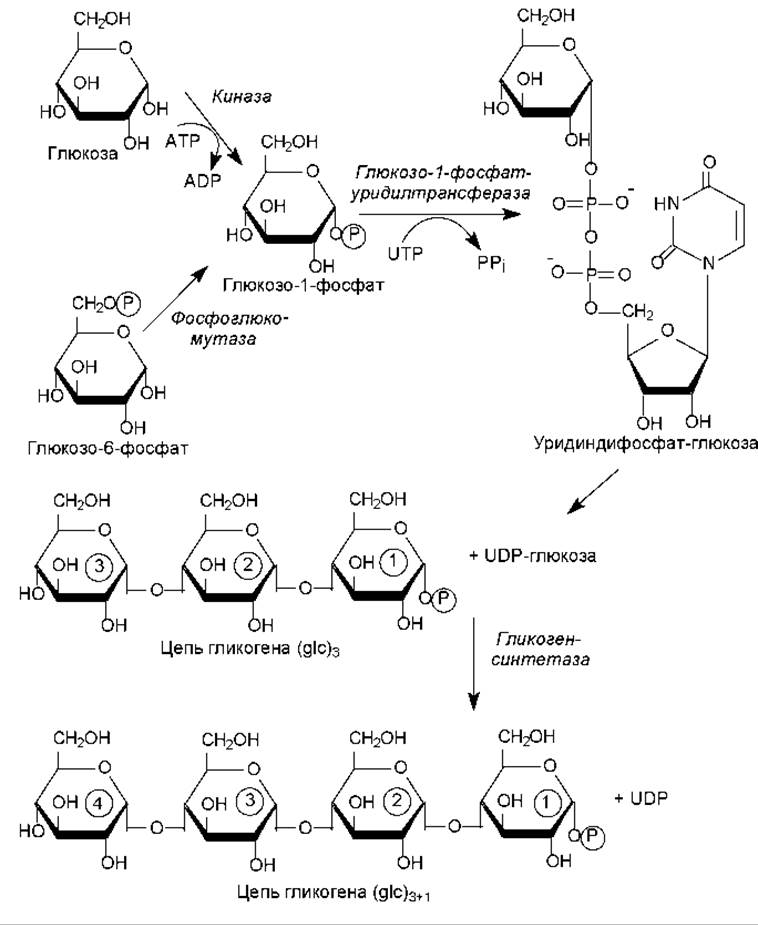
Fig. 14.3. Biosynthesis of linear glycogen chains
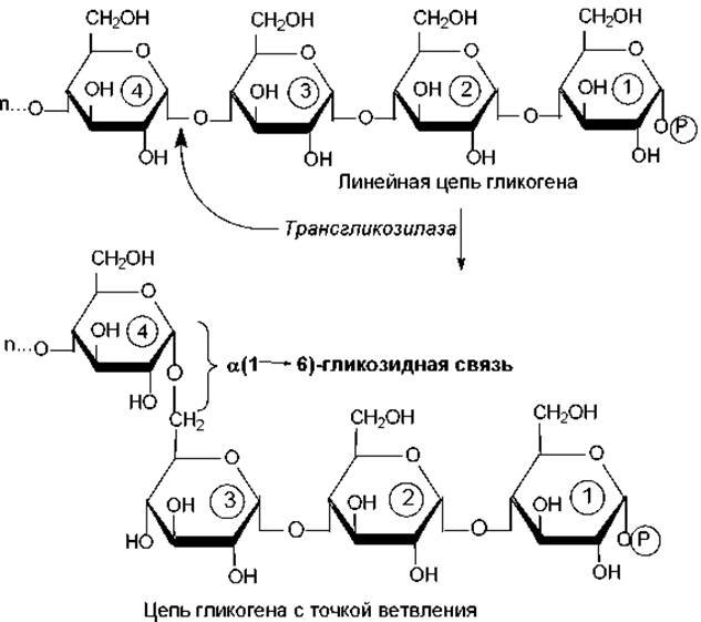
Fig. 14.4. Formation of glycogen branch points
In the next step, UDP-NAG is transformed into the second component of this heteropolymer: UDP-N-acetylmuramic acid (UDP-NAM). To achieve this, a lactic acid residue is attached to the UDP-NAG molecule via interaction with phosphoenolpyruvate (PEP). Subsequently, five amino acid residues are sequentially added to UDP-NAM, forming UDP-N-acetylmuramyl pentapeptide (Fig. 14.5).
The pentapeptide chain within UDP-N-acetylmuramyl pentapeptide is assembled in an unusual way: peptide bonds are formed here using ATP energy with the participation of specialized Enzymes. Such a peptide (containing D-Amino Acids and a y-peptide bond) cannot be synthesized via the standard ribosomal pathway.
The formed N-acetylmuramyl pentapeptide is transferred by an enzyme from UDP to a membrane-bound lipid carrier—a hydrophobic molecule capable of shuttle movements across the membrane. Next, NAG (donated by the previously formed UDP-NAG) is attached to the NAM-peptide unit linked to the lipid carrier, forming a disaccharide unit. The Structure of the P(1→4)-glycosidic bond between the monosaccharide units is shown in Fig. 5.4. Finally, a pentaglycine bridge is built onto the ε-amino group of the Lysine residue in the pentapeptide (Fig. 14.6).
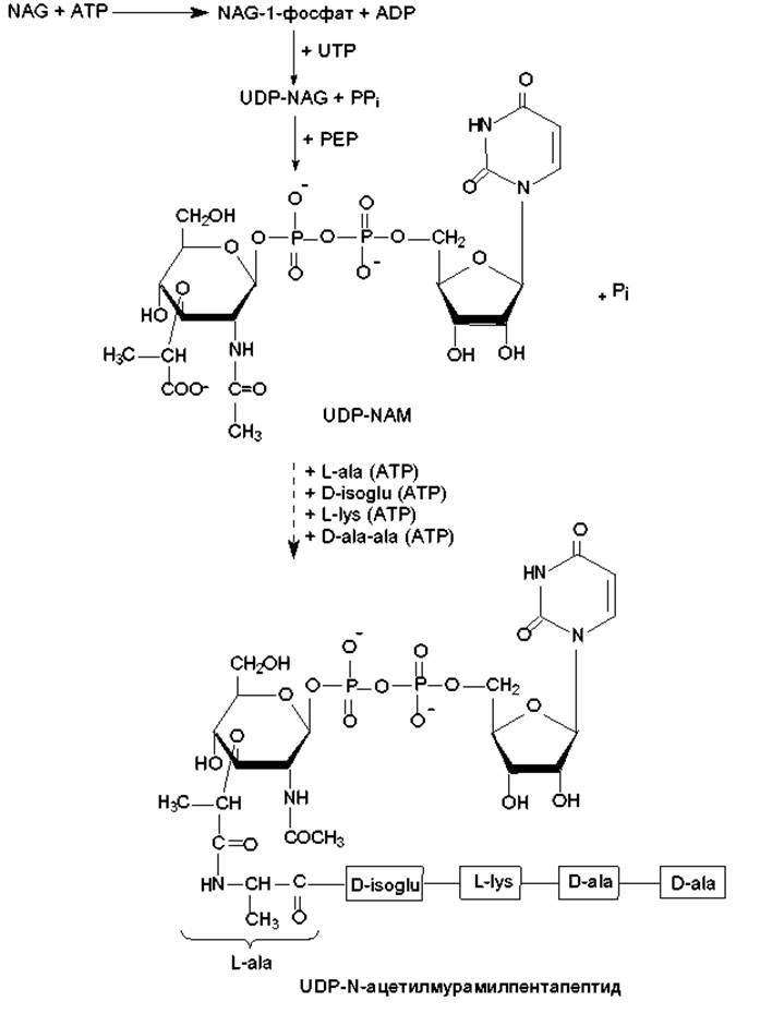
Fig. 14.5. Murein biosynthesis. Stages leading to the formation of UDP-N-acetylmuramyl pentapeptide (details in text).
The resulting disaccharide-peptide unit is transferred from the lipid carrier to the growing polysaccharide chain (Fig. 14.6). At The final stage, individual polysaccharide chains are cross-linked by pentaglycine bridges (a transpeptidation reaction catalyzed by the enzyme glycopeptide transpeptidase). During this reaction, the terminal amino group of the pentaglycine bridge attacks the peptide bond between two D-Alanine residues in the pentapeptide of another disaccharide unit. This creates a peptide bond between the terminal Glycine residue and the D-ala residue, while the second (terminal) D-ala residue is released. This reaction takes place entirely outside the cell and does not require ATP, as it is driven by the energy of the hydrolyzed D-ala—D-ala bond. The resulting structure is illustrated in Fig. 5.6.
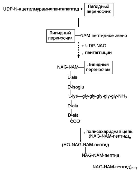
Fig. 14.6. Murein biosynthesis. Formation of the disaccharide-peptide unit and chain elongation (details in text)
The murein biosynthesis process is subject to the inhibitory Action of Certain Antibiotics. Specifically, ß-lactam antibiotics covalently bind to the Active Site of glycopeptide transpeptidase, causing its irreversible inhibition (Chapter 18). In this case, the final stage of biosynthesis—the formation of cross-links between tetrapeptides—cannot take place. Bacitracin blocks the regeneration of the lipid carrier, thereby halting cell wall synthesis.
Most exopolysaccharides are synthesized following the patterns described for murein biosynthesis: participation of nucleoside diphaccharides, Lipid carriers, and the assembly of polysaccharide molecules from disaccharide units on the cell surface with the aid of extracellular enzymes.
Last update: 06/08/2026
Editorial and Educational Adaptation: This material has been compiled based on the primary/original source text. The project team performed an editorial review, corrected technical inaccuracies, structured sections, and adapted the content for an educational format.
What was processed:
- elimination of formatting defects (OCR errors, structural breaks, corrupted characters);
- editorial organization of content;
- standardization of terminology in accordance with academic sources;
- verification of factual statements against the original source text.
All mentions of the author, publication year, and origin of the primary text have been preserved in accordance with the source.