ECOLOGICAL BIOCHEMISTRY - Study Guide - V. M. Isaienko 2005
Chapter 6. ECOLOGICAL AND BIOCHEMICAL INTERACTIONS AMONG ANIMALS
6.3. Interactions between animals of different species
6.3.1. Allomone poisons as a chemical means of defense and predation
As noted previously, the substances involved in these interactions can act either as allomones or kairomones.
The defensive action of poisonous substances produced by animals (esotoxins), as well as their role in predation, is realized through A wide variety of mechanisms—repellent (deterrent), nociceptive (pain-inducing), paralytic (immobilizing), and others.
All venomous and poisonous animals can be divided into two groups: primarily poisonous and secondarily poisonous (Fig. 6.9).
Class="center">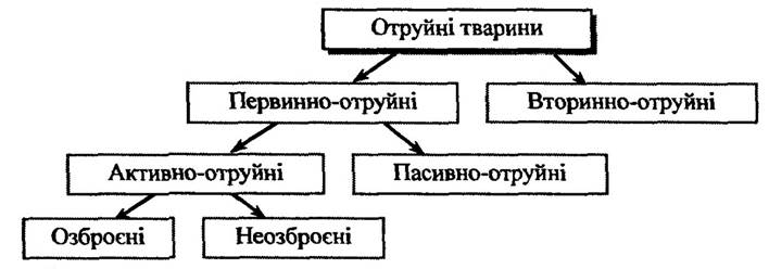
Fig. 6.9. Classification of poisonous animals
Primarily poisonous animals include those that synthesize venom in specialized glands or contain toxic metabolic products. Based on how the poison is produced and utilized, these animals are subdivided into actively poisonous and passively poisonous.
Actively poisonous animals equipped with a specialized venom apparatus for inflicting injury are referred to as armed. A typical apparatus of this type—consisting of a venom gland with an excretory duct and an offending Structure—includes the venomous fangs of snakes, the stingers of insects, and the spines and stingers of fish. The secretion of the venom glands is injected into the victim's body parenterally, that is, outside the digestive tract.
Another group of actively poisonous animals consists of those whose venom apparatus lacks a specialized wounding structure; these are unarmed actively poisonous animals. Examples include the dermal poison glands of amphibians, the anal glands of insects, and others. The toxic secretions of such glands produce a toxic effect upon contact with the integument of the victim.
In passively poisonous animals (such as fish, Mollusks, and insects), the toxin is synthesized within the Organism and accumulated in various Organs and Tissues.
A major category of poisonous animals comprises secondarily poisonous organisms, which accumulate exogenous toxins and exhibit toxicity only when ingested. Examples include fish and mollusks that accumulate cyanobacterial toxins in their bodies, or insects that feed on plants poisonous to other animals. As a rule, secondary toxicity does not provide individual protection, but at the cost of the death of individual specimens, the survival chances of Selection/30.html">The population as a whole are increased.
The route by which toxins enter the victim's body is of great significance. Typically, protein-based toxins (from snakes, arachnids, etc.) are injected parenterally using an armed apparatus, as many of them are degraded by the Enzymes of the gastrointestinal tract. At the same time, non-protein toxins are effective when introduced orally (such as amphibian Alkaloids and toxins from certain species of fish and mollusks). Some animals defend themselves by spraying their toxins in the form of an aerosol (e.g., the bombardier beetle). The effectiveness of such toxins depends on the condition of the victim's integument and the local concentration of the toxic substance.
Once inside the organism, a toxin is distributed unevenly, as its distribution is influenced by various barriers—capillary walls, Cell membranes, Blood-Brain and placental barriers, and others. From the site of inoculation, the toxins enter the BLOOD AND LYMPH. Most animal toxins undergo biotransformation within the organism, which largely accounts for the natural resistance of certain animals to specific venoms.
The Liver and Kidneys bear the primary burden of detoxification and elimination of toxins from the body. Consequently, these organs are typically damaged during poisoning. To some extent, toxic substances and their metabolites may also be excreted through other pathways, such as via the Skin or in breast milk.
The issue of how animals remain resistant to their own toxins is of considerable importance. Several key mechanisms ensure the resistance of such organisms to these venoms. Perhaps the most effective is the localization of the toxin in specialized organs (glands), the walls of which prevent the poison from spreading throughout the body. The glandular Cells are insensitive to the action of the toxins, making them inherently resistant. Cytolytic Components of the venoms (mainly enzymes) remain inactive due to the presence of specific inhibitors. Upon entering the victim's body, the venom is diluted by Body Fluids, the concentration of inhibitors drops, and the enzyme
constituting the venom is activated. Glands that produce protein toxins generally contain proteinase inhibitors that protect them against proteolysis (found in snakes, scorpions, cnidarians, and hymenopterans).
Humoral defense mechanisms against endogenous toxins also exist. For instance, the blood of certain snakes contains protein factors that inactivate the venom.
Furthermore, the absence or restricted availability of receptors on The Plasma Membrane of cells in certain poisonous animals (such as specific amphibians and fish) protects these animals against the components of their own venom.
Allomone poisons of Protozoa. Free-living protozoa belonging to the order Dinoflagellata (class Phytomastigina) have been found to produce potent toxins, the primary ones being saxitoxin, gonyaulotoxins, gymnodimines, and others.
Dinoflagellates are associated with an ecological phenomenon known as "red tides." This phenomenon is characterized by a red discoloration or "bloom" of the Water driven by the intensive proliferation of dinoflagellates. Within the affected zone, massive mortality of fish and other aquatic organisms is observed.
It has been established that "red tides" involve different species of dinoflagellates depending on the geographic region. For example, along the Atlantic coast of Canada and the Pacific coast of the USA, the most prevalent species is *Gonyaulax catenella*; along the Atlantic coast of the USA, it is *Gonyaulax tamarensis*; along the Atlantic coast of Spain, *Gonyaulax excavata* and *Noctiluca miliaris*; in the English Channel, *Gymnodinium verruculosum*; in Hong Kong, *Pyrodinium bahamense*; and in the Gulf of Mexico, *Gonyaulax monilata* and *Karenia brevis* (formerly *Gymnodinium breve*), among others.
Saxitoxin, as already mentioned, is one of the toxins produced by dinoflagellates. Its molecule consists of a tetrahydropurine ring fused with two guanidine moieties (Fig. 6.10).
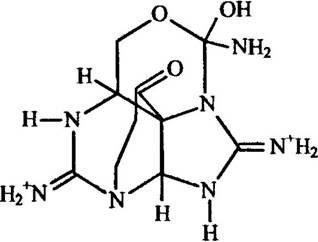
Fig. 6.10. Structure of saxitoxin
In addition to saxitoxin, dinoflagellates such as Gonyaulax tamarensis, Gonyaulax excavata, and Gonyaluax cateneila produce gonyautoxins, which are structurally and functionally similar (Fig. 6.11).
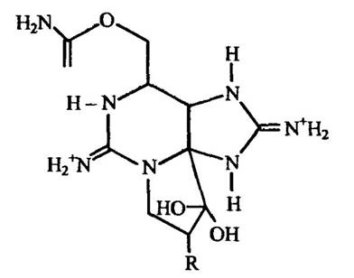
Fig. 6.11. Structure of gonyautoxin
Note. In gonyautoxins II and III, the radical (R) is an α-OH or β-OH group, respectively.
The Toxic effects of saxitoxin and its analogs are related to neurotropic activity and direct suppression of the respiratory center, manifesting in cardiovascular and respiratory dysfunctions. The lethal dose for a 70 kg human is estimated at 0.3 — 1.0 mg according to various sources. Death results from respiratory arrest.
Species sensitivity to saxitoxin varies: fish, mollusks, and amphibians are more resistant than warm-blooded animals. By MECHANISM OF ACTION, this toxin selectively blocks sodium channels in electrically excitable membranes.
Besides dinoflagellates, primary producers of saxitoxin and its analogs include the cyanobacterium Aphanizomenon flos-aquae and potentially others. Secondary poisonous animals that accumulate saxitoxin through food chains include mollusks (Saxidomus giganteus, Mytilus californianus, and many others) and coral reef crabs (Zosimus aeneus, Atergatas floridus, Platypodia granulus, and several others).
Some dinoflagellates, notably Gymnodinium brevis, produce gymberotoxins, which are also neurotoxins that cause inflammation, convulsions, paralysis, as well as cardiovascular and respiratory disorders.
Dinoflagellates of the species Gymnodinium brevis produce lipophilic toxins that cause Muscle paralysis, hypersalivation, and cardiovascular and respiratory disorders. They also exhibit cytotoxic effects and are characterized by high toxicity (0.01 µg/kg).
Ciguatoxin and maitotoxin, which cause severe joint pain, loss of motor coordination, diarrhea, nausea, as well as cardiovascular and neurological disorders, are produced by the dinoflagellate Gambierdiscus toxicus.
VENOMS-ALLOMONES OF MARINE INVERTEBRATES. Invertebrates make up a significant portion of marine and oceanic fauna. Among them are numerous venomous species belonging to Sponges, Coelenterates, worms, mollusks, and echinoderms.
Sponges. Sponges (Spongia) are typical passive-poisonous animals. They contain a range of substances with antibiotic, cytostatic, and toxic activities, including sesquiterpenoids, sterols, biogenic amines, and toxic Proteins.
A protein called suberitin has been isolated from the sponge Suberites domuncula. It exhibits neurotoxic activity, hemolyzes erythrocytes, and hydrolyzes ATP.
Extracts from the sponge Suberites inconstans exhibit cytotoxicity accompanied by cytoplasmic and karyoplasmic fragmentation, as well as rupture of cell and nuclear membranes. Bromophenols from Dysidea herbacea possess cytostatic activity. A number of sponge-derived compounds, particularly from Cryptotethya crypta, show antitumor effects. Overall, more than 50 furan, hydroquinone, and isonitrile sesquiterpenoids have been isolated from sponges.
Some of these sponge toxin substances are shown in Fig. 6.12.
Coelenterates. A characteristic feature of coelenterates (Coelenterata), which comprise up to 9,000 species (jellyfish, sea anemones, polyps, etc.), is the presence of stinging cells (nematocysts) that produce venoms used for defense against predators, as well as for paralyzing and killing prey. All coelenterates are carnivorous, feeding on a wide variety of organisms ranging from planktonic crustaceans to fish.
The venom of the stalked jellyfish Gonionemus vertens blocks N-cholinergic receptors in neuromuscular synapses and parasympathetic ganglia. This venom triggers the release of histamine and serotonin in the victim's body.
The venom of cubozoans (order Cubomedusae), particularly Chironex fleckeri, is characterized by hemolytic and dermonecrotic properties and causes burning pain. It contains proteins, CARBOHYDRATES, and indole derivatives.
Envenomation by scyphozoans (order Semeostomea), notably the lion's mane jellyfish Cyanea capillata, causes burning pain, erythema development, and edema. The venom contains a mixture of toxic proteins. The venom of the sea nettle jellyfish Chsaora quinquecirrha contains enzymes (DNases, RNases, ATPases, hyaluronidase, acid and alkaline Phosphatases, collagenase), a number of Peptides, amines, and Other Compounds. It exhibits cardiotoxic, hemolytic, and dermonecrotic actions, causing burning pain.

Fig. 6.12. Selected sponge venom-allomones:
1 — sterol sulfates from the sponge Toxadocia zumi; 2 — latrunculins from the sponge Latruncula magnifica:
2a — latrunculin A, 2b — latrunculin B; 3 — axisonitrile-1 from the sponge Axinella cannabina; 4 — sesterpenes from the sponge Cacospongia; 4a — scalaradial; 4b — furoscalarol; 4c — deoxoscalarin; Ac — acetyl
Rootmouth jellyfish (order Rhizostomida), particularly of the genus Rhizostoma, produce a toxic protein called rhysostamin, which causes burning pain and respiratory paralysis.
Coral polyps (Class Anthozoa) lead a sedentary lifestyle. Horny corals (order Gorgonaria, specifically the genus Lophogorgia) produce lophotoxin, which causes muscular paralysis, ataxia, and Respiratory system damage.
Sea anemones (order Actiniaria) are mostly solitary animals resembling flowers in shape. Equinatoxin, a protein with cytotoxic action that causes bradycardia, erythrocyte hemolysis, etc., has been isolated from the venom extract of the sea anemone Actinia equina.
Mat anemones (Zoantharia), particularly Palythoa toxica, produce palytoxin, which is a modified fatty acid that promotes histamine release, erythrocyte hemolysis, and the contraction of unstriated Muscles.
The venom of stony corals (order Madreporaria), particularly Goniopora, contains a polypeptide neurotoxin that causes muscle rigidity and respiratory disorders.
In ribbon worms (phylum Nemertea), the venom apparatus is located within the proboscis, the epithelium of which secretes the toxin. Paranemertes peregrina produces the toxin anabaseine, while Amphiporus angulatus produces its derivatives 2,3'-bipyridyl and nemertilen (Fig. 6.13), which are similar in action to nicotine.
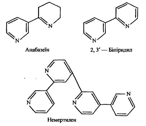
Fig. 6.13. Structure of nemertean toxins from Amphiporus and Paranemertes
These toxins cause paralysis in polychaetes and crustaceans.
The nemertean Cerebratulus lacteus secretes a poisonous mucus produced by Skin glands. It contains Polypeptides with cytolytic and neurotoxic effects.
Some marine segmented worms (phylum Annelida) also produce venom, especially polychaetes (Class Polychaeta). They possess chitinous jaws at the border of the Pharynx and buccal region. A pair of venom-producing glands opens into the anterior section of the Esophagus located behind the pharynx. For instance, the bloodworm Glycera convoluta produces the neurotoxin α-glycerotoxin, which causes cardiac arrest in daphnia. The venom of the polychaete Lumberineris heteropoda contains nereistoxin (Fig. 6.14), a potent insecticide that primarily affects the insect Nervous system.
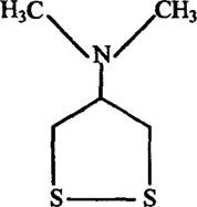
Fig. 6.14. Structure of nereistoxin
Cartap (Bancol), an organophosphorus derivative of nereistoxin, is an effective agent for controlling the Colorado potato beetle and is nearly harmless to birds, fish, rodents, and bees.
Molluscs. Among molluscs (phylum Mollusca), both passively venomous and actively venomous species occur.
Passively venomous gastropods (Class Gastropoda) with toxic digestive glands include molluscs of the families Buccinidae, Turbinidae, Aplysiidae, and Achatinidae, as well as certain sea slugs of the order Nudibranchia. The venom of Buccinum japonicum causes visual disturbances. It contains suratoxin (Fig. 6.15), which consists of bromoindole, pteridine, and myo-Inositol fragments and acts as a potent ganglionic blocker.
The venom of Turbo argyrostoma and Turbo marmorata is similar in action to saxitoxin and ciguatoxin—dinoflagellate toxins—and results from their accumulation in molluscs along the food chain.
Sea hares (family Aplysiidae) contain aplysin, a toxin that causes convulsions, ataxia, and respiratory arrest. This toxin and its analogues are produced by red Algae of the genus Laurencia, upon which sea hares feed. Additionally, Other toxins have been discovered in these molluscs—aplysiatoxin (Fig. 6.16) and debromoaplysiatoxin—which cause hyperemia of the nasal and oral mucosa, edema, and slow healing of skin ulcers.
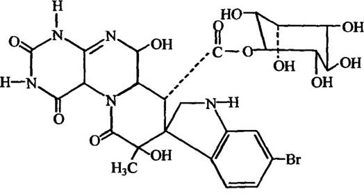
Fig. 6.15. Structure of suratoxin
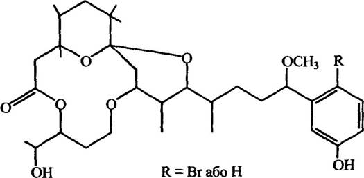
Fig. 6.16. Structure of aplysiatoxin
Toxins from the digestive glands of certain Nudibranchia (Cadlina flavomaculata, Aldisis nobilis, Doriopsilla albopunctata, etc.) induce bradycardia and hypotension.
Toxins from Achatinidae, specifically Achatina fulica, cause a decrease in catecholamines and induce narcotic Sleep.
Molluscs with poisonous hypobranchial Glands of the genus Murex, particularly Murex brandaris, produce the toxin murexine (Fig. 6.17).
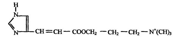
Fig. 6.17. Structure of murexine
Senecioylcholine has been isolated from the hypobranchial glands of Thais floridana, and acrylocholine from Buccinum undatum (Fig. 6.18).
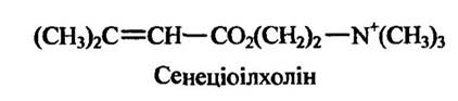

Fig. 6.18. Structure of senecioylcholine and acrylocholine
Murexine and its analogues produce effects similar to those of acetylcholine, inducing bradycardia, stimulating respiration and intestinal motility, and enhancing salivation, among other responses.
Opisthobranch molluscs (group Opisthobranchia) secrete a mucus containing a sesquiterpene that causes convulsions and loss of coordination.
A characteristic representative of actively venomous gastropods is the genus of cone snails (Conus). These are predatory species that feed on polychaetes and molluscs, while certain species, such as Conus geographus, prey on fish.
The venom apparatus of these molluscs consists of a venom bulb, a venom duct, a radula with Teeth, and a proboscis. The mobile proboscis forms the anterior end of the HEAD, holds one of the anterior radular teeth, and is used to strike prey. The remaining teeth are arranged in two rows on either side of the radula. These teeth contain channels through which venom flows, supplied by the venom duct where it is secreted. The venom bulb serves to pump the venom into the channels of the teeth.
The venom of Conus striatus is a glycoprotein known as striatoxin. It induces paresis, Skeletal Muscle spasms, and respiratory failure leading to cardiac arrest. It exhibits neurotropic properties. By its mechanism of action, this venom blocks sodium and calcium channels. The active principle of Conus magus venom is a heat-labile protein, which causes contractions in both striated and smooth muscles. The Mechanism of action involves increasing the permeability of membranes to sodium ions.
The venom of Conus geographus differs in its action from the venoms of other cone snails. It does not affect the contraction of smooth or cardiac muscle. The active components of this venom are proteins. At relatively high concentrations (12 mg/kg), it exhibits a postsynaptic mode of action, and death results from respiratory failure.
In Conus californicus, the venom contains a high-molecular-weight protein component that causes the death of vertebrates, and a low-molecular-weight component with cholinomimetic properties. The primary manifestations of this venom's action are the inhibition of heart contraction and the hyperpolarization of Central Nervous System Neurons.
The venom of Conus achatinus contains a protein component that induces cell membrane depolarization and Conformational Changes in muscle cell Membrane Proteins, resulting in increased membrane permeability to sodium and potassium.
An individual toxin named eburnetoxin has been isolated from the venom of Conus ebraeus. It causes vascular smooth Muscle contraction. The mechanism of action of this toxin involves increasing calcium influx across smooth muscle cell membranes, thereby triggering their contraction.
The venoms of Conus arenatus, Conus lividus, and Conus quercinus exhibit proteolytic activity. The proteases they contain cause local necrotic lesions.
Actively venomous molluscs also include those whose venom is contained in salivary gland secretions, notably Neptunea arthritica and Neptunea intersculpta. The saliva of these molluscs contains the toxin tetramine, which causes impaired motor coordination, nausea, general weakness, photophobia, and convulsions.
The saliva of molluscs belonging to the genera Thais and Cassis contains acids that dissolve mollusc shells and echinoderm tests, whereas the paralytic activity of their venom is associated with other components. These induce bradycardia, central nervous system depression, and vasodilation.
Among molluscs (phylum Mollusca), the cephalopods (Class Cephalopoda) are the most highly organized. In these animals, the muscular pharynx contains a chitinous beak capable of piercing fish skin, crab carapaces, or mollusc shells. The cuttlefish Sepia officinalis injects venom into the body of its prey, which is produced in the posterior Salivary Glands.
The venom of the octopuses Octopus dofleini and Octopus vulgaris, as well as the cuttlefish Sepia officinalis, contains biogenic amines—serotonin, Histidine, tyramine, dopamine, and noradrenaline—along with a toxic glycoprotein called cephalotoxin, which acts as a paralytic poison. Another proteinaceous toxin produced by the posterior salivary glands of the octopuses Eledone moschata and Eledone aldrovandi is eledoisin. The posterior salivary glands of the blue-ringed octopus Hapalochlaena maculosa produce the non-protein neurotoxins maculotoxin and hapalotoxin.
Echinoderms. Among echinoderms (phylum Echinodermata), sea urchins (Class Echinoidea), starfish (Class Asteroidea), and sea cucumbers (Class Holothuroidea) are venomous to varying degrees.
The venom apparatus of sea urchins comprises spines and pedicellariae. The spines are covered with a Glandular Epithelium that produces venom. When a victim is struck, the tip of the spine breaks off, and the venom is discharged into the victim's body. The spines are capable of moving in various directions thanks to muscles at their base. Pedicellariae are modified spines with a more complex structure. The venom of the pedicellariae of the sea urchin Toxopneustes pileolus contains proteins, a mixture of which is known as urchinitoxins. These compounds affect the amplitude of heart contractions, vascular tone and permeability, and the contraction of smooth muscle.
A protein toxin has also been isolated from the pedicellariae of Toxopneustes gratilla. It promotes the release of histamine from The Heart, Lungs, and intestine, and causes the degradation of serum α2-globulins. Its action results in smooth muscle contraction.
The venom of sea urchin spines exhibits certain differences from that of pedicellariae. In addition to proteins that trigger smooth muscle contraction, it also contains noradrenaline.
Starfish, notably Asterias amurensis, produce asterosaponins A and B, which feature a specific set of sugars. Asterosaponin A is linked via a glycosidic bond to D-quinovose and D-fructose, whereas asterosaponin B is bound to D-quinovose, D-fructose, D-xylulose, and D-galactose (Fig. 6.19).
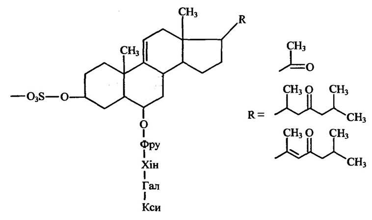
Fig. 6.19. Structure of asterosaponin B; Fru — fructose, Qui — quinovose, Gal — galactose, Xyl — xylose
Asterosaponins exhibit hemolytic activity and block Nerve Impulse transmission in vertebrate muscles.
A steroid saponin has also been isolated from the starfish Marthasterias glacialis; it acts as a surfactant and toxin on erythrocytes, inducing their hemolysis.
Holothurians of the order Aspidochirota possess specialized Cuvierian organs—glandular adhesive tubules opening into the cloaca. Through the cloaca, these tubules can be forcefully ejected into the surrounding water as sticky white threads that entangle and frequently immobilize prey. Holothurian toxin is present not only in the Cuvierian organs but also in the body wall.
The sea cucumbers Stichopus japonicus, Cucumaria japonica, and Cucumaria fraudatrix produce cytotoxic triterpene Glycosides known as holotoxins, stichoposides, and cucumariosides. The Chemical Structure of holotoxin B is shown in Fig. 6.20.
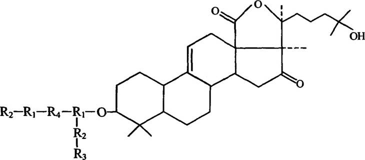
Fig. 6.20. Structure of holotoxin B (R1 = D-xylose, R2= D-glucose,
R3= 3-O-methyl-D-glucose, R4 = D-quinovose)
Holotoxins and stichoposides exhibit fungicidal activity, whereas cucumariosides inhibit nucleic acid and METABOLISM/35.html">Protein Biosynthesis in sea urchin eggs and Saccharomyces Yeast cells, as well as affecting cell membrane permeability.
ARTHROPOD ALLOMONE VENOMS. Among venomous Representatives of the phylum Arthropoda, the most thoroughly studied are arachnids (class Arachnida)—including scorpions, spiders, and mites—insects (class Insecta), and myriapods (group Myriapoda).
Scorpions. The order Scorpions (Scorpiones) comprises over 15,000 species. Their segmented metasoma ("tail") terminates in an anal lobe, or telson, equipped with a venomous sting. A pair of venom glands is housed within the telson, with their ducts opening near the tip of the sting via two orifices.
Scorpion venom induces severe pain, hyperpathia, hyperemia, and edema. Systemic toxic manifestations include thermoregulatory dysfunction, convulsions, muscle tremors, tachycardia, respiratory distress, nasal mucus hypersecretion, bronchial hypersecretion, and altered mental status, frequently accompanied by myocarditis, pancreatitis, and pulmonary edema.
Several dozen individual polypeptide toxins have been isolated from scorpion venoms. Depending on their specific biological activity, they can be classified into those affecting mammals, insects, and crustaceans.
Mammal-specific toxins consist of 65–80 amino acid residues, forming compactly folded structures stabilized by Disulfide Bonds. The venom of the Central Asian scorpion Buthus eupeus contains the so-called toxin M10 (Fig. 6.21), which serves as its principal active component.
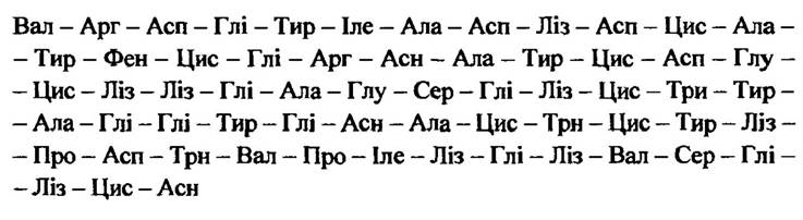
Fig. 6.21. STRUCTURE OF THE M10 polypeptide from the scorpion Buthus eupeus
In terms of its Amino Acid Sequence, toxin M10 is homologous to the neurotoxin A-II from the venom of Androctonus australis and L-V from the venom of Leiurus quinquestriatus.
A mammal-specific toxin comprising only 32 amino acid residues has been isolated from the venom of the scorpion Scorpio maurus palmatus.
Scorpion insect toxins are categorized into "short" toxins, consisting of 35–36 amino acid residues, and "long" toxins, comprising 67–70 residues. Similarly, crustacean-active toxins include a "long" toxin with 69 amino acid residues found in Androctonus australis, and "short" toxins containing 31–34 residues, such as those from Scorpio maurus palmatus.
Scorpion venoms contain enzymes characteristic of other venomous animals: Hydrolases (phospholipases A and B, acetylcholinesterase, acid phosphatase, 5'-nucleotidase, phosphodiesterase, hyaluronidase, and Ribonuclease).
The most venomous scorpions belong to the families Buthidae, Scorpionidae, Vejovidae, and Diplocentridae. Scorpion venom neurotoxins exhibit selective toxicity toward animals of different systematic groups. Insect-specific toxins display the highest degree of Specificity and do not affect other
animals, whereas mammalian and crustacean toxins possess a lower relative specificity.
A hallmark of venom action from buthid scorpions (genera Buthus, Androctonus, Leiurus, Centruroides, Tityus) is the massive release of Neurotransmitters from postganglionic nerve terminals. This results in cardiac and respiratory dysfunction, along with alterations in blood levels of BIOLOGICALLY ACTIVE SUBSTANCES. Hyperglycemia subsequently develops, which promotes Insulin secretion while suppressing Glucagon production.
Scorpion venom also exerts a direct effect on the Pancreas, leading to enhanced secretion of amylase and Trypsin.
Scorpion neurotoxins can directly affect the central nervous system. For instance, α-toxins inhibit the inactivation processes of fast Na+ channels in excitable membranes, thereby triggering membrane depolarization. The binding of α-toxins to the membrane is potential-dependent and decreases upon membrane depolarization. Scorpion venom β-toxins also modify the activation mechanisms of Na+ channels; unlike α-toxins, their binding to membranes is not potential-dependent.
The venom of Centruroides noxius contains noxiustoxin, a polypeptide K+ channel blocker. Scorpion venom from the family Scorpionidae contains a cytotoxin known as the "direct lytic factor," which exerts a hemolytic effect that is enhanced by phospholipase A2.
Certain species of these scorpions (specifically, Heterometrus fulvipes) possess a neurotropic venom that induces hyperglycemia associated with increased epinephrine production, while others (Heterometrus gravimanus) stimulate acetylcholine production, and still others (Pandinus exitialis) trigger catecholamine release.
The venom of scorpions from the family Scorpionidae (specifically, Palamneus gravimanus) contains histamine, hyaluronidase, and alkaline phosphatase.
Venom from scorpions of the family Diplocentridae (notably Nebo hierochonticus) is toxic to both invertebrates and vertebrates, and its action is independent of proteolytic activity. The effects of this venom manifest as severe burning pain, erythema, edema, and necrotic changes at the site of the sting.
Spiders. The order Araneae comprises over 27,000 species, the majority of which are venomous. Spiders of the suborders Mygalomorphae and Araneomorphae are venomous to humans. Spiders seize their prey using chelicerae—the anterior pair of appendages situated in front of the Mouth on the ventral side of the cephalothorax. The ducts of a pair of venom glands, located within the basal segments or the cephalothorax, open at the tips of the claw-like segments positioned at the apex of the outer margin of the main cheliceral segment.
Within the Mygalomorphae, the families Aviculariidae and Dipluridae are the most venomous. Among tarantulas of the family Aviculariidae, representatives of the genera Avicularia, Acanthoscurria, Lasiodora, Pterinochilus, and Poecilotheria pose a significant hazard. For example, the venom of Poecilotheria fasciata causes severe pain and involuntary skeletal muscle contractions, and it contains both histamine and serotonin.
The venom of Pterinochilus sp. also causes intense pain and exhibits a neurotoxic effect. A toxic protein component consisting of 77 amino acid residues has been isolated from it.
The family Dipluridae includes spiders such as Atrax robustus, whose venom induces pain, salivation, lacrimation, tachycardia, Hypertension, acetylcholine release, and muscle contractions. It contains a toxic polypeptide, atraxin, as well as γ-aminobutyric acid, spermine, and hyaluronidase.
Among the Araneomorphae, there are numerous venomous spiders, particularly those belonging to the families Sicariidae, Theridiidae, Araneidae, Lycosidae, Clubionidae, and Eresidae. Within the Sicariidae family, representatives of the genus Loxosceles are the most thoroughly studied. Their venom causes hemolytic anemia, thrombocytopenia, blood clotting disorders, and renal failure. The venom of Loxosceles species (including Loxosceles reclusa, Loxosceles laeta, and Loxosceles rufescens) contains hyaluronidase, 5'-nucleotidase, sphingomyelinase, as well as low- and high-molecular-weight factors. The presence of insectotoxins accounts for the venom's activity against insects. The enzymes present in the venom induce the lysis of Blood Cells, adipose tissue, and Muscle tissue in invertebrates. In vertebrates, it triggers dermonecrosis and causes platelets to release serotonin, leading to their aggregation.
The family Theridiidae includes the Mediterranean black widow, Latrodectus tredecimguttatus. Its venom causes severe pain, Autonomic nervous system overactivation, and psychomotor agitation that can progress to depression, loss of consciousness, and delirium; death may result from bronchospasm and pulmonary edema. In invertebrates, black widow venom induces paralysis. Its composition includes protein-based neurotoxins and enzymes such as hyaluronidase, phosphodiesterase, cholinesterase, and kinase. Among the neurotoxins, α-latrotoxin is the primary active principle. It acts as a presynaptic toxin, forming channels for Ca2+ and certain other ions, and promotes neurotransmitter release. Black widow venom is toxic to cattle, horses, camels, rodents, and humans, whereas dogs, hedgehogs, bats, amphibians, and reptiles exhibit high resistance to it.
Spiders of the species Lityphantes paykulliana produce a neurotropic venom containing an oligomeric toxin that binds to presynaptic terminals, alongside another toxin that forms channels for Ca2+ ions.
Spiders of the family Araneidae include the European garden spider (Araneus diadematus), the bolas spider (Mastophora gasteracanthoides), and orb-weaver spiders of the genus Nephila.
The venom of Araneus diadematus is toxic to insects and arachnids. In vertebrates, it induces local inflammatory responses, hindlimb muscle paresis, respiratory distress, and subcutaneous hemorrhages at the bite site. This venom contains hemolysin, a neurotoxin that blocks synaptic transmission via acetylcholine and glutamatergic synapses in both vertebrate and invertebrate animals.
The venom of Mastophora gasteracanthoides causes severe pain and hemorrhagic edema. A potent insecticide is found in the venom of Nephila spiders, particularly Nephila clavata, which blocks glutamatergic synapses. A similar action is exhibited by the venom of the spider Argiope lobata.
Tarantulas (family Lycosidae) produce venom that is toxic to both invertebrates and vertebrates. In Arthropods, it causes paralysis resulting from disrupted synaptic transmission and membrane depolarization. In mammals, it induces pain, edema, hyperemia, and increased vascular permeability. The venom of tarantulas, particularly Lycosa singoriensis, contains sermin, spermidine, putrescine, cadaverine, and a range of polypeptides and enzymes (hyaluronidase, proteases, esterases, and Kinases). Tarantula toxicity is seasonal, reaching maximum toxicity from May to August.
The venom of spiders of the family Clubionidae (such as Chiracanthium japonicum and Trachelas volutus) causes burning pain, local erythema, edema, necrosis, respiratory impairment, and paralysis. The venom of Clubiona spiders is toxic to arthropods. In humans, it leads to lymphadenitis and lymphangitis, cardiac disorders, asphyxia, and other complications.
The venom of spiders of the family Eresidae, particularly Eresus niger, causes pain, numbness, and restricted mobility.
Ticks. The saliva of certain ticks (order Parasitiformes), particularly Ixodidae and Argasidae, contains venom. For instance, the venom of the castor bean tick (*Ixodes ricinus*), which parasitizes livestock, dogs, and hares and frequently attacks humans, causes epidermal necrosis, lymphocyte infiltration, an elevated basophil count, and exhibits anticoagulant properties.
The venom of Ixodes holocyclus causes paralysis that can be fatal. Species sensitivity to this venom varies: rats are almost insensitive to the action of these tick venoms, whereas dogs are highly sensitive. It poses a serious threat to humans.
Toxic substances from various ticks (Amblyomma hebraeum, Boophilus decoloratus, Boophilus microplus, Rhipicephalus evertsi) produce similar histopathological changes in host tissues, namely The formation of exudate resulting from the interaction between salivary toxins and host tissues, which serves as a food source for the ticks.
Tick toxins are polypeptides. They are capable of inducing hyperesthesia, anorexia, Conjunctivitis, rhinitis, and hemorrhagic diarrhea. Local necrosis may occur in the liver, along with edema of the Urinary Bladder mucosa and circulatory disturbances in the lungs.
Tick venom, much like that of insects, anemones, and snakes, contains protease inhibitors. These compounds protect protein toxins from degradation by host proteinases and also provide an anticoagulant effect.
Insect allomone venoms. Among the approximately 1,500,000 known insect species, there are both actively and passively venomous forms. An armed venom apparatus in the form of an ovipositor or stinger is possessed by members of the order Hymenoptera—parasitoid wasps, bees, wasps, hornets, and bumblebees. Most beetles (Coleoptera) contain toxic substances in their hemolymph, which they can eject over a certain distance. Some Lepidoptera species possess a primitive wounding apparatus (typically in the caterpillar stage), whereas adult butterflies are predominantly passively venomous. Among Diptera, there are species that inject venom during a bite, such as horseflies, blackflies, and mosquito larvae.
Insects produce venoms that vary widely in Chemical Composition and action. When threatened, they release these venoms onto The surface of their bodies or eject them over a distance. Certain beetles, bugs, cockroaches, stick insects, and earwigs are capable of directing a stream of defensive venom toward an enemy and can adjust the direction of this stream to some extent.
Hymenoptera include many venomous insects, such as bees (Apoidea), wasps (Vespoidea, Sphecoidea, etc.), bumblebees (Bomboidae), parasitic wasps (Ichneumonidae), and ants (Formicoidea).
The stinging apparatus in various Hymenoptera families shares common features. In parasitic wasps, the ovipositor serves both to deposit eggs into the body of other arthropods and to inject venom to paralyze them. In bees, wasps, and certain other stinging Hymenoptera, the ovipositor is modified into a stinger used for defense and attack. When stinging, a bee bends the tip of its abdomen downward and drives the stinger into the victim's body. Along with the venom, the bee injects a mixture of isoamyl acetate, isoamyl propionate, and isoamyl butyrate into the victim, which act as attractants that lure other bees to the target.
Bees. After delivering a sting, the bee instinctively tries to fly away, but the stinger—along with the venom glands and the last ganglion of the abdominal nerve cord—remains embedded in the victim's skin and continues to function automatically for some time. A bee that has lost its stinger dies. Thus, at the cost of the death of individual insects, the efficiency of the sting is increased, which benefits the bee colony as a whole.
The effects of bee venom can be of an intoxication nature, caused by multiple stings, as well as allergenic, which occurs in 2% of people. In such individuals, even a single sting can trigger anaphylactic Shock.
The honey bee Apis mellifera produces venom containing numerous components, the primary ones being enzymes (phospholipase A2, hyaluronidase, phosphatases, α-glucosidase, β-galactosidase), polypeptides (melittin, apamin, tertiapin, secapin, procamine, cardiopep, mast cell degranulating peptide—MSD peptide), and biogenic amines (serotonin, histamine, catecholamines). The chemical composition of the venom changes as the bee ages. The unique Chemical composition of bee venom determines its wide range of physiological effects—affecting nerve cell membranes and The Cardiovascular system, while also producing analgesic and anti-inflammatory effects.
The main component of bee venom, accounting for up to 50%, is melittin. It is a polypeptide consisting of 23 amino acid residues (Fig. 6.22).

Fig. 6.22. Structure of bee melittin
Its primary action is the Modification of the lipid matrix of membranes and, consequently, its effect on membrane-bound enzymes. It is also capable of causing erythrocyte hemolysis and releasing histamine from mast cells.
While different bee species show some variations in the Introduction/19.html">Primary Structure of melittin, overall, the N-terminal sequence between the 1st and 20th amino acid residues is hydrophobic, whereas the C-terminal sequence between the 21st and 26th is hydrophilic.
Apamin is another component of bee venom that exhibits neurotoxic properties (Fig. 6.23).

Fig. 6.23. Structure of apamin
The MSD peptide (Mast Cell Degranulating Peptide) consists of 22 amino acid residues and two disulfide bonds (Fig. 6.24).

Fig. 6.24. Structure of the bee MSD peptide
Tertiapin exhibits a pronounced presynaptic effect (Fig. 6.25) and consists of a sequence of 21 Amino Acids.

Fig. 6.25. Structure of tertiapin
Tertiapin inhibits the Ca2+-binding protein calmodulin and regulates The activity of several Ca-dependent enzymes.
Bee venom also contains many other substances, notably secapin, which exhibits sedative effects, hypothermia, and piloerection, as well as cardiopep, which significantly enhances Cardiac Activity.
The venom of bumblebees (genus Bombus), which make up nearly half of all apids, is also complex in composition. It contains phospholipases A and B, acetylcholine, histamine, serotonin, and other substances, and exhibits cardiotropic effects.
Wasps. The superfamily Sphecoidea includes the beewolf (Philanthus triangulum), which preys on honey bees. Its venom contains acetylcholine, glutamate, and α-, β-, γ-, and δ-philanthotoxins, which together exert pre- and postsynaptic effects that paralyze honey bees.
Solitary wasps also include spider wasps (Pompiloidea), whose stings, much like those of digger wasps, cause mild pain and The Development of erythema.
Hornets of the genus Vespa belong to the superfamily Vespoidea, or social paper wasps. They build nests out of paper that they manufacture themselves by chewing wood and mixing it with water and sticky saliva.
Their venom causes pain at the sting site, Swelling, general malaise, fever, dizziness, elevated heart rate, and nausea. Allergic reactions may also occur.
The venom of these wasps contains phospholipase A2, lysophospholipase, histidine decarboxylase, hyaluronidase, DNases, proteases, toxic polypeptides, acetylcholine, histamine, dopamine, noradrenaline, and other substances.
Among the venom components of wasps are kinins similar to bradykinin. Some of them are shown in Fig. 6.26.
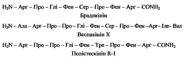
Fig. 6.26. Structure of certain kinins from Polistes wasps
Kinins cause increased vascular permeability and reverse contraction of smooth muscles.
The presence of Biogenic Amines and acetylcholine in the venom explains its effect on the cardiovascular system.
MSD peptides, which cause mast cell degranulation and are similar to those found in bees, have been detected in wasp venom. For instance, the mastoparan MSD peptide was found in the venom of Vespula lewisii, and mastoparan X in the venom of Vespa xanthoptera (Fig. 6.27).
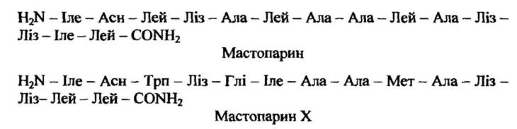
Fig. 6.27. Structure of mastoparan and mastoparan X
A number of neurotoxins have also been observed in wasp venom. For example, the neurotoxin from the venom of Vespa insularis, known as neurotoxin E, hyperpolarizes the postsynaptic membrane by increasing its chloride conductance. Mandarotoxins produced by Vespa mandarina are also typical neurotoxins.
Parasitoids. These insects of the family Braconidae, which belong to endophages, paralyze their victims with venom for only a short time. Soon, the paralyzed insects (caterpillars) begin to move again and resume normal life activity, but the parasitoid larvae, developing from eggs introduced simultaneously with the venom, ensure their own further survival. At the same time, certain parasitoids, particularly of the genus Microbracon, paralyze their prey completely or for a prolonged period.
As an active principle, parasitoid venom contains polypeptides. In particular, Microbracon hebetor contains proteinaceous components A and B, which exhibit presynaptic activity. Their Site of Action is the glutamatergic synapses of Lepidoptera and locusts.
Ants. Stinging ants belong to the families Myrmicidae and Poneridae. Relatively potent venom is produced by ants of the genera Solenopsis, Pogonomyrmex, and Myrmecia.
2,6-dialkylpiperidines, whose derivative methyl-2-nonylpiperidine (Fig. 6.28) is produced by ants of the genus Solenopsis, possess hemolytic, insecticidal, and antibiotic properties.
This venom exhibits neurotoxic activity, blocking The Effect of acetylcholine at the Neuromuscular Junction. It induces non-specific histamine release from mast cells via a lytic mechanism.
Solenopsis fugax ants also synthesize trans-2-butyl-5-heptylpyrrolidine, which acts both as a defensive toxin and as a repellent to deter other ant species. This compound is also synthesized by some other ant species, notably Monomorium pharaonis.
The venom of Pogonomyrmex ants, specifically Pogonomyrmex badius, is among the most toxic found in insects. It contains a hemolytic polypeptide called barbatolysin, which consists of 34 amino acid residues. Its composition also includes phospholipases A and B, hyaluronidase, acid phosphatase, lipase, and several esterases.
Stings from Myrmecia ants, particularly Myrmecia pyriformis, cause more intense pain than those of bees. Severe itching and erythema progressing to edema also occur.
The venom of these ants contains phospholipase A, hyaluronidase, histamine, hemolysins, and a smooth muscle-contracting factor.
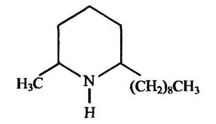
Fig. 6.28. Structure of methyl-2-nonylpiperidine
It should be noted that the ant venoms discussed do not exhaust all those produced by these insects. The venoms of Myrmecaria natalensis ants contain monoterpenes, Solenopsis punctaticeps — trans-dialkylpyrrolidines, Atta sexdens — 3-ethyl-2,5-dimethylpyrazine, and many others. Some of them, such as anabaseine in Aphanogaster fulva, have lost their significance as toxins and are instead used by ants as pheromones. Dendrolaszin from Dendrolasius fuliginosus ants exhibits insecticidal properties, while iridomyrmex has antibiotic properties as well (Fig. 6.29).
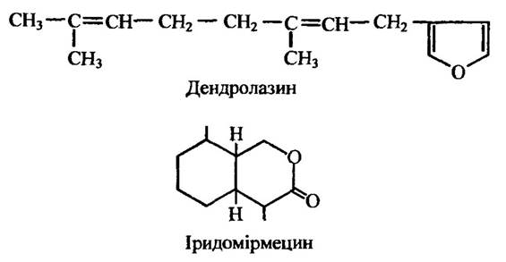
Fig. 6.29. Structure of dendrolasin and iridomyrmecin from ants
Beetles. There are about 25,000 known species of beetles (Coleoptera), including several venomous ones. The hemolymph of most venomous beetles exerts an intimidating, irritant, or toxic effect. When threatened, it emerges as droplets on specific areas of the body surface or is occasionally even sprayed over a distance.
The most venomous among the studied beetles belong to the families Meloidae (Mylabris, Meloe, Lytta, Epicauta) and Staphylinidae (Paederus).
Blister beetles of the genus Mylabris contain the deterrent cantharidin in their hemolymph (Fig. 6.30).
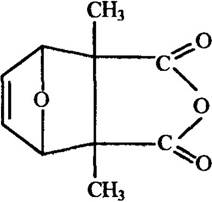
Fig. 6.30. Structure of cantharidin
This toxin induces hyperemia of the mucous membranes, liver, and kidneys, disrupts conditioned reflex activity, and leads to paralysis. Contact with the skin causes hyperemia, dermatitis, and blistering.
Cantharidin has also been detected in the blood of the Spanish fly, Lytta vesicatoria. In addition to the properties mentioned above, this substance causes severe irritation and serves as the active ingredient in "Spanish fly aphrodisiac preparations," whose effect on human sexual intercourse is driven by its irritant action on the urogenital organs. Using it is extremely dangerous because it is highly toxic—the lethal dose for humans is 0.5 mg/kg.
The active component in the venom of the blue-winged rove beetles Paederus is the terpenoid pederin (Fig. 6.31), which is capable of inhibiting Protein Synthesis. Ingestion of the venom into the digestive tract results in enteritis.
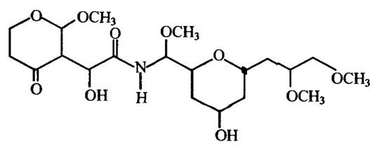
Fig. 6.31. Structure of pederin
The hemolymph of the Colorado potato beetle Leptinotarsa decemlineata (family Chrysomelidae) contains the toxic protein $\beta$-leptinotarsin, which affects neuromuscular transmission in vertebrates, inhibits cell growth, and exhibits blister-causing properties. Besides hemolymph, this protein is also present in the eggs.
When threatened, ladybird beetles (family Coccinellidae) secrete droplets of hemolymph from their leg joints. This hemolymph contains the alkaloids adaline and concinineline (Fig. 6.32), which taste bitter to humans and effectively protect these insects against ants and quail.
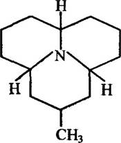
Fig. 6.32. Structure of concinineline
In some cases, beetles (as well as other arthropods) contain substances of diverse biosynthetic origin. For instance, the rove beetle Staphylinus alens secretes the terpenoid iridodial and 4-methylenehexan-3-one, which is formed during the FATTY ACID BIOSYNTHESIS pathway in the beetle's body. This mixture has a highly unpleasant odor.
Insects with toxic hemolymph frequently possess defensive glands whose secretions contain the same toxic compounds found in the hemolymph. For example, the grasshopper Poekilocerus bufonis features a gland on its abdomen that secretes histamine and a series of glycosides.
Diving beetles of the genus Ilybius fenestratus synthesize the alkaloid methyl 8-hydroxyquinoline-2-carboxylate (Fig. 6.33), which induces convulsions in mice but is nontoxic to amphibians and fish.
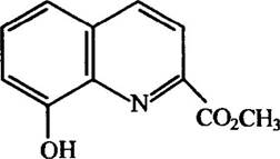
Fig. 6.33. Structure of methyl 8-hydroxyquinoline-2-carboxylate
Toxic properties are also exhibited by the larvae of leaf beetles such as Diamphidia locusta and Diamphidia nigroornata (family Chrysomelidae). Their venom contains a polypeptide known as diamphidiotoxin, which ranks among the most potent natural toxins.
As a defense mechanism, arthropods (beetles, arachnids, myriapods, termites) widely utilize phenols and Quinones. A striking example of this is The Use of benzoquinone by bombardier beetles (Brachinus). Upon the approach of a predator, these beetles discharge a hot cloud of toxin that causes severe irritation, primarily to the eyes. The Temperature of the toxin reaches 100°C. Its discharge involves a reaction driven by hydroquinone, H2O2, and catalase. During this exothermic reaction, hydroquinone is oxidized to benzoquinone, the primary defensive substance. The reaction occurs explosively and is accompanied by a loud sound.
Lepidopterans. In certain species of butterflies and moths (Lepidoptera), adults and larvae (caterpillars) possess stinging hairs that secrete venom produced by glandular cells.
The most characteristic symptoms of exposure to lepidopteran venom are dermatitis and conjunctivitis. However, occasionally—particularly following envenomation by caterpillars of Megalopyge urens—severe pain, bradycardia, convulsions, and nausea may occur.
Based on their origin, the toxic substances of lepidopterans can be divided into two groups: 1) compounds synthesized by the insects themselves at various stages of their life cycle; and 2) plant-derived substances accumulated in the tissues of larvae and imagos without structural modification, or metabolites of plant compounds. Thus, among lepidopterans, there are both Primary and secondary venomous species.
Primary venomous lepidopterans produce a wide variety of toxic compounds. For instance, Parasa consocia and several other moths produce histamine, while Zygaena species produce hydrogen cyanide in addition to histamine. In Euproctis chrysorrhoea and certain other moths, the venom has been found to contain polypeptides exhibiting trypsin-like, Chymotrypsin-like, kallikrein-like, and phospholipase activities. The venom of Parasa consocia contains proteins that cause severe pain and smooth muscle contraction.
Polypeptide toxins have been detected in lepidopterans even at the adult stage. For instance, the toxic polypeptide cain is isolated from the abdomen of female garden tiger moths, Arctia caja (family Arctiidae). It affects both other insects (such as locusts, cockroaches, and small white butterflies) and mammals, acting as a neuromuscular poison that can cause convulsions and respiratory arrest.
Many lepidopterans are classified as secondarily toxic. Some of these are danaine butterflies, notably Danais chrysippus. Cardiotropic substances of the cardenolide-lipid group have been isolated from adult specimens (both males and females) as well as from eggs. One of these is uzarigenin, which is toxic to birds that prey on danaines (Fig. 6.34).
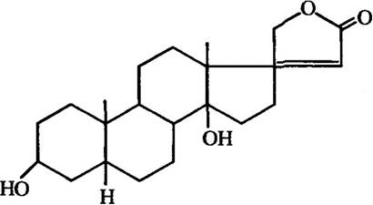
Fig. 6.34. Structure of uzarigenin
The cardenolides found in danaines are also present in the sap of plants such as Calotropis procera and Asclepias curassavica, which serve as food for these butterflies. Consequently, toxic cardenolides are produced by plants and accumulated by danaines through food chains.
Insects that inject venom during a bite. These insects lack a stinging venom apparatus. Instead, their venom is introduced into the victim's body
during a bite along with digestive enzymes contained in saliva. There is no clear boundary between the toxic and digestive components of saliva; for instance, hydrolytic enzymes facilitate the access of toxic substances to cells, while cytotoxins promote their Enzymatic Hydrolysis. The production of venom by salivary glands or their homologs occurs in many animals and is most perfectly developed in snakes.
Among dipterans (order Diptera), the toxicity of salivary gland secretions is most highly developed in larvae. For example, mosquito larvae of the families Cecidomyiidae (genera Platyura and Ceroplatus) and Macroceridae (genus Macrocera) paralyze their prey with venom containing a relatively high amount of oxalic acid (up to 0.15%), which is toxic to many other insects.
The venom of Tetanocera plebea and Tetanocera elata larvae causes paralysis in slugs. Another example of paralytic venom is found in the saliva of robber fly larvae (family Asilidae), specifically Philonicus dorsiger, which causes respiratory failure and death in other insects, such as the migratory locust (Locusta migratoria).
Blood-sucking blackflies (family Simuliidae) inject toxic substances into the wound along with an anticoagulant that prevents blood clotting, causing a burning sensation, itching, pain, and swelling. Repeated bites can lead to systemic poisoning. Painful bites are also inflicted by adults and larvae of horseflies (family Tabanidae) and robber flies (family Asilidae). The venom of horseflies and robber flies causes paralysis in invertebrates.
The saliva of true bugs (Hemiptera) also possesses toxic properties. For instance, the venom of assassin bugs (family Reduviidae) paralyzes invertebrates, and some species contain venom that is hazardous to mammals. Painful bites are inflicted by bugs belonging to the families Notonectidae (the backswimmer Notonecta glauca) and Nepidae (the water scorpion Nepa cinerea). Giant water bugs of the family Belostomatidae feed on fish fry, tadpoles, and occasionally small fish. Their salivary secretion not only paralyzes the prey but also liquefies the victim's Internal Organs. Some bugs also contain defensive venoms in their hemolymph, typically consisting of several unsaturated aldehydes and n-tridecane. For example, the venom of the burrowing bug Scaptocoris divergens contains propenal, propanol, octenal, furan, methylquinoline, and other quinones.
The venoms of certain bugs, particularly Triatoma, cause allergic reactions in humans.
Net-winged insects (Neuroptera) also produce venoms, but unlike bugs, these are produced by digestive glands. For instance, the venom of spoon-winged lacewing larvae (family Osmylidae) paralyzes a much larger midge larva in just 10 minutes, whereas the venom of
antlions (family Myrmeleontidae) immobilizes their victims within 2 to 4 minutes. The venom of owlflies (family Ascalaphidae), specifically Ululodes mexicanus, paralyzes cockroaches in 1 second.
MYRIAPOD ALLOMONE VENOMS. Myriapods (Myriapoda) comprise 4 superclasses, of which the representatives of millipedes (Diplopoda) and centipedes (Chilopoda) are venomous.
When threatened, millipedes coil into a spiral on their ventral side and secrete a venom from numerous pores located on the lateral PARTS OF THE tergites of each body segment, which has a distinct unpleasant odor for humans.
In centipedes, the venom apparatus consists of poison claws (forciputes), each composed of several segments. The terminal segment is pointed and claw-like, curved inward. A fine venom gland canal runs through this segment, originating within it and partially in the preceding segment, and in some centipede species, even in the basal segment.
The venom of the banded centipede Scolopendra cingulata (superclass Chilopoda) contains acetylcholine, histamine, serotonin, hyaluronidase, cholinesterase, kinase, esterase, and other compounds. Its paralytic effect is due to its action on presynaptic nerve terminals.
The centipede Scolopendra morsitrans produces venom that induces severe hyperglycemia by enhancing Glycogenolysis in the liver and muscles. The hyperglycemic effect of this venom may be associated with the presence of serotonin, which enhances the release of adrenaline from the Adrenal Glands, thereby promoting Glycogen breakdown.
The European pill millipede Glomeris marginata (superclass Diplopoda) synthesizes the quinazolines glomerine and homoglomerine, which cause death in spiders and mice that consume this millipede. Polyzonimine, synthesized by the millipede Polyzonium rosalbum, acts as a local irritant to predatory insects, causing itching in cockroaches, for example.
Some myriapods secrete defensive venoms. For instance, Fontinaria gracilis, Fontinaria virginica, and certain others release hydrocyanic acid when threatened, which is formed from a lowly toxic precursor. Millipedes of the species Apheloria corrugata possess 22 pairs of defensive glands containing mandelonitrile. When threatened, these millipedes contract a muscle that pushes the venom from the gland 'reservoir' into another section—the 'vestibule'—where mandelonitrile is converted by an enzyme into hydrocyanic acid and benzaldehyde. These substances are released externally to affect predators. In the millipede Glomeris marginata, the defensive venom contains the quinazolines glomerine and homoglomerine (Fig. 6.35), which cause par-
alysis or even death in spiders. Other defensive allomones of myriapods include a series of quinones, p-cresol, iodine, and various other substances.
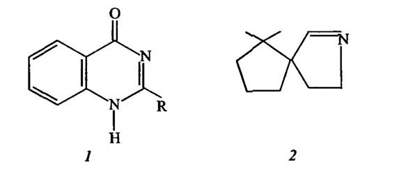
Fig. 6.35. Structure of:
1 — glomerine (R = СН3) and homoglomerine (R = С2Н5); 2 — polyzonimine
The millipede Apheloria corrugata, aside from soluble toxins, releases a pair of hydrogen cyanide streams for several minutes after being disturbed. This acid, along with benzaldehyde, is produced during the enzymatic breakdown of a substance called mandelonitrile right before it is expelled from the poison gland.
FISH ALLO-POISONS. Among the cartilaginous fish (Chondrochthyes), which include about 630 species, and bony fish (Osteichthyes), numbering over 20,000 species, there are venomous varieties. They can be divided into actively venomous and passively venomous. The former possess both mucous skin poison glands and a wide variety of spines and prickles, which often contain specialized venomous glands that likely originated from mucous poison glands.
Other venomous fish harbor toxins in their muscles, skin, and internal organs, particularly the reproductive organs. It is hardly a coincidence that toxin concentration in such fish peaks during the spawning season. Thus, passively venomous fish defend themselves through toxins at the cost of the sacrifice of individual specimens to predators in order to preserve the population.
A representative of cartilaginous actively venomous fish is the spiny dogfish, or spiny shark Squalus acanthias. The tip of the spine is bare, while the lower part is covered by a skin sheath beneath which the venom glands are located. The venom is proteinaceous in nature and causes skeletal muscle paralysis and paresis in animals and humans.
Cartilaginous venomous fish also include stingrays, in particular the common stingray Dasyatis pastinaca, the giant freshwater stingray Urolophoides giganteus, and others. The venom glands are located in the grooves On the surface of the spine, which is situated in the middle part of the tail. The venom is of a protein nature and causes edema, convulsions, respiratory and cardiac disorders,
a drop in blood pressure, and impaired cardiac activity. Typically, an affected person recovers within 5 to 7 days, but punctures to the chest or abdomen can be fatal.
An actively venomous bony fish is the greater weever Trachinus draco, in the grooves of whose dorsal fin spines venom glands covered with a sheath at the base are located, while the sharp tips remain bare. The venom composition has been found to include albumins, Polysaccharides, glycoprotein complexes, serotonin, histamine, cholinesterase, and other compounds. At the site of the puncture, edema appears, the wound reddens, and tissue necrosis develops. A burning sensation is felt, accompanied by headaches, heavy sweating, impaired heart function, and breathing difficulties; limb paralysis may occur, and in the most severe cases, death. In most cases, poisoning symptoms subside after 2 to 3 days.
Rockfishes, in particular the yellow Irish lord Sebastes trivittatus, the Pacific ocean perch Sebastes alutus, and the golden redfish Sebastes marinus, have a venom apparatus structure similar to that of the greater weever Trachinus draco, but their dorsal fin is T-shaped. The venom is proteinaceous and causes disorders in movement coordination, respiration, and cardiac activity.
Another venomous bony fish is the black scorpionfish, or sea ruffe Scorpaena porcus. The venom is injected during a puncture by the spines of the dorsal, ventral, and anal fins. It is protein-based, causes severe pain, impairs breathing and heart function, and sometimes leads to limb muscle paresis.
Passively venomous fish include, in particular, representatives of the carp family (Cyprinidae), including marinkas (Schizothorax), osmans (Diptychus), and barbels (Barbus), in which the caviar and milt are toxic. They contain so-called ichthyotoxins, the component of which is cyprinidin. These poisons cause adynamia, hypothermia, hypotension, cardiovascular and respiratory disorders, skeletal muscle paralysis, and, at high doses, cardiac arrest.
From the ichthyotoxins of Stichaeus grigorjewi, the toxic lipoprotein dinoineulin (also known as liposticharin) has been isolated. The roe of many other fish also contains toxic substances.
The Bile of the grass carp Ctenopharyngodon idella also exhibits toxic properties, inducing diuresis and bradycardia.
Fish of the pufferfish family Tetraodontidae, which comprises about 90 species and are also called toadfish or fugu, contain tetrodotoxin (Fig. 6.36). This compound consists of an aminoperhydroquinazoline with a guanidine group. The lethal dose of tetrodotoxin for humans is 1 • 10 mg/kg. In its potency, this toxin exceeds curare by 10 times and strychnine by more than 400 times.
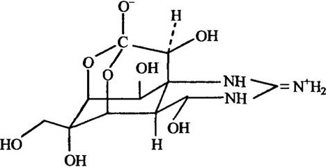
Fig. 6.36. Structure of tetrodotoxin
Despite its toxicity, fugu is considered a delicacy in Eastern countries (primarily in Japan), and specific preparation Methods exist. The most toxic parts are the Gonads and liver, with the skin and intestines being somewhat less toxic. The toxicity of this fish exhibits seasonal fluctuations and peaks during the spawning season—from May to July.
Tetrodotoxin is rapidly absorbed into the bloodstream, penetrates various body barriers, and accumulates primarily in the kidneys and heart.
The first symptoms of tetrodotoxin poisoning appear anywhere from a few minutes to 3 hours after consuming the fish. Sometimes death can occur within the first hour, but typically within 4 to 6 hours. The first sign of poisoning is tingling and numbness of the Tongue, loss of its mobility, as well as numbness in the fingertips and toes. Numbness can spread to the entire body. Then pain occurs in the head, abdomen, and limbs. This is followed by a progressive decrease in vascular tone and blood pressure, respiratory depression, skeletal muscle paralysis, a drop in body temperature, and so on. The patient falls into a coma, and shortly after losing consciousness, respiratory arrest and death ensue.
The toxic action of tetrodotoxin is based on its ability to block Nerve Impulse Conduction in excitable tissues. The uniqueness of its action lies in its ability, even at low concentrations (10−7 mol/L), to block the outward sodium current during the Development of the Action Potential.
At certain low doses, tetrodotoxin is used as a potent analgesic for severe forms of leprosy, inoperable Cancer, and other conditions.
Tetrodotoxin has been detected in the tissues of a number of invertebrates, including the mollusk Charonia sauliae; sea stars Astropecten polyacanthus and Astropecten scoparius; crabs Atergatis floridus and Zosimus aeneus; the blue-ringed octopus Hapalochlaena maculosa; and certain other marine invertebrates. It has also been found in newts of the genus Taricha (e.g., Taricha torosa) and frogs of the genus Atelopus.
It is believed that this toxin enters the bodies of herbivorous fish, such as Scarus gibbus, and crabs via the red alga Tania sp., which they feed on. Additionally, tetrodotoxin is contained in Bacteria of the genus Pseudomonas present on this alga. Therefore, this toxin may be of bacterial origin.
Passively venomous fish also include members of the boxfish family (Ostraciontidae), which comprises 7 genera and about 20 species. Typical representatives include boxfishes such as Acanthostracion quadricornis, Ostracion lentiginosum, Lactophrys gibbosus, and others.
If boxfishes are placed in an aquarium, the toxins they release into the water are capable of killing other fish. Their armor and toxins do not fully protect them, as they are frequently found in the stomachs of predatory fish.
A number of other toxins have been discovered in fish. For instance, goatfishes such as Upeneus arge and Mulloidichthys samoensis contain a poison that causes hallucinations, nightmares, muscle weakness, and partial leg paralysis in humans. Symptoms appear within a few minutes to 2 hours and can last from 5 to 24 hours. Poisoning is caused by both raw and cooked fish. Toxicity peaks during the spawning season in July and August.
Hallucinations also occur after eating the mullets Mugil cephalus and Neomyxus chaphalli.
Consuming so-called "sleeper fish," particularly Kyphosus vaigiensis, also induces hallucinations and nightmares in humans, followed by prolonged sleep.
AMPHIBIAN ALLOMONE POISONS. This class of vertebrates comprises over 4,000 species, among which several representatives of tailless (Anura) and tailed (Caudata) amphibians are venomous. These animals are classified as unarmed actively venomous species. Their venom apparatus lacks delivery devices (such as spines or stingers), and they also lack venom glands associated with the oral apparatus, unlike what is observed in snakes.
The production of poisons by amphibians serves a defensive function. The poison is synthesized by specialized alveolar mucous glands of the skin.
The secretion from these glands exerts a toxic effect upon contact with the skin. The primary active ingredients of the venom are steroidal alkaloids, which are not broken down by digestive enzymes when ingested orally.
The venom of the tailed amphibian, the fire salamander (Salamandra), includes steroidal alkaloids such as samandarin (Fig. 6.37), samandaron, cycloneosamandaron, etc., as well as serotonin and hemolytic proteins.
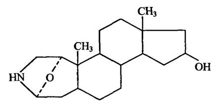
Fig. 6.37. Structure of samandarin
Salamander venom is capable of being absorbed through intact mucous membranes. It exhibits neurotoxic, cardiovascular, bactericidal, and fungicidal activities.
Toads of the genus Bufo belong to the tailless amphibians. The most common species is the common toad, or European toad (Bufo bufo). Smaller in size are the European green toad (Bufo viridis) and the natterjack toad (Bufo calamita). Toad skin contains numerous poison glands, among which two large suprascapular glands (parotoid glands) stand out.
Toad venom contains a number of compounds (about 100), the most effective of which are bufotenin, a dimethyl derivative of tryptamine (N,N-dimethyl-5-oxytryptamine) (Fig. 6.38); bufadienolides (derivatives of cyclopentanoperhydrophenanthrene containing a lactone ring in the protein chain); and batrachotoxin (the ester of 2,4-dimethylpyrrole-3-carboxylic acid).
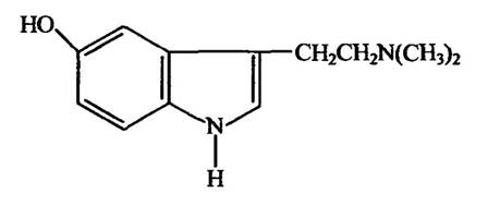
Fig. 6.38. Structure of bufotenin
In addition to these, toad venom contains several other compounds—tryptamine, serotonin, bufotenin ester, bufotenidine, bufadienolide homologs—cardiolipins, catecholamines, endorphins, and phospholipases A2.
The venoms of different toads have distinct compositional features. For instance, the venom of the green toad contains resibufogenin, but lacks bufotemidin, which is found in the venom of the common toad.
Toad venom has a wide spectrum of action, causing circulatory and respiratory disorders, convulsions, and limb paralysis (in mice, rats, and rabbits); cardiovascular system disturbances (in dogs); and limb paralysis (in amphibians).
Fire-bellied toads (specifically the European fire-bellied toad Bombina bombina and the yellow-bellied toad Bombina orientalis) belong to the order of tailless amphibians. Their venom contains bufotenin and bufotenidine, the polypeptide bombesin (Fig. 6.39), a hemolytic protein, and possibly other compounds.

Fig. 6.39. Structure of bombesin
Fire-bellied toad venom causes erythropenia, a decrease in Hemoglobin concentration, and a strong stimulating effect on gastrin secretion.
Tailless amphibians also include the common spadefoot toad (Pelobates fuscus), whose skin glands secrete a venom with a garlic-like odor. It is toxic to small animals and causes irritation of mucous membranes in humans.
SNAKE ALLOMONE POISONS. The total number of snake species is about 3,000, and among them nearly 230 species are venomous.
The venom glands in most snakes are modified salivary glands—the upper labial and temporal glands. These venom glands open into specialized teeth located at the anterior or posterior end of the upper jaw. The venom enters grooves or canals in these "fang" teeth. During a bite, the venom flows down the teeth into the victim's body. Some snakes are capable of forcefully exhaling air over a considerable distance, carrying along the venom that flows down their teeth.
Snake venom is a complex mixture of substances. Among them are pre- and postsynaptic toxins, cardiotoxins and myotoxins, hemorrhagic toxins, and Proteolytic Enzymes, which are highly toxic compounds for humans. Less toxic components include phospholipases, anticoagulants, and a number of enzymes such as phosphodiesterases, acetylcholinesterases, and NAD-nucleosidases. Among the proteolytic enzymes, endopeptidase, isopeptidase, collagenase, Elastase, and others have been identified.
Some components of snake venom, particularly certain enzymes (phospholipase A2, hyaluronidase, oxidase, etc.), are common to the venoms of snakes from different families. At the same time, others are characteristic of a specific family. For example, the venom of elapids and sea snakes contains neurotoxins that disrupt excitation transmission at neuromuscular synapses, thereby causing paralysis of the skeletal and respiratory musculature. These venoms contain acetylcholinesterase. Conversely, the venom of vipers and pit vipers lacks acetylcholinesterase, but is rich in enzymes with trypsin-like, Thrombin-like, and kallikrein-like activities.
Proteroglyphous snakes ("poisonous" fangs located on the anterior edge of the Maxilla) include the Central Asian cobra, vipers (common, steppe, Caucasian, and sand vipers), pit vipers (mamushi, eastern pit viper), and others.
The venom of the Central Asian cobra Naja oxiana (family Elapidae) contains neurotoxins (Fig. 6.40), cytotoxins, proteins with specific Functions (nerve growth factor, anticomplementary factors, etc.), and a number of enzymes—acetylcholinesterase, phospholipase A2, endoribonuclease, deoxyribonuclease, phosphodiesterase, 5'-nucleotidase, L-Amino Acid Oxidase, hyaluronidase, and others.
Allomone venoms of lizards. Among lizards (Sauria), which comprise about 3,500 species, only 2 families are venomous: the Gila monster family (Helodermatidae) and monitor lizards (Varanidae).
Venomous lizards are represented by 2 species: the Gila monster (Heloderma suspectum) and the Mexican beaded lizard (Heloderma horridum). Their venom is produced by sublabial glands located on the lower sides of the anterior half of the Mandible. Through several ducts, it flows to the outer side of the largestgrooved teeth of the lower jaw.
The venom composition includes L-amino acid oxidase, hyaluronidase, phospholipase A2, Arginine esterase, serotonin, and other substances.
The bite site develops edema, weakness, dizziness, respiratory disorders, lymphadenitis, and thrombocytopenia.
Monitor lizards include 30 species. Their bite is accompanied by an inflammatory reaction and severe pain. There is evidence that venom is present in their saliva and is capable of immobilizing prey.
Allomone venoms of birds. Until recently, the existence of birds capable of producing and releasing venom was doubted. However, in New Guinea, birds such as Pitohui dichrous, Pitohui ferrugineus, Pitohui kirhocephalus, and Ifrita kowaldi have been discovered that produce the steroidal alkaloid batrachotoxin. As already noted, this is one of the most potent natural non-protein toxins. It is also produced by certain animals, notably frogs of the genus Phyllobates.
The venom is secreted by birds from glands located on the ventral side of the body. To get rid of parasites and predators, the bird crawls around its nest and smears it with venom. A bird weighing 60–65 g produces up to 15–20 mg of venom, with 2–3 mg contained in the plumage. The lethal dose of batrachotoxin for a 20 g mouse is 0.03 mg. The secretion of venom by the aforementioned birds is clearly an adaptation for defense against predators.
ALLOMONE VENOMS OF MAMMALS. The presence of venom is not characteristic of mammals (Mammalia). This can be explained by the fact that the high development of The Nervous System and effective defense mechanisms made the production and secretion of venom unnecessary. At the same time, among egg-laying mammals (monotremes) Prototheria and live-bearing Theria, there are individual venomous representatives.
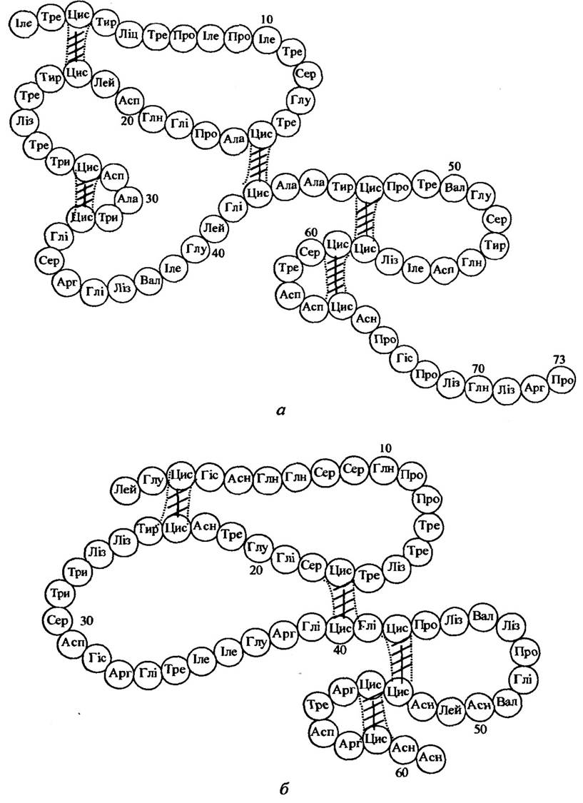
Fig. 6.40. Structure of neurotoxin I (a) and neurotoxin II (b) of the Central Asian cobra
Egg-laying mammals are grouped into the families: platypuses (Ornithorhynchidae) with a single genus Ornithorhynchus, and echidnas (Tachyglossidae) with five species. In the platypus and echidna, the venom apparatus is localized in the femoral gland, which is connected by a duct to a spur located on the inner side of the hind limb. Only males have a developed spur; in young females, the spur is in a rudimentary state, and in sexually mature females, it is absent.
Venom toxicity is seasonal in nature, reaching its maximum in June before the onset of mating activity. It is proteinaceous in nature, thermolabile, causes Blood Plasma coagulation in the absence of calcium, and lacks proteolytic and hemolytic enzymes. Human envenomation results in edema and cardiovascular disorders. No fatal cases in humans have been reported from this venom.
Among live-bearing mammals, venomous species belong to the Order Insectivora, specifically two families: solenodons (Solenodontidae) and shrews (Soricidae). The saliva of certain species of these animals exhibits neurotoxic effects.
Solenodons are represented by 2 species: Solenodon paradoxus and Solenodon cubanus. They feed mainly on various invertebrates and small vertebrates, and are also capable of eating certain plants.
Shrews are represented by approximately 250 species. Their primary food is insects. The saliva of some of their species is toxic, particularly to amphibians. It is possible that the toxicity of the salivary gland secretion in solenodons and shrews is an adaptation for hunting prey that is mobile and large relative to these animals.
Venom has been discovered on the quills of porcupines (Hystricidae), which apparently performs a protective function.
BIOLOGICAL BEHAVIOR OF ANIMALS REGARDING THE VENOMS THEY PRODUCE. Predatory animals generally possess an armed venom apparatus (snakes, scorpions, spiders, sea anemones, wasps, etc.). Their venom has a pronounced neurotropic effect, as it is primarily intended to immobilize prey. Often, the neurotropic action of such venoms is characterized by high selectivity. For example, scorpion venom contains species-specific neurotoxins that selectively affect insects, crustaceans, and mammals, while braconid parasitoid wasp venom is mainly active against lepidopterans. The venom of predatory animals is also used for active defense.
In animals with an unarmed venom apparatus, venom is produced primarily for defense, and one such method is deterrence. For example, repellents are produced by amphibians, centipedes, beetles, ants, and others. Among mammals, some badgers, the striped skunk (Mephitis mephitis), and the hog-nosed skunk (Conepatus suffocans) use foul-smelling compounds to deter enemies. When they see an enemy, they turn their backs to it and spray a stream of liquid containing foul-smelling mercaptans through the anus.
Note that the action of allomone venoms is relative. It does not manifest against all animal species, but only specific ones. Snakes, scorpions, and spiders possess venoms with the broadest range of action. At certain doses, they can rapidly kill most vertebrates, while only individual animal species are weaker or slower to succumb to such venoms.
Last update: 06/08/2026
Editorial and Educational Adaptation: This material has been compiled based on the primary/original source text. The project team performed an editorial review, corrected technical inaccuracies, structured sections, and adapted the content for an educational format.
What was processed:
- elimination of formatting defects (OCR errors, structural breaks, corrupted characters);
- editorial organization of content;
- standardization of terminology in accordance with academic sources;
- verification of factual statements against the original source text.
All mentions of the author, publication year, and origin of the primary text have been preserved in accordance with the source.