STRUCTURE AND PROPERTIES OF BIOMOLECULES - A. E. Zemlyakov - 2017
17. THE NERVE CELL. NEUROTRANSMITTERS
❖ The Nerve Cell. The Nervous system coordinates The activity of specialized Cells in the Organism. It transmits nerve impulses from one part of the body to another, enabling the body to respond to stimuli as a single integrated whole.
Centralized control of the organism is carried out by the Central Nervous System, which includes the Brain AND SPINAL cord. Organs and tissues are connected to the central nervous system via the Peripheral Nervous System through the transmission of nerve impulses.
Nervous Tissue is built of Nerve Cells, or Neurons. The human Cerebral Cortex contains 10–20 billion neurons. A neuron consists of a cell body ranging from 3 to 100 µm in diameter, which contains The Nucleus and other Organelles, and processes. Two Types of processes are distinguished: the axon, a long process that conducts excitation away from the neuron body, and dendrites, which are usually short and highly branched processes. A neuron may have several dendrites and typically only one axon.
Class="center">
The nervous system converts physical stimuli (light, sound, Temperature, pressure, Touch) or chemical stimuli (from substances present in the air, food, or internal bodily fluids) received through Sense Organs into nerve impulses.
These transformations are underpinned by the chemical conversions of Biomolecules. The stimulating signal is perceived by a highly specific receptor protein located in the excitable membrane. As a result of this interaction, the protein conformation, membrane permeability, the activity of membrane-bound Enzymes, and Ion transport across the membrane all change, leading to a significant Amplification of the response to the initial stimulus. The functional Changes in the receptor are reversible. The secondary signal travels via nerve fibers to the central nervous system, where it is processed, and a response impulse is subsequently sent to the periphery.

✵ Synapses. Neurons are characterized by a very large number of intercellular contacts (up to 20,000). Communication between nerve cells is carried out by specialized structures known as synapses.
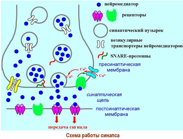
The nerve terminal of The Cell transmitting the Nerve Impulse contains the presynaptic membrane. This nerve signal is received by the membrane of another cell, the postsynaptic membrane. A synaptic cleft approximately 50 nm wide lies between the two membranes.
Chemical messengers of signal transmission, known as Neurotransmitters, are produced in the synaptic terminal region with the help of specific enzymes. Neurotransmitters are stored (depleted) in specialized organelles called synaptic vesicles, up to 50 nm in diameter. Special transmembrane channels, known as vesicular neurotransmitter transporters, facilitate their entry into the storage depot across the lipid membrane.
Polarization of the nerve cell biomembrane during signal propagation leads to the activation of specialized calcium channels. The concentration of Ca2+ ions increases within the neuron terminal. The binding of Calcium Ions to Proteins on the synaptic vesicle membrane facilitates their fusion (exocytosis) with the presynaptic membrane. The anchoring of vesicles to The cell membrane is ensured by the intermolecular interaction of specialized proteins (SNARE proteins).
As a result of exocytosis, a portion of the neurotransmitter is released into the synaptic cleft. Diffusing across the cleft, the signaling molecules bind to specific receptors on the postsynaptic membrane, which then propagate the signal further.
Receptors located on the presynaptic membrane participate in regulating synaptic activity. Unbound neurotransmitter molecules are either taken back up into the neuron by neurotransmitter transporters or chemically inactivated by specific enzymes.
✵ Botulinum Toxin. The Gram-positive bacterium Clostridium botulinum belongs to anaerobic microorganisms. Human poisoning from home-canned mushrooms, meat, or fish is very frequently associated with the ingestion of these Bacteria, which release toxins as they multiply. Botulinum toxin is the most toxic chemical substance known, with a lethal dose of 1-10-5 µg/kg.

The toxin Structure comprises two domains linked by a disulfide bridge. The larger domain (~100 kDa) is responsible for binding the toxin to the SNARE proteins of acetylcholine synapses (see below), while the smaller domain (~50 kDa), which exhibits protease activity, disrupts The structure of these proteins. This blocks the release of acetylcholine from synaptic vesicles and halts the transmission of nerve signals.
❖ Neurotransmitters. Nerve impulses are transmitted from one synapse to another with the aid of chemical substances called neurotransmitters.
The first chemical compound whose role as a mediator in nerve signal transmission was identified is acetylcholine. This discovery was made in 1921 by the Austrian-born American pharmacologist O. Loewi. In 1936, together with the English neurobiologist H. Dale, he was awarded the Nobel Prize in Physiology or Medicine "for their discoveries relating to Chemical Transmission of nerve impulses."

To date, the neurotransmitter function has been established for more than a dozen chemical compounds. A number of other substances are currently being investigated as potential neurotransmitters.
Based on their chemical nature, neurotransmitters are subdivided into:
♦ Amino Acids — Glycine, y-aminobutyric acid (GABA), glutamic acid;
♦ Peptides, such as N-acetylaspartylglutamate;
♦ biogenic amines—acetylcholine, serotonin, histamine, as well as the catecholamine subgroup (dopamine, norepinephrine).
Depending on The Nature of the neurotransmitter, several types of synapses are distinguished. Let us examine the structure and signal transmission mechanisms for some of them.
Cholinergic synapse
In the presynaptic axon terminal, The Biosynthesis of acetylcholine takes place in the Cytoplasm with the participation of acetyl-CoA and the enzyme Choline acetyltransferase.

Acetylcholine released into the synaptic cleft binds to specific cholinergic receptors on the postsynaptic membrane. Subsequent Hydrolysis of acetylcholine into choline by the action of the enzyme acetylcholinesterase restores the receptors to their initial state. Meanwhile, acetylcholine is resynthesized from the choline entering the synapse.

❖ Cholinergic receptors are complex glycoprotein complexes. Depending on the activating substances, they are subdivided into nicotine-sensitive (or nicotinic) receptors and muscarine-sensitive (or muscarinic) receptors.
The former are typically located at the contact sites between axons and skeletal Muscles or in the electric organ of rays (Muscle-type receptors), as well as in the superficial layers of the cerebral cortex (neuronal-type receptors). The latter are concentrated in the brain, secretory cells, smooth muscle, and cardiac muscle.
The similarity in the Spatial Structure of acetylcholine and protonated nicotine molecules, as well as acetylcholine and muscarine, explains the action of these Alkaloids on their respective receptors.

✵ Muscarinic receptors are subdivided into five subtypes:
♦ M1 are located in CNS cells;
♦ M2 is the main muscarinic receptor subtype in The Heart, alongside presynaptic M2 receptors that function to reduce acetylcholine release;
♦ M3 are present in smooth muscle and most exocrine glands;
♦ M4 have been found in the heart, walls of pulmonary alveoli, and the CNS;
♦ M5 are identified in the CNS, Salivary Glands, and the iris of the eye. Receptors of this type belong to metabotropic receptors. M1, M3, and M5 receptors are coupled to Gq proteins, whereas M2 and M4 receptors are coupled to Gi proteins. The interaction of acetylcholine with muscarinic receptors causes a decrease in Blood pressure, a slowing of heart rate, pupillary constriction, and enhanced glandular secretion.
✵ Nicotinic receptors. The nicotinic receptor (e.g., from an electric ray) is an ion channel composed of five subunits of four types (2α, β, γ, δ) with a total molecular mass of approximately 290 kDa. The height of the receptor is about 12 nm, and its diameter is ~9 nm.
Acetylcholine molecules bind to the α-subunits, causing a conformational change in the receptor structure and opening the ion channel, which has a diameter of about 2 nm. The permeability to Na+ ions entering the cell and K+ ions leaving the cell increases, leading to depolarization of the postsynaptic membrane and ultimately triggering muscle cell excitation and, consequently, skeletal Muscle contraction.

❖ Acetylcholinesterase is a glycoprotein in which the carbohydrate moiety accounts for about 8% of the mass. Three isoforms of the enzyme have been identified: AChT (the principal form), AChER (erythrocytic), and AChEH (found in electric rays and mice). The enzyme from the electric ray and bovine erythrocytes consists of 4 subunits pairwise linked by disulfide bridges. Each subunit (molecular mass 70–80 kDa) possesses a single Active Site. In humans, membrane-bound tetrameric and soluble monomeric forms of the enzyme predominate.

✵ MECHANISM OF ACTION of acetylcholinesterase. Excess acetylcholine molecules, as well as acetylcholine molecules that have dissociated from receptors into the synaptic cleft following signal transmission, are cleaved by acetylcholinesterase within 2 milliseconds.
Acetylcholine binds to the Active Site of the enzyme through The formation of an ionic bond between the carboxylate ion and the quaternary nitrogen atom. Cleavage begins with the protonation of the ester oxygen atom by a proton from the hydroxyl group of a Tyrosine residue. The hydroxyl group of a Serine residue dissociates, and its nucleophilic oxygen atom attacks the carbonyl carbon of the acetyl group. A Histidine residue acts as a proton shuttle. Subsequently, the acyl-enzyme intermediate is hydrolyzed with the release of acetic acid.

✵ Organophosphorus chemical warfare agents. The Toxic effects of organophosphorus chemical warfare agents (such as sarin, soman, and tabun) and organophosphorus insecticides (such as chlorophos and dichlorvos) are associated with the phosphorylation of serine residues in the active center of acetylcholinesterase. Such a phosphorylated acyl-enzyme analogue is unable to undergo hydrolysis, thereby blocking the hydrolysis of acetylcholine. An increase in the concentration of this neurotransmitter leads to overexcitation of the nervous system.

❖ Cholinoceptor Agonists and Antagonists. Alkaloids frequently exhibit The properties of both receptor agonists and antagonists (see the following chapter). Nicotine and muscarine have already been mentioned as agonists of nicotinic and muscarinic acetylcholine receptors, respectively. Nicotinic receptor agonists also include lobeline and anabasine, which are used in smoking cessation medications. The alkaloid pilocarpine is a muscarinic receptor agonist.

The alkaloids platyphylline, atropine, and scopolamine block muscarinic receptors, whereas d-tubocurarine chloride acts as a nicotinic receptor antagonist. When α-tubocurarine chloride enters the bloodstream, it blocks nerve signal transmission in skeletal muscles, leading to respiratory paralysis. In low doses, it induces muscle relaxation. In surgery, both α-tubocurarine chloride and its synthetic analogues, such as terkuronium, are employed as muscle relaxants.
✵ Drugs acting on cholinergic processes. From a pharmaceutical standpoint, medicinal products that affect cholinoceptors can exert either a stimulating (cholinomimetic) or an inhibitory (cholinolytic) effect.

Cholinergic processes can be influenced by substances that target not only the receptors themselves, but also Other components of this system. For instance, carbachol enhances the release of acetylcholine at presynaptic terminals, whereas botulinum toxin, conversely, inhibits neurotransmitter release.
Reversible acetylcholinesterase blockers (physostigmine, galantamine) constrict the pupil and lower intraocular pressure, making them useful in the Treatment of glaucoma, as well as in overcoming motor disorders and a number of other pathologies.

Table 13. Drugs acting on cholinergic processes
Drug Type |
Drugs |
Pharmacological Effect |
Muscarinic and nicotinic cholinomimetics |
carbachol |
decreases intraocular pressure, increases intestinal muscle tone |
Muscarinic cholinomimetics |
pilocarpine |
decreases intraocular pressure |
aceclidine |
increases the tone of internal organ muscles, constricts the pupil, decreases intraocular pressure |
|
Nicotinic cholinomimetics |
anabasine, lobeline |
stimulate Respiration, aid in smoking cessation |
Muscarinic and nicotinic cholinolytics |
cyclodol |
reduces tremor in the treatment of parkinsonism |
Muscarinic cholinolytics |
atropine |
dilates the pupil, decreases glandular secretion, increases heart rate |
platyphylline scopolamine |
relieves spasms of The Stomach, intestinal, bronchial, and blood vessel muscles; dilates the pupil; exhibits antiemetic and sedative effects |
|
Nicotinic cholinolytics |
pentamin, higronium |
relieve vascular muscle spasms, lower blood pressure |
tubocurarine chloride, terkuronium |
muscle relaxants |
Adrenergic synapse
❖ Adrenergic synapse. In 1946, the Swedish physiologist U. von Euler established that noradrenaline, much like acetylcholine, Functions as a neurotransmitter. In 1970, he shared the Nobel Prize in Physiology or Medicine with the German-British biophysicist B. Katz and the American biochemist J. Axelrod "for their discoveries concerning the humoral Transmitters in the nerve terminals and the mechanisms for their storage, release, and inactivation."
During adrenergic signal transmission, the biosynthesis of noradrenaline takes place at the nerve cell terminal using the amino acids Phenylalanine and Tyrosine. The synthesized noradrenaline is stored in synaptic vesicles. Its passage across the membrane is facilitated by the vesicular monoamine transporter. When a nerve impulse travels down the axon, noradrenaline is released into the synaptic cleft. This process, as in the case of the cholinergic synapse, is regulated by calcium ions.

Noradrenaline molecules cross the synaptic cleft and bind to specific adrenoceptors on the postsynaptic membrane. Such receptors are located both on peripheral nerve terminals and in central nervous system synapses. Noradrenaline predominantly acts as the "neurotransmitter of arousal."
Neurotransmitter molecules are also capable of activating receptors on presynaptic membranes, thereby reducing the release of noradrenaline. A fraction of these molecules is transported back into the nerve terminal via noradrenaline transporters.

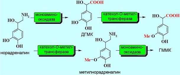
Two enzymes are involved in the degradation of noradrenaline. Under the action of monoamine oxidase, it is oxidized to 3,4-dihydroxymandelic acid (DHMA). Catalyzed by catechol-O-methyltransferase, the inactivation of excess noradrenaline in the synaptic cleft occurs by converting it into normethanephrine (normetanephrine), and DHMA into 4-hydroxy-3-methoxymandelic acid (HMMA).
❖ Adrenoceptors. The Study of the effects of various adrenaline derivatives revealed the heterogeneity of adrenoceptors. Initially, two types were identified—α and β-receptors—which are further subdivided into α1-2 and β1-3 subtypes. All of them belong to metabotropic receptors, albeit of different types.
✵ α1-Receptors are located, in particular, on Blood Vessels and the dilator pupillae muscle of the eye. Upon activation, they cause smooth muscle contraction and an increase in blood pressure or pupillary dilation. The biological signal is transmitted via Gq protein participation through the activation of phospholipase C (PL-C), which leads to an increased concentration of intracellular mediators such as diacylglycerol (DAG), Inositol trisphosphate (IP3), and Ca2+ ions, ultimately resulting in the activation of protein kinase C.
✵ α2-Receptors are situated on the presynaptic membranes of adrenergic synapses and control the release of noradrenaline. When the concentration of noradrenaline in the synaptic cleft rises, a portion of the neurotransmitter molecules reaches the presynaptic receptors, which inhibit the enzyme adenylate cyclase (AC) via the α-subunit of the Gi protein. Concurrently, the interaction of the β,γ-subunit complex with cAMP phosphodiesterase (cAMP-PDE) leads to its activation and, consequently, the hydrolysis of the intracellular messenger cAMP. The ultimate outcome is a decreased concentration of cAMP and the inhibition of noradrenaline secretion.
✵ β-Receptors. The primary localization of β1-receptors is on the postsynaptic membranes of heart muscle cells. Their activation leads to an increased heart rate.
Unlike the adrenoceptors discussed above, β2-receptors are activated not by the neurotransmitter norepinephrine, but by the hormone epinephrine (see below). They are located On the surface of cells in the Bronchi, Kidneys, Liver blood vessels, brain, and other organs, as well as on the presynaptic membranes of adrenergic synapses. Stimulation of these receptors results in accelerated heartbeat, dilation of the bronchi and blood vessels, increased renal renin production, and activation of norepinephrine secretion.
Relatively recently discovered, β3-adrenoceptors are located in adipose tissue cells, where their activation stimulates lipolysis and, consequently, energy release.
The binding of norepinephrine and epinephrine to β-receptors activates the adenylate cyclase system, leading to an increased production of the "secondary" messenger cAMP.

❖ Adrenomimetics and Adrenoblockers. Medicine widely utilizes both epinephrine itself and various substances that either stimulate (adrenomimetics) or inhibit (adrenoblockers) adrenoceptor activity. From the perspective of receptor interaction, adrenomimetics act as agonists, whereas adrenoblockers function as antagonists of adrenoceptors.
Adrenomimetics are subdivided into systemic agents, such as ephedrine, and those acting selectively on vascular α-adrenoceptors and cardiac β-adrenoceptors. The first group includes: mezaton (phenylephrine), a more stable analogue of epinephrine with a longer duration of action; naphazoline (sanorin), which exerts a decongestant effect upon contact with mucous membranes; and clonidine along with its analogue tramazoline, both of which lower blood pressure.
Representatives of the second group include: isadrine (isoprenaline), which—despite minor structural differences from epinephrine, namely the replacement of a methyl group with an isopropyl group—does not raise blood pressure but stimulates the heartbeat; salbutamol, which produces a bronchodilatory effect without affecting blood pressure or causing tachycardia (elevated heart rate); and dobutamine, a cardiac stimulant.

Compounds that block adrenoceptors are also employed as Pharmaceuticals, for example, ergot alkaloids (including the synthetic derivative dihydroergotamine), which act as α-adrenoblockers; the non-selective β-adrenoblocker propranolol; or the cardioselective β-adrenoblocker atenolol, which, unlike propranolol, does not affect the bronchi.

Neurotransmitters - Biogenic Amines
❖ Catecholamines are natural amines derived from pyrocatechol. They are also referred to as biogenic amines because they are synthesized within the body from the amino acids phenylalanine and tyrosine. In the organism, they perform the functions of neurotransmitters and Hormones.
The Biosynthesis of Catecholamines initially proceeds via the hydroxylation of phenylalanine to tyrosine, and subsequently to 3,4-dihydroxyphenylalanine (DOPA, derived from the obsolete name dihydroxyphenylalanine).

The oxidation of the C-H bond in the benzene rings of these amino acids is mediated by specialized enzymes known as hydroxylases. Hydroxylases share an identical tetrameric architecture while differing in the structure of their catalytic domain.


This oxidation process involves the coenzyme tetrahydrobiopterin (BH4), which donates two hydrogen atoms as a result of the reaction and is converted into dihydrobiopterin (BH2).

Subsequently, DOPA undergoes decarboxylation to yield dopamine.

Subsequent benzylic oxidation and N-methylation sequentially produce norepinephrine and epinephrine.

✵ Dopamine is capable of acting on specific dopamine receptors:
♦ D1 - Cells of the central nervous system, kidneys, and parathyroid gland;
♦ D2 - cells of the central nervous system, kidneys, Pituitary Gland, and Hypothalamus;
♦ D3 - cells of the olfactory center;
♦ D4 - cells of the olfactory center and hypothalamus;
♦ D5 - cells of the central nervous system.
D1 and D5 receptors are coupled to GS proteins, which activate adenylate cyclase. The binding of dopamine to D2 through D4 receptors stimulates Gi proteins and decreases cAMP concentration. At high concentrations, dopamine also activates adrenergic receptors, though not directly, but by promoting the release of norepinephrine. The binding of dopamine to specific renal receptors leads to the dilation of renal blood vessels and increases renal filtration. By acting on hypothalamic D2 receptors, it inhibits the secretion of several pituitary Neuropeptides.
✵ Norepinephrine is one of the primary substances involved in the transmission of nerve signals (see above).
✵ Epinephrine is produced by the Adrenal Glands and regulates Cardiac Activity and Carbohydrate METABOLISM. It is often called the "stress hormone" because it is released during stressful situations, increasing heart rate and respiration.
❖ Serotonin (5-hydroxytryptamine) is a biogenic amine formed in the body through the decarboxylation of 5-hydroxytryptophan, which in turn is produced from Tryptophan via hydroxylation by the enzyme 5-tryptophan hydroxylase.

Acting as both a hormone and a neurotransmitter, it regulates a range of physiological and behavioral processes, including Sleep, appetite, memory, sexual activity, aggression, and hallucinations. Serotonin is also a key mediator in allergic and inflammatory processes, as well as a regulator of motility and secretion in the gastrointestinal tract.

Serotonin is well known as the "feel-good hormone." Light plays a crucial role in serotonin production, which is why a lack of sunlight during the winter months often triggers seasonal depression. Consuming foods high in tryptophan (such as figs, dates, bananas, and chocolate) is also believed to help improve mood.
Like other neurotransmitters, serotonin acts on specific (serotonergic) receptors located on The surface of smooth muscle cells in blood vessels, bronchi, the gastrointestinal tract, and the central nervous system, among others. To date, seven types of these receptors have been identified, designated as 5-HT1 through 5-HT7. Several subtypes have been discovered for some of them:
♦ 5-HT1A,B,D,E,F; 5-HT3; 5-HT4; 5-HT5AB; 5-HT6; 5-HT7 — central nervous system cells;
♦ 5-HT2A — smooth muscle cells and central nervous system cells;
♦ 5-HT2B - cells of the intestine, heart, kidneys, and Lungs;
♦ 5-HT2C - cells of the Spinal Cord and brain.
The 5-HT1 and 5-HT5 receptors are coupled to Gi proteins, 5-HT4, 5-HT6, and 5-HT7 to Gs proteins, and 5-HT2 to Gq proteins. The 5-HT3 receptors function as Ion Channels.
It has been shown that presynaptic 5-HT1A receptors are involved in regulating serotonin production, while 5-HT7 receptors are implicated in the body's thermoregulation. Stimulation of 5-HT3 receptors leads to nausea and intestinal irritation, whereas 5-HT4 improves mood and appetite. Activation of 5-HT1B triggers aggression, 5-HT1D causes anxiety and vasoconstriction, 5-HT1F is linked to migraines, and 5-HT2C induces hypoactivity.
The Mechanism of action of psychotropic drugs—including antidepressants and hallucinogens (which act on 5-HT2A receptors)—as well as antiemetic medications (acting on 5-HT3 receptors) is closely tied to their interaction with serotonin receptors. The blockade of 5-HT2A, 5-HT2C, 5-HT6, and 5-HT7 receptors is associated with Schizophrenia.
❖ Melatonin is a derivative of serotonin. This hormone is produced by pinealocytes in the epiphysis (Pineal Gland) and plays a key role in sleep regulation. Approximately 30 µg of melatonin is synthesized in The Human Body daily (70% of it at night). With age, the activity of the pineal gland declines, leading to a drop in hormone production, which causes poorer sleep quality and the onset of insomnia.
Melatonin receptors have been identified: MTNR1A, located in pituitary and hypothalamic cells, and MTNR1B, found in cells of the central nervous system, retina, and lung tissue. Both receptor types are coupled to Gi proteins. Additionally, melatonin reception is mediated by intracellular retinoid-type receptors.
The conversion of serotonin into melatonin is catalyzed by two enzymes that can act sequentially in either order.

Ramelteon, a drug structurally similar to melatonin, is used as a sleeping aid. This compound acts as a specific melatonin receptor agonist.

Recently, interest in melatonin has grown due to its potential as an immune booster and antitumor agent. This is believed to be linked to its interaction with intracellular retinoid-type receptors. In addition, melatonin is a potent endogenous free radical scavenger.
❖ Histamine is another biogenic amine. It is produced in the body through the decarboxylation of The amino acid histidine and serves, among other functions, as a mediator of allergic reactions.

Under normal physiological conditions, histamine is present in a bound form. However, various pathological processes (such as Burns, allergies, and frostbite) trigger the release of large amounts of histamine, which causes tissue edema and smooth muscle spasm, including in the bronchi.
The Development of allergic responses is associated with the release of IgE Antibodies by plasma cells. These IMMUNOGLOBULINS, in turn, act on specific mast cell receptors and, in the presence of an allergen, stimulate histamine production.

Several types of histamine receptors (H1 to H4) have been identified:
♦ H1 — localized in the cells of the central nervous system, bronchi, blood vessels, and gastrointestinal tract;
♦ H2 — found in the cells of the central nervous system, heart, blood vessels, and gastrointestinal tract;
♦ H3 — located in central nervous system cells;
♦ H4 — identified on immune cells.
All of these belong to metabotropic receptors. H1 receptors activate Gq proteins, H2 activate Gs proteins, while H3 and H3 activate Gi proteins. Activation of H1 receptors causes contraction of the bronchial and intestinal muscles, whereas stimulation of H2 receptors excites certain Regions of the central nervous system and myocardial cells, as well as increases gastric acid secretion. Presynaptic H3 receptors in corresponding synapses are involved in regulating histamine production.
✵ Drugs acting on histamine receptors. Depending on which type of histamine receptor a drug targets, different therapeutic effects are achieved. Drugs that block H1 receptors are primarily used as anti-allergic antihistamines. These include diphenhydramine, tavegyl, suprastin, and claritin, among others. Because they interact with central nervous system H1 receptors, most of these drugs exhibit a sedative side effect.

Conversely, drugs such as ranitidine and cimetidine, which act as H2 receptor blockers, are widely used as anti-ulcer medications.

Glutamatergic synapse
Glutamic acid is one of the most abundant amino acids in nature. Along with other functions, it acts as a neurotransmitter in nerve impulse transmission, making it the primary excitatory neurotransmitter in the vertebrate nervous system.
Glutamic acid has standard biosynthetic pathways, such as the Transamination of α-ketoglutarate, which proceeds with the coenzyme participation of Pyridoxal phosphate.

This acid is also formed through the deamination of glutamine via the enzyme glutaminase.

Glutamate reception is mediated by metabotropic glutamate receptors mGluR1 – mGluR8 and ionotropic receptors.
✵ Metabotropic receptors. Receptors of the mGLuR1 and mGLuR5 subtypes are coupled with Gq-proteins and also interact with Na+ and K+ channels. They are predominantly localized on postsynaptic membranes. The remaining subtypes of these receptors are typically found on presynaptic membranes and mediate biological signals via Gi/0-proteins.

N-acetylaspartylglutamate (NAAG) also acts as an agonist for mGLuR2 and mGLuR3 receptors. This dipeptide exhibits all the properties of a neurotransmitter: it is stored in synaptic vesicles, released in portions into the synaptic cleft during nerve signal transmission, activates receptors, and is cleaved by specific enzymes.
✵ The NMDA receptor is an ionotropic glutamate receptor. N-methyl-D-aspartic acid (NMDA) serves as the characteristic Ligand for this type of receptor.

The ion channel is formed by two blocks containing two subunits each of NR1 and NR2, which in turn have several subtypes. A magnesium ion participates in regulating receptor function; in the resting state, it blocks the channel, but in the presence of glutamate, it is displaced, allowing sodium and calcium cations to enter the cell and potassium ions to exit through the open channel.
Receptors of this type also possess binding sites for glycine, which acts as a co-agonist to glutamate. Additionally, L-aspartic acid exhibits the properties of a natural NMDA receptor agonist.
✵ AMPA receptor. The principal type of glutamate receptor. α-3-amino-5-methyl-4-isoxazolepropionic acid (AMPA) is a specific agonist for this receptor type. Four protein subunits form the ion channel. Typically, two dimer blocks are assembled, each comprising a GluR2 subunit and one of the following subunits: GluR1, GluR3, or GluR4. Homotetrameric channels consisting of GluR1 or GluR2 subunits are significantly less common.

Each subunit has three transmembrane segments and one intramembrane domain. Two ligand-binding sites, S1 and S2, are formed by extracellular polypeptide chains. Upon interaction with glutamate, these segments move closer together, conformational changes are transmitted to the intramembrane chains, and the channel opens.
The selectivity of the AMPA receptor is influenced by the Nature of the amino acid at the Q/R site of the TM2 domain. The receptors most prevalent in the nervous system contain an Arginine residue and are impermeable to calcium ions.

✵ Kainate receptor. Similar in Organization to the AMPA receptor. It is significantly less abundant in the central nervous system compared to the ionotropic receptors discussed above. Kainic acid is its specific agonist.
The ion channel may incorporate GluR5–GluR7 subunits, which are capable of forming both homo- and heteromeric structures. KA1 and KA2 subunits, which are part of heterotetrameric receptors, have also been identified. The kainate receptor preferentially allows the passage of sodium and potassium ions across the membrane, and only to a minor extent Ca2+ cations.
Glycine–GABAergic Synapse
γ-Aminobutyric acid (GABA) is a biogenic amino acid that functions as an inhibitory neurotransmitter. It is synthesized through the decarboxylation of glutamic acid mediated by the enzyme glutamate decarboxylase.


Inhibitory signal transmission by GABA is carried out in conjunction with glycine, which also acts as a neurotransmitter in this context. The postsynaptic membrane of the GABA-glycine synapse features receptors for both GABA and glycine. Their activation leads to coordinated signal transmission into the cell.
The mechanism of GABA action is associated with its effect on specific GABAergic receptors. GABAA, GABAB, and GABAC receptors are distinguished. GABAA and GABAC receptors, much like the glycine receptor, are anion channels. When GABA binds to sites located in the contact region between the α and β subunits, a conformational change occurs in the protein subunits, forming an ion channel. Chloride ions flow through this channel into the cell, triggering the inhibitory process.
The GABAB receptor is structurally similar to α2-adrenergic receptors and is capable of inhibiting adenylate cyclase as well as regulating the activity of K+ and Ca2+ channels.
The stimulation or blockade of these GABA receptors forms The basis of the biological action of various pharmaceuticals (such as hypnotics, anticonvulsants, and tranquilizers). GABA itself is a medication that enhances brain function and activates memory and cognition.
The GABAA receptor possesses binding sites for barbituric acid and benzodiazepine derivatives, which exhibit hypnotic and sedative properties. This same receptor is also capable of binding ethanol molecules. Therefore, it is strictly forbidden to use these types of medications concurrently with alcoholic beverages, as alcohol drastically potentiates the effects of the drugs.

✵ Barbiturates. Barbituric acid derivatives are widely used as hypnotics (veronal), sedatives (phenobarbital), and anesthetics (thiopental sodium). Currently, their use is limited because they do not promote normal physiological sleep and also lead to habituation.

✵ 1,4-Benzodiazepine tranquilizers. Tranquilizers are substances that reduce feelings of fear, anxiety, and nervous tension. Drugs of this group have practically replaced barbiturates as sedatives because they have fewer side effects.

Last update: 06/08/2026
Editorial and Educational Adaptation: This material has been compiled based on the primary/original source text. The project team performed an editorial review, corrected technical inaccuracies, structured sections, and adapted the content for an educational format.
What was processed:
- elimination of formatting defects (OCR errors, structural breaks, corrupted characters);
- editorial organization of content;
- standardization of terminology in accordance with academic sources;
- verification of factual statements against the original source text.
All mentions of the author, publication year, and origin of the primary text have been preserved in accordance with the source.