PLANT BIOTECHNOLOGY AND BIOSAFETY - A. P. Ermishin - 2015
CHAPTER 3. MAIN TYPES OF PLANT CELL CULTURES: CALLUS, SUSPENSION, AND PROTOPLAST CULTURES. BASIC PRINCIPLES OF IN VITRO PLANT CELL, TISSUE, AND PROTOPLAST CULTURE
This chapter examines the MAIN TYPES OF plant Cell cultures—callus, suspension, and protoplast cultures—along with Methods FOR CULTIVATING isolated single Cells and protoplasts. Considerable attention is devoted to the fundamental processes involved in establishing cell cultures and regenerating whole plants from cultured cells. This Background is essential for a thorough understanding of the subsequent material, which covers the Structure/179.html">Practical Applications OF PLANT CELL CULTURE methods.
3.1. Dedifferentiation as a Prerequisite for Specialized Cells to Resume Division and Form Callus Tissue. Hormones as Dedifferentiation Inducers
Cells of a developing plant embryo are undifferentiated. They divide intensively and are capable of giving rise to various Plant Tissues and Organs (i.e., they are totipotent). During differentiation, a cell goes through three phases: 1) division; 2) elongation; 3) differentiation. A characteristic feature of the final phase of cell development is the thickening of the secondary Cell wall and the loss of The Cell's ability to divide. The embryo develops a meristem—a tissue that retains the capacity for division and The formation of new cells throughout the plant's life. Some meristematic cells (initials) remain in an undifferentiated state and, through division, ensure the continuous growth of the plant. Other cells gradually differentiate, forming permanent tissues (dermal, vascular, mechanical, etc.).
There are two main types of Meristems: apical and lateral. Apical meristems are located at the tips of shoots and roots, driving their elongation. This type of growth is termed primary growth, and the meristems themselves are called primary meristems. Lateral meristems (cambium and phellogen) lie parallel to the lateral surfaces of axial organs, enabling these organs to increase in girth. Additionally, during growth, meristematic tissue is retained in certain PARTS OF THE plant: in roots (as the pericycle), in SHOOT nodes, and in the pith rays of the stem. The so-called intercalary meristem is characteristic of cereal crops, located within the stem internodes and at the bases of leaf petioles.
Plants can also form new, secondary meristems, which typically occurs in response to wounding. Wound meristems give rise to callus—a specialized tissue consisting of uniform parenchymal cells that divide intensively to cover the wound site. Through secondary differentiation, damaged tissues and organs are repaired. The natural ability of plants to form callus and subsequently regenerate tissues and organs is utilized in horticulture for grafting (fusion of rootstock and scion) and vegetative propagation via stem cuttings (rooting of cuttings).
It is important to note that cells from various specialized plant organs (leaves, stems, roots, cotyledons) and tissues (meristematic, dermal, vascular, ground, secretory, mechanical) possess the capacity for callus formation. The initial prerequisite for this process is dedifferentiation: the loss of cellular specialization and the restoration of mitotic capability. In other words, during callus formation, specialized cells effectively revert to a meristematic state. Research has established that cell dedifferentiation and callus formation are triggered by specific phytohormones.
Placing a fragment of specialized plant tissue (an explant) onto a nutrient medium supplemented with the phytohormones required for callus induction yields an in vitro callus culture—the primary type of plant cell culture. The dedifferentiation of specialized cells is induced by Auxins. However, for these dedifferentiated cells to actually begin dividing, Cytokinins are also required. Auxins stimulate the synthesis of DNA, RNA, and Proteins necessary for progression through the Cell Cycle AND drive the synthetic processes of the G1 phase in preparation for division. Cytokinins act downstream of auxins, lifting the blocks on syntheses required to assemble the mitotic apparatus during the transition from the S to the G2 phase of the cell cycle. Auxins induce the synthesis of key protein Kinases involved in Cell Division, whereas cytokinins induce cyclins. Thus, during dedifferentiation, the action of these two phytohormones unfolds sequentially: auxins are engaged first, followed by cytokinins. Consequently, nutrient media for callus culture must contain both auxins and cytokinins. Depending on the explant used (and thus its endogenous phytohormone levels), this rule may not always strictly apply. For instance, mature and immature embryos of wheat and barley are capable of callus formation on nutrient media containing only auxins (such as 2,4-D), which is presumably due to an adequate supply of endogenous cytokinins.
While the dedifferentiation of specialized cells is triggered by division induced by phytohormones, the dedifferentiation of actively dividing meristematic cells involves a temporary cessation of division, dedifferentiation, and subsequent re-induction of division leading to callus formation.
3.2. Cytomorphological, Physiological, and Biochemical Features of Callus Cultures
In vitro callus tissue lacks a defined Anatomical Structure. Depending on the explant and cultivation conditions, its consistency can vary: it may be friable, consisting of highly vacuolated cells that easily dissociate into small aggregates; or it can range from moderately dense to compact, often exhibiting differentiating vascular elements and meristematic foci. When grown in the dark, the tissues are yellowish, whereas exposure to light can induce a light-green coloration, either uniformly distributed or appearing as distinct patches (Fig. 3.1).
Class="center">Fig. 3.1. Calli of varying consistency (explant: potato anthers): a - friable, highly hydrated callus breaking down into small aggregates; b - moderately dense callus with meristematic foci and shoot regeneration; c - finger-like callus: 1 - etiolated, friable consistency; 2 - high and medium density

The transition of a cell in vitro from a differentiated state to dedifferentiation and active cell division is driven by alterations in Gene Expression. In dicotyledonous plants, the processes of gene repression and depression underlying dedifferentiation occur more readily than in monocots. Consequently, establishing callus cultures from dicots is significantly easier and can be achieved from a much wider range of explant types compared to monocots. The best explants for initiating callus cultures are those containing weakly differentiated cells (such as immature embryos) or meristematic tissues.
During dedifferentiation, specific proteins appear in callus cells while other proteins characteristic of the original explant cells disappear. Explant cells deplete their reserves of Lipids, starch, and proteins. Photosynthetic cells lose chlorophyll and chloroplast lipids. Simultaneously, the number of amyloplasts increases, the Golgi apparatus degrades, and the Endoplasmic reticulum and cytoskeletal elements undergo reorganization. The METABOLISM/26.html">Energy Metabolism of callus cells largely resembles that of meristematic cells. Their Mitochondria are poorly developed with few cristae, which negatively impacts aerobic Respiration; consequently, they consume less oxygen than specialized plant cells. A metabolic shift occurs, favoring Fermentation over respiration. There is an increased consumption of CARBOHYDRATES due to enhanced aerobic Glycolysis, alongside a shift in Carbohydrate Metabolism toward the Pentose Phosphate Pathway, which supplies the pentoses essential for dividing cells.
At the same time, in vitro callus cells can retain certain PHYSIOLOGICAL AND BIOCHEMICAL traits of the donor plant. For example, nutrient media for callus cultures derived from plants that prefer acidic soils (such as rhododendrons, blueberries, and lingonberries) must have a more acidic pH than media for plants thriving in neutral or alkaline soils. Callus cultures of thermophilic plants grow better at higher temperatures than those of temperate plant species. Under specific conditions, callus cells retain The ability to synthesize secondary metabolites characteristic of the donor plants (such as panaxasides in ginseng). Furthermore, callus cells can exhibit resistance to toxins, herbicides, Antibiotics, and salinity, as well as cold and heat tolerance, provided these traits were present in the intact source plants.
3.3. Heterogeneity of Callus Tissue. Genetic and Epigenetic Cellular Changes in In Vitro Culture
Unlike meristematic tissues, which display high uniformity and genetic stability, the growth and secondary differentiation of callus cells occur in an unorganized and asynchronous manner. Callus tissue may comprise parenchymal, meristematic, necrotic, and vascular elements. Cells at various Stages of the mitotic cycle coexist within the callus, and their cell cycle is generally longer than that of meristematic cells.
Callus tissue cultures exhibit heterogeneity not only in the degree and direction of cellular differentiation but also in their cytogenetic profiles. Most notably, callus cultures often contain cells of varying ploidy levels (including high ploidy levels of 6–8x or more), even when initiated from meristematic tissues that were exclusively diploid. As cultivation proceeds, the proportion of polyploid cells typically increases.
Callus cell cultures are characterized by a high frequency of mitotic irregularities, leading to numerous Chromosomal aberrations. Specialized genetic studies have also demonstrated that various point Mutations arise with a relatively high frequency during callus cultivation.
Crucially, many of these genetic alterations can be "transferred" to the whole-Organism level by regenerating plants from genetically altered cells. This phenomenon of genetic Variability in plants regenerated through in vitro cell culture (encompassing callus cultures, cell Suspensions, and protoplast cultures) is known as somaclonal variation. Somaclonal variation is of great practical importance and can be harnessed to produce polyploids and novel mutant genotypes. The causes of somaclonal variation and its practical applications will be discussed in Chapter 6.
3.4. The phenomenon of Habituation (Hormone Independence)
Maintaining the proliferation of callus cells in in vitro culture typically requires the presence of auxins and cytokinins in the nutrient medium. However, prolonged cultivation can lead to The Emergence of cell clones capable of growing on media devoid of growth regulators. This phenomenon is known as habituation. Tissues formed by such habituated cells (which are autonomous regarding exogenous auxins and cytokinins) are termed hormone-independent. They are sometimes referred to as "chemical tumors" because, alongside habituated callus tissues, naturally occurring plant tumors also display hormone independence in vitro. The most thoroughly studied among these are crown galls—tumors induced in dicotyledonous plants by agrobacteria.
A shared property of both Artificial and natural plant tumors is hormone independence. In habituated tissues, hormone independence is achieved through altered activity of genes that encode Enzymes involved in phytohormone Biosynthesis. Thus, the metabolic shifts in callus cells related to the synthesis of their own phytohormones are primarily epigenetic in nature, although the appearance of specific mutations cannot be entirely ruled out. In tumor tissues, hormone synthesis is linked to The transfer of bacterial genes responsible for this process into The plant cell genome, resulting in cellular transformation from healthy to tumorous states.
Work on the in vitro culture of plant tumors and the discovery of hormone independence in them sparked a surge of scientific interest in uncovering its underlying mechanisms (the mechanisms of Agrobacterium-mediated transformation will be discussed below in Chapter 9). This research into The Nature of Agrobacterium-mediated transformation ultimately led to The Development of the primary and most effective method for introducing in vitro constructed genes into plant cells to produce Transgenic Plants.
3.5. Plant Cell Suspension Culture
Much like microorganisms, plant cells can be cultivated in vitro not only on semi-solid (agarized) nutrient media but also in liquid media as cell suspensions. In such cultures, an adequate oxygen supply is achieved by continuous agitation of the medium using orbital shakers, roller apparatuses, or other specialized devices.
To obtain a cell suspension, callus tissue is placed into a vessel containing liquid nutrient medium (approximately 2 g of tissue per 100 ml of medium). It is preferable to use friable calli that readily dissociate into cell clusters and small aggregates. The suspension is agitated on a shaker at 100-120 rpm. Unlike microorganisms, obtaining a suspension of single plant cells is rather challenging. Therefore, after approximately two weeks, the suspension is fractionated into single cells, small clusters, and large callus aggregates. The simplest way to achieve this is by allowing the suspension to settle for 1-2 minutes. Large aggregates precipitate to the bottom, while single cells and small cell clusters remain in the upper phase. Filtration through 1-2 layers of gauze or nylon sieves is also used to remove large cell aggregates from the suspension.
The disaggregation of callus cells during the establishment of a cell suspension culture can be improved by pre-culturing the callus on a nutrient medium lacking Calcium Ions and containing 2,4-D as an auxin. A beneficial effect is also achieved by supplementing the medium with the enzymes pectinase (about 2 mg/L) and cellulase (0.01 mg/L). In such media, a cell suspension can even be obtained directly from the explant. To sustain division of the suspension cells, auxins and cytokinins must be present in the nutrient medium.
Like callus cultures, cell suspensions are periodically transferred to fresh nutrient medium (subcultured). To do this, the suspension is filtered through a nylon sieve, and a portion of the culture volume containing single cells or small aggregates—known as the inoculum—is transferred under sterile conditions into freshly prepared nutrient medium. Each plant cell culture line is characterized by a minimum inoculum volume below which the culture fails to resume growth (the smaller this volume, the better the line). Other indicators of a robust cell line include a high growth rate under specific cultivation conditions, a high degree of disaggregation (no more than 5-10 cells per group), morphological uniformity of the cells (small size, spherical or oval shape, dense Cytoplasm), and the absence of Vascular System elements (tracheary elements).
Cell growth in suspension cultures is assessed using one or more of the following parameters:
• packed cell volume (PCV). A small volume of the cell suspension is transferred into a 15 ml graduated centrifuge tube (preferably conical) and centrifuged for 5 min at 200 g. The PCV is The ratio of the sediment volume to the total suspension volume, expressed as a percentage;
• cell number per unit volume of nutrient medium, counted using a Fuchs-Rosenthal hemocytometer;
• fresh and dry weight. The cell suspension is filtered through a pre-wetted and weighed filter placed in a Buchner funnel under mild vacuum. The cells are washed with distilled Water, excess water is drained under vacuum, and they are weighed again along with the filter; dry weight is determined similarly, except using a pre-dried filter, with the cells and filter dried together in an incubator at 60 °C to a constant mass;
• protein content. To determine protein content, cells are collected on a fiberglass filter, washed twice with boiling 70% ethanol solution, dried with acetone, hydrolyzed in 1M NaOH at 85 °C for 90 min, and then filtered and quantified using the Lowry assay;
• medium conductivity is measured using a conductometer and is inversely proportional to the cell mass;
• cell viability is assessed by observing cytoplasmic streaming under a Microscope, as well as by vital staining (0.5% Evans Blue solution or 0.01% fluorescein diacetate solution): viable cells remain unstained because their intact cell membranes are impermeable to the Dyes.
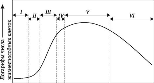
The growth curve of cell cultures is typically sigmoid (Fig. 3.2). During the initial lag phase, there is no increase in cell number or mass. During this time, cells are preparing for division. The subsequent logarithmic phase (exponential growth) is characterized by the highest mitotic activity and the greatest increase in cell mass. The third phase is linear growth, during which cell mass increases at a constant rate. This is followed by a deceleration phase, and finally, the growth curve reaches a plateau. During this period, The rate of callus cell death is still balanced by The production of new cells through division. However, if the culture is not subcultured into fresh nutrient medium at this stage, cell degradation can become irreversible, leading to culture death.
Fig. 3.2. Phases of the growth curve of a plant cell suspension culture: I - lag phase; II - exponential phase; III - linear growth phase; IV - deceleration phase; V - stationary phase; VI - death phase
3.6. Plant Protoplast Culture
An isolated protoplast is the portion of a plant cell that remains after the complete removal of The cell wall. E. Cocking (1960) from the University of Nottingham pioneered the method of enzymatic cell wall removal, which makes it possible to obtain large quantities of protoplasts and cultivate them in vitro. Currently, cell wall degradation is achieved using cellulases, hemicellulases, and pectinases derived from Fungi such as Myrothecium, Aspergillus, and Trichoderma, as well as from the digestive juice of the Roman snail Helix pomatia.
Protoplasts can be isolated from various plant tissues, including callus and suspension cultures. To obtain a high yield of uniform protoplasts from dicotyledonous plants, the mesophyll of young leaves is typically used. Isolation and culture methods are most well-established for Representatives of the Solanaceae family and certain Brassica species. Difficult objects for obtaining protoplasts capable of expressing totipotency include monocotyledonous plants (such as cereals) and conifers.
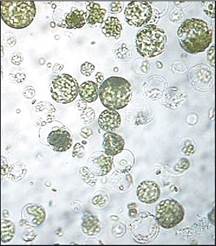
The Regulation of cellular water balance is closely tied to the presence of a cell wall. The survival of a cell stripped of its wall is only possible if the osmotic Properties of the isolation and culture media are strictly optimized. To achieve this, cells and isolated protoplasts are maintained in a plasmolyzed state by adding 0.3-0.8 M osmotic stabilizers, such as sugars (mannitol, glucose, sorbitol, xylose) or ionic osmotics (CaCl2, KCl). Under The Influence of osmotica, the protoplast decreases in volume and assumes a spherical shape (Fig. 3.3). The composition and concentration of osmotic agents are selected individually for each plant species. Similarly, the specific enzymes, their combinations, and concentrations are chosen based on the plant origin and the source tissue used for protoplast isolation. A typical protocol for isolating protoplasts from tobacco leaf mesophyll is outlined below.
Fig. 3.3. Protoplast culture from tobacco leaf mesophyll
Leaves from young, sterile-grown plants are surface-sterilized for 1 min in 70% ethanol, followed by 20 min in a 2% sodium hypochlorite solution (NaOCl • 5H2O). After rinsing the leaves with sterile water, the lower epidermis and large Veins are removed, and the remaining tissue is cut into strips approximately 5 mm wide. These leaf fragments are placed in Petri dishes containing a mixture of pectinase and cellulase enzymes. For instance, for tobacco leaves, a mixture of 0.5% pectinase, 2% cellulase, and 13% sorbitol at pH 5.4 is commonly used. The leaf fragments are incubated in the enzyme mixture in an incubator at 25 °C for 15-18 hours. Following incubation, the protoplasts are purified from cell wall debris by filtration through a nylon mesh. The protoplasts are washed free of enzymes by three consecutive centrifugations at 170g for 2 min each. The washed protoplasts are then resuspended in a culture medium containing 13% mannitol to a final concentration of 4 × 10⁵ protoplasts per 1 mL. The protoplast density should be optimized for each specific culture. The protoplast suspension is transferred into Petri dishes containing liquid or Agar-solidified medium, or cultivated using the micro-drop method (described below).
Immediately after the protoplast suspension is washed free of the enzyme solution, the protoplasts begin to regenerate their cell walls. The appearance of cellulosic microfibrils can be observed, which gradually organize into a typical cell wall. Within just 2-4 days, the protoplasts lose their spherical shape, indicating the complete restoration of the cell wall. Protoplasts that fail to regenerate a cell wall lose their capacity for normal mitotic division and colony formation. Such protoplasts either never divide or undergo abnormal divisions resulting in multinucleated structures, as karyokinesis is not accompanied by cytokinesis.
However, Cell wall formation is not always sufficient on its own to trigger cell division. Depending on the species and culture conditions, anywhere from 0.1% to 80% of protoplasts that successfully regenerate a cell wall will proceed to divide. The first divisions typically occur within 2-10 days after the initiation of protoplast culture. Once transferred to a regeneration medium, the resulting cell colonies can give rise to Plant Regeneration in the same manner as conventional callus cultures.
3.7. Single Cell and Protoplast Culture. Obtaining Colonies from Cell Suspensions
The primary application of cell suspensions in biotechnology is the production of secondary metabolites, many of which are valuable Pharmaceuticals (this topic will be discussed in Chapter 4). Cell suspensions are also of significant interest for the spatial Separation of various cell variants present in a cell culture, with the ultimate goal of obtaining modified regenerated plants from them. Unlike microorganism cultures, plating a cell suspension onto The surface of an agar-nutrient medium does not always yield separate colonies from individual plant cells. These difficulties arise because single cells, or even relatively large numbers of plant cells in a highly diluted suspension, lose their ability to divide upon plating on an agar medium while remaining viable. The same phenomenon occurs in cell suspensions if the inoculum size falls below a certain minimum threshold during subculturing—in other words, when the suspension is overly diluted. To achieve successful results, specialized single-cell culture methods must be employed. These are particularly crucial for protoplast cultures, specifically for obtaining colonies and subsequently regenerating plants from somatic hybrids (in cases of visual Selection of hybrid protoplasts under a microscope, see Chapter 8).
The most widespread technique is plating, originally proposed by L. Bergmann (1960). The starting material is a rapidly growing plant cell suspension at twice the concentration intended for plating onto the agar medium. First, the suspension is filtered through a nylon mesh to retain only single cells or small cell aggregates. A 0.6–1% agar medium is prepared with the same composition used to maintain the growth of the suspension, heated to dissolve the agar, and, after cooling to 35 °C, mixed with an equal volume of the cell suspension. The mixture is poured into Petri dishes to a depth of about 1 mm, and the dishes are sealed with Parafilm (a specialized elastic film that protects the cultures from contamination while ensuring normal gas exchange). Using an inverted microscope, the positions of individual cells are identified and marked with a marker on the dish lids. Cultivation is carried out in an incubator, periodically monitoring cell division under the microscope using the reference marks.
The conditioning factor plays a crucial role in ensuring the successful division of plant cells either in suspension or when plated on agar media. It has been hypothesized that actively dividing cells secrete certain substances (the conditioning factor) into the nutrient medium, which stimulate the division of neighboring cells. When the cell density per unit volume of the nutrient medium is low, the concentration of these substances is insufficient to induce division. In L. Bergmann's experiments, about 20% of single tobacco callus cells divided and formed colonies. This is attributed to the fact that, using the described technique, not only single cells but also small clusters are plated onto the agar medium. The division of cells within these clusters may be accompanied by the release of conditioning factors into the medium, which stimulate the division of single cells.
The chemical nature and MECHANISM OF ACTION of the conditioning factor remain to be elucidated. It has been established that it is thermolabile, contains a low-molecular-weight fraction, and cannot be replaced by phytohormones. There is reason to believe that it is not a single compound, but rather a complex of substances secreted by the cells.
Several methods have been proposed to condition the nutrient medium for single-cell cultivation. The simplest of these is supplementing the cell culture medium with spent medium from a rapidly growing cell suspension in the exponential growth phase (after filtration through a fine filter). Alternatively, a "feeder layer" can be used for medium conditioning—an agar-supported layer of irradiated cells that have lost their capacity for division but are still capable of supporting the growth of other cells (D. Raveh, E. Galun, 1975).
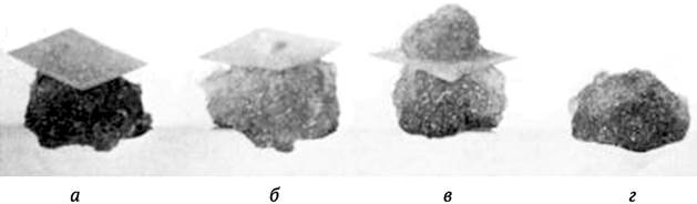
W. H. Muir et al. (1954) proposed the "nurse" culture method. A piece of filter paper is placed On the surface of an actively growing callus, and a suspension containing predominantly single cells is applied to it. The colonies that develop on the filter paper can subsequently be transferred to an agar nutrient medium (Fig. 3.4). A simpler approach can also be used: a thin layer of cell suspension is plated onto the surface of an agar medium containing several actively growing calluses. As a rule, cells located in the immediate vicinity of the callus cultures are the first to begin dividing and forming colonies.
Fig. 3.4. Stages of cell culture growth using a "nurse culture": a — single cells placed on filter paper over an actively growing callus culture; b — initiation of a new cell colony; c — actively growing new cell colony; d — newly obtained colony ready for transfer to an artificial nutrient medium
Some researchers believed that single cells in suspension could be induced to divide by using nutrient media enriched with various Vitamins, Amino Acids, sugars, or plant extracts (coconut milk, casein hydrolysate). While these approaches yielded good results for certain cultures, their scope proved to be quite limited. For instance, the enriched 8p Kao-Michayluk nutrient medium (K. N. Kao, M. R. Michayluk, 1975) produced favorable results only in the cultivation of diluted bean and soybean protoplast suspensions, yet it failed to support the culture of single protoplasts.
M. Caboche (1980) demonstrated that if tobacco mesophyll protoplasts are cultured for the first few days on a nutrient medium containing high concentrations of auxin and then transferred to a medium with a lower auxin content, the cells retain the capacity for division even at a plating density of 1–2 cells/mL.
Another potential approach involves drastically reducing the volume of the nutrient medium during single-cell cultivation: As a result, the ratio of cell volume to nutrient medium volume remains comparable to that of conventional cultures at low dilutions. Specialized devices are used for this purpose. L. E. Jones et al. (1960) proposed cultivating single cells in a specialized microchamber, which is easily assembled as follows. Two coverslips are affixed a short distance apart on a microscope slide using paraffin oil, a square of paraffin oil is applied between them, and a drop of conditioned nutrient medium containing an isolated cell is placed inside it. A coverslip is placed on top, supported at two
edges by the underlying coverslips. This creates a cavity beneath the coverslip where cell cultivation takes place. Desiccation of the nutrient medium is prevented because the chamber is sealed from the environment by paraffin oil, while normal gas exchange is still maintained. Cell division can be monitored under a microscope. Once the colony reaches a sufficient size, it is transferred to fresh nutrient medium (liquid or agar). Using the methodology described above, V. Vasil and A. Hildebrandt (1965) successfully obtained not only a colony from a single cell but also regenerated a normal flowering tobacco plant from the callus. However, unlike L. E. Jones, they used a fresh nutrient medium enriched with calcium pantothenate and coconut milk.
Yu. Yu. Gleba (1978) developed a method for culturing protoplasts in microdrops of nutrient medium with volumes up to 1 µL. In microdrops of this volume containing a single cell, the ratio of cell volume to nutrient medium volume is identical to that in a macroculture with a density of 103 cells/mL. This method has found widespread application for the mechanical isolation of individual fused protoplasts using a micromanipulator, followed by their cloning and the regeneration of somatic hybrid plants.
Experiments on plant regeneration from cultured single somatic cells provided direct proof of G. Haberlandt's hypothesis regarding plant cell totipotency.
3.8. Plant Cell Totipotency
Unlike animals, in which Cell Differentiation is irreversible, plants retain the ability of even differentiated, mature cells of specialized tissues to revert to a meristematic state (provided they possess a viable nucleus and membrane system). Under certain conditions (in vivo or in vitro culture), dedifferentiated cells can redifferentiate, making it possible to regenerate tissues, organs, or even an entire plant. The capacity of individual plant cells to alter their developmental program, undergo dedifferentiation, redifferentiation, and consequently give rise to a whole plant—thereby realizing their inherent Genetic information—is termed totipotency (from Latin *totus* — all, whole, and *potentia* — power).
In nature, cell totipotency provides plants with the ability to heal wounds (mediated by rapidly dividing undifferentiated callus cells) and facilitates vegetative propagation. This property varies in expression among different plant species: it is stronger in dicots and weaker in monocots. There are also significant differences in the totipotency of cells from various plant tissues (embryonic and meristematic tissues are the most totipotent).
The development of *in vitro* plant cell culture methods has made it possible to manifest totipotency even in cells where it is practically unexpressed in the intact plant, such as microspores. Initially, it was believed that this property could be realized by any plant cell under defined culture conditions. However, it has now been experimentally proven that even in plant species responsive to *in vitro* culture, not every explant cell is totipotent. Nevertheless, the APPLICATION OF PLANT cell culture techniques allows for the targeted induction of dedifferentiation and redifferentiation in cells of many plant species, thereby significantly expanding the practical applications of plant cell totipotency.
3.9. Secondary Cytodifferentiation: Histogenesis, Organogenesis, Embryogenesis
Following dedifferentiation into a callus, the development of a specialized tissue cell may proceed in the following directions:
✵ the cell undergoes redifferentiation and ceases division, transforming into a differentiated callus cell. This represents the normal developmental cycle of a callus cell, culminating in Senescence and Death;
✵ stable dedifferentiation of the callus cell, whereby it acquires the ability to grow on hormone-free media, transforming into a tumor-like state (characteristic of certain cells from old long-term cultures);
✵ secondary differentiation accompanied by histogenesis (the formation of various tissues within the callus) or morphogenesis (the emergence of organized structures). Histological differentiation of callus cells can lead to the formation of laticifers, fibers, trichomes, vascular elements—xylem tracheae and tracheids, phloem sieve tubes, and companion cells. Morphogenesis occurs via organogenesis or somatic embryogenesis. Organogenesis is the regeneration of individual plant organs in *in vitro* cell cultures: stems, roots, and more rarely floral elements or leaf primordia. Somatic embryogenesis is the formation of embryoids—structures resembling zygotic embryos. The phenomenon of somatic embryogenesis was first discovered by F. Steward et al. (1958) and J. Reinert (1959) in plant cell suspension cultures. However, it was later established that embryoid formation can also occur in callus cultures, during anther cultivation, and in other contexts. Organogenesis and somatic embryogenesis can take place not only in cell cultures but also *in vivo* in plants (e.g., adventive polyembryony in citrus species) and *in vitro* directly from the explant (direct morphogenesis).
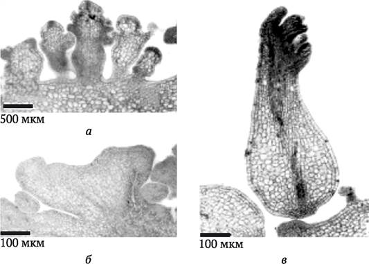
From a practical standpoint, shoot organogenesis and somatic embryogenesis are of the greatest interest, as they allow an entire plant to be regenerated from a cell culture or directly from an explant. Shoot organogenesis results in the formation of a unipolar structure containing a shoot apical meristem. The developing shoot maintains vascular connection with the maternal tissue (Fig. 3.5, a, b). Progenitor plantlets formed in culture can be rooted (*in vitro* or *in vivo*), thereby yielding a complete plant. Somatic embryogenesis yields a bipolar structure (an embryoid) containing both shoot and ROOT meristems (Fig. 3.5, c). Embryoids lack vascular connections with maternal tissues. Under specific *in vitro* conditions, embryoids are also capable of developing into complete plants.
Fig. 3.5. Morphogenic structures of Melia azedarach: a and b — unipolar structures containing apical meristems and maintaining connection with The vascular system of the maternal tissue (organogenesis; at different magnifications); c — bipolar structure with shoot and root system primordia (embryoid) lacking connection with the maternal tissue (after S. Vila et al., 2010)
Already in the early 1990s, it was reported that plant regenerants had been obtained in cell cultures of more than 1,000 plant species. As a result, general patterns governing this process have been established. In dicotyledonous plants, it is easier than in monocots to achieve not only callus formation from explants, but also morphogenesis (organogenesis or embryogenesis) from the callus. Among dicots, species belonging to Solanaceae, Brassicaceae, and Apiaceae exhibit higher regenerative capacity, whereas Fabaceae show a lower capacity. Cereals possess the lowest regenerative capacity. Within individual species, genotypes with relatively high and low capacities for morphogenesis can be found.
The capacity of isolated plant cells for morphogenesis is influenced by various factors associated with both the plant itself and the cultivation conditions. Of great importance are the plant's age (especially for woody crops and cereals), its physiological state and health status, the conditions under which it was grown, as well as the plant part (organ) from which the explant is taken, the physiological and ontogenetic age of the organ, the season when explantation is performed, the size of the explant, and even its orientation and degree of immersion when placed onto the nutrient medium. As a rule, the highest regenerative capacity is exhibited by cell cultures derived from explants originating from young, healthy plants originating from embryonic (mature or immature embryos) or meristematic tissues. During subculturing, the regenerative capacity of a cell culture gradually decreases (initially for shoot organogenesis, later for root organogenesis) and in many cases is lost entirely. Hormone-independent cultures and tumor cell cultures are generally incapable of organ regeneration and embryoid formation. If shoot primordia do form, they most often exhibit aberrant shapes, and obtaining a healthy plant from them proves unsuccessful.
Among the factors related to plant cell cultivation, the COMPOSITION OF THE nutrient medium is of paramount importance for morphogenesis, particularly the composition and ratio of phytohormones (growth regulators), its consistency (liquid or agar-solidified), as well as cultivation conditions: Temperature, dark or light incubation, illumination characteristics (light spectral features, light intensity, and photoperiod length), the application of stress factors to the cultures (e.g., temperature drops), exposure of cultures to electric current, and others.
3.10. Induction of Morphogenesis. Hormones as Morphogenesis Inducers
Under the influence of a particular morphogenic stimulus, a callus cell becomes determined, i.e., it commits to the developmental pathway it will follow. However, not all cells, but only one out of 400–1000, enter The pathway of secondary differentiation leading to morphogenesis. Consequently, an inducer (stimulus) alone is insufficient for the transition to morphogenesis; the cell must also be primed to respond to it. The ability to perceive an inducing stimulus and specifically react to it by altering the developmental program is referred to as cell competence.
The most powerful inducer of organogenesis, commonly referred to as a morphogenesis stimulus or signal, is a shift in the ratio between the concentrations of cytokinins and auxins included in the nutrient media. In their now-classic paper, F. Skoog and C. Miller (1957) demonstrated that when tobacco stem parenchyma callus is cultured on a nutrient medium containing an auxin (IAA) and a cytokinin (kinetin) at approximately equal molar concentrations, intensive cell proliferation occurs; when cytokinins predominate over auxins, shoot organogenesis is frequently initiated, whereas auxin predominance leads to root organogenesis (Fig. 3.6).
Fig. 3.6. Shoot and root organogenesis in tobacco cell culture depending on the ratio of auxin (indole-3-acetic acid, IAA) and cytokinin (kinetin) concentrations in the nutrient medium
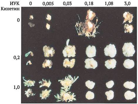
The application of exogenous hormonal preparations remains one of the primary approaches for obtaining plant regenerants across various plant species. At the same time, the range of applied auxins, cytokinins, and their analogs has expanded significantly, and Other growth regulators—such as Gibberellins, Abscisic acid, and Ethylene—have also come into common use for these purposes. It has been found that the regularity established by F. Skoog and C. Miller is not observed in many cases. For instance, species such as alfalfa have been discovered where, unlike tobacco, the predominance of auxins over cytokinins leads to shoot organogenesis. This is explained by the varying levels of endogenous phytohormones in the explant tissues and the differing sensitivities of competent callus cells to growth regulators. Therefore, in each specific case, to effectively induce morphogenesis and obtain plant regenerants, it is necessary to find the optimal phytohormone ratio and maximally account for other factors that may influence this process, as well as their potential interactions.
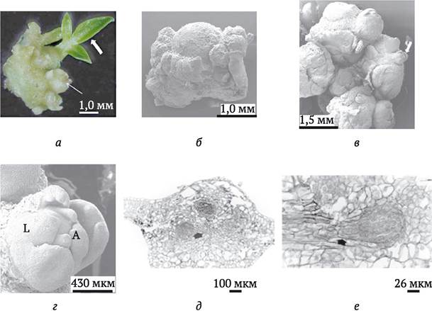
Organogenesis in callus tissue begins when, under the influence of appropriate stimuli (primarily through Changes in the ratio of auxin and cytokinin concentrations in the nutrient medium), a competent cell or a group of competent cells separates from the surrounding callus cells, forming zones of heightened mitotic activity—the so-called meristemoids (J. G. Torrey, 1966) (the term "initial" is sometimes used). Meristemoids are typically located in the lower part of the callus, near tracheid-like elements, which ensures contact with the nutrient medium and the perception of the hormonal stimulus. Meristemoid cells differ from other callus cells by their small size, isodiametric shape, thin walls, and numerous small vacuoles. A large nucleus usually occupies a central position. Subsequently, primordia of the shoot, root, leaf, or flower bud differentiate within the meristematic focus, leading to shoot, root, leaf, or floral organogenesis, respectively (Fig. 3.7). Meristemoid formation is preceded by starch accumulation in the zones of their Location. The organogenesis process itself is characterized by significant Energy Expenditure (enhanced glycolysis and the Pentose Phosphate Pathway of Carbohydrate Oxidation). It has been shown that morphogenesis in tobacco callus tissue cultures is characterized by the activation and Repression of the Synthesis of specific marker proteins. Induced determination of callus tissue cells is associated with the appearance of an antigen that serves as a marker for stem meristem cells.
Fig. 3.7. Appearance and Developmental Stages of a Kidney-shaped meristemoid forming in an in vitro culture of a Sclerocarya birrea leaf explant (a–d) (after M. Moyo et al., 2009); Anatomical structure of a meristemoid developing in an in vitro culture of a Passiflora edulis leaf explant (e and f) (after B. Gloria et al., 1999): a — appearance of a meristemoid (thin arrow) and a shoot-like structure (bold arrow) forming in the leaf explant culture; b — scanning electron micrograph of the globular stage of meristemoid development; c — scanning electron micrograph of a cluster of differentiating meristemoids, with the arrow pointing to one of the initiating vegetative buds; d — micrograph of a formed vegetative bud with a differentiated shoot apical meristem (A) and leaf primordia (L); e and f — histological cross-section of a Passiflora edulis leaf explant on the 21st (e) and 28th (f) day of explant culture on Murashige-Skoog medium supplemented with 1 mg/L naphthaleneacetic acid; the arrow indicates the site of formation of a root meristemoid developing within the vascular System of the leaf explant
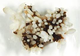
To induce somatic embryogenesis, the presence of an auxin (most commonly 2,4-D) in the nutrient medium is usually required. It is believed that auxin, along with other embryogenesis inducers (such as an electric field) applied to induce somatic embryogenesis, alters the polarity of competent cells, leading to the formation of gradients of endogenous auxin, bioelectric potentials, and calcium. In addition, cytoskeletal elements undergo reorganization within the cells, which is associated with their polarization and subsequent asymmetric divisions. It is precisely the small daughter cells resulting from asymmetric division that are embryogenic (determined for embryogenesis). Predetermined embryogenic cells may also be initially present in the explant tissues, making it possible to obtain somatic embryoids directly from explant cells (direct somatic embryogenesis) (Fig. 3.8) or to derive an "embryogenic callus" from such explants, from which large numbers of embryoids can easily be produced (Fig. 3.9).
Fig. 3.8. Somatic embryogenesis from a Coffea canephora Pierre leaf explant (after M. Priyono et al., 2010)
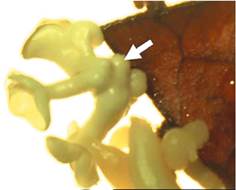
Fig. 3.9. Somatic embryogenesis from grape callus (www.itg.beckman.illinois.edu)
Embryogenic cells differ from other callus cells by their small size, angular shape, denser cytoplasm, small vacuoles (about 30% of the cell volume), and numerous starch grains. They possess a large nucleus and an Abundance of Ribosomes. Their microtubules exhibit a linear orientation parallel to the cell axis. Plasmodesmata, which are virtually absent in the bulk of callus cells, are re-established between embryogenic cells.
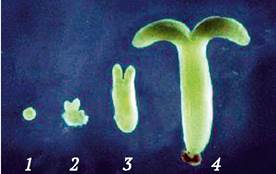
The division of embryogenic cells results in the formation of a globular proembryo. The condition for its further development is the transfer of the cultures to a nutrient medium containing reduced auxin concentrations or to a completely hormone-free medium. The removal of the dedifferentiating factor (auxin) from the nutrient medium causes the proembryo to transform into a globular embryo, which subsequently passes through the stages characteristic of zygotic embryo development: Heart, torpedo, and mature somatic embryoid (Fig. 3.10). Somatic embryoids possess both shoot and root meristems. A plantlet develops from the shoot apex. The "root" end of the embryoid is typically oriented toward the callus, into which the newly formed roots "grow" and from which the developing plant receives essential nutrients.
Fig. 3.10. Stages of histodifferentiation of soybean somatic embryoids: 1 — globular embryo; 2 — heart stage; 3 — torpedo stage; 4 — mature somatic embryoid
(www.parrotlab.uga.edu)
A comparative analysis of somatic and sexual embryo development reveals a parallelism in their development, manifested in the fundamental patterns of morphogenesis: bipolarity, Symmetry, and cellular and tissue differentiation. Several genes expressed during somatic embryogenesis have been isolated. They are classified into three categories: 1) genes involved in cell division (21D7 and CEM1); 2) genes associated with embryo organ formation (CAR3, CAR4, and CHB4 — hypocotyl formation; CAR5, CAR6, and CHB — hypocotyl and/or root formation at the globular and torpedo embryo stages); 3) embryo-specific genes (CHB2 and CEM6) (L. Komamine, 2001).
3.11. Regeneration of Plants
The induction of stem organogenesis or somatic embryogenesis makes it possible to obtain regenerated plants. Through stem organogenesis, plants regenerate from callus cultures as well as from explant cells by inducing adventitious buds or from the axillary buds of stem cuttings. Adventitious buds in intact plants are formed not at the shoot apex or in the leaf axil, but on various plant organs (roots, leaves, stems). Unlike apical meristems, they are not connected to them, but instead form from various tissues, both superficial and deeply situated. Cell culture techniques allow the production of adventitious buds from leaf, tuber, stem, and other plant explants in large quantities. The same applies to the induction of shoots from the axillary buds of stem explants. Obtaining regenerated plants from axillary and adventitious buds represents the Main methods of vegetative (clonal) plant propagation in vitro. Shoots formed from buds are separated from the explant, rooted on a nutrient medium containing auxins (or lacking growth regulators), and then transferred to in vivo conditions where they are grown into mature plants.
Somatic embryogenesis is induced in cell suspension cultures, callus cultures, and anther cultures. The resulting embryoids are transferred to a hormone-free nutrient medium where, when cultivated in the light, they develop into test-tube plants that are subsequently planted in soil after a hardening-off period. Somatic embryogenesis is also of great interest for clonal plant propagation. The methods of in vitro clonal plant propagation will be discussed in detail in Chapter 5.
The cells of a developing plant embryo are undifferentiated. They divide intensively and can give rise to various plant tissues and organs. During differentiation, cells become specialized, concurrently losing their capacity to divide. Only meristematic cells retain an embryonic state. However, under certain conditions—such as wounding—plants can develop new, secondary meristems. Wound meristems give rise to callus, a specialized tissue consisting of undifferentiated parenchymal cells that divide intensively to cover the wound site. Through their secondary differentiation, damaged tissues and organs are repaired.
The process of callus formation and the regeneration of tissues, organs, and whole plants from differentiated, specialized cells can be reproduced in plant cell cultures on artificial nutrient media containing specific phytohormones or growth regulators. The process of specialized cell dedifferentiation is induced by auxins. However, for dedifferentiated cells to resume division, cytokinins are required. The initial prerequisite in this process is dedifferentiation: the loss of tissue cell specialization and the restoration of their division capacity. Dedifferentiation results in a substantial shift in cellular metabolism. Explant cells lose stored lipids, starch, and proteins. Photosynthetic cells lose chlorophyll and chloroplast lipids, while the number of amyloplasts increases, the Golgi apparatus breaks down, and The endoplasmic reticulum and Cytoskeleton elements undergo reorganization. The energy metabolism of callus cells largely resembles that of meristematic cells. Their mitochondria are poorly developed with few cristae, which negatively affects aerobic respiration activity. Consequently, they consume less oxygen compared to specialized plant cells, and their respiratory-fermentative balance shifts toward fermentation.
Unlike meristematic cells, which are characterized by high uniformity and genetic stability, the growth and secondary differentiation of callus cells occur in an unorganized and asynchronous manner. Callus tissue simultaneously contains cells at various stages of the mitotic cycle. Furthermore, callus tissue culture is characterized by cytogenetic instability, including changes in cellular ploidy, chromosomal aberrations, and point mutations.
Cell division in in vitro culture requires the presence of phytohormones or synthetic growth regulators in the nutrient medium. However, prolonged cultivation can lead to the emergence of cells with persistent dedifferentiation, a characteristic feature of which is hormone independence. Hormone independence is also typical of crown gall tumor cell cultures. It has been established that this disease is caused by agrobacteria capable of transforming plant cells by transferring and integrating bacterial DNA fragments carrying genes that encode auxin and cytokinin biosynthesis into the plant genome.
During cultivation, cells may either maintain persistent dedifferentiation or undergo secondary differentiation accompanied by histogenesis (the formation of various tissues within the callus) or morphogenesis (the emergence of organized structures). Morphogenesis occurs via organogenesis or somatic embryogenesis. Organogenesis is the regeneration of individual plant organs (stems, roots, and more rarely, floral elements or leaf primordia) in in vitro cell culture. Somatic embryogenesis is the formation of embryoids, i.e., structures resembling zygotic embryos. Both organogenesis and somatic embryogenesis can occur not only in cell culture, but also in plants in vivo, as well as directly from the explant in in vitro culture (direct morphogenesis).
The ability of individual plant cells to alter their developmental program, undergo dedifferentiation and secondary differentiation, and consequently give rise to an entire plant—thereby realizing their inherent genetic information—is termed totipotency.
The most potent inducer of organogenesis is A change in the ratio of cytokinin to auxin concentrations in the nutrient media. F. Skoog and C. Miller demonstrated that cultivating callus on a nutrient medium containing auxin and cytokinin in approximately equal molar concentrations leads to intense cell proliferation; when cytokinins predominate over auxins, stem organogenesis is frequently initiated, whereas auxin predominance favors root organogenesis. Organogenesis in callus tissue begins when, under the influence of appropriate stimuli (primarily changes in the auxin-to-cytokinin ratio in the nutrient medium), a competent cell or a group of competent cells separates from the surrounding callus cells to form zones of heightened mitotic activity known as meristemoids. Subsequently, stem, root, leaf, or floral buds differentiate within this meristematic focus, resulting in stem, root, leaf, or floral organogenesis, respectively.
Inducing somatic embryogenesis typically requires the presence of an auxin (most commonly 2,4-D) in the nutrient medium. It is believed that auxin alters the polarity of competent cells, leading to subsequent asymmetric divisions. The small daughter cells produced by asymmetric division are embryogenic (determined for embryogenesis). The division of these embryogenic cells leads to the formation of a globular proembryo. A prerequisite for its further development is the transfer of the cultures to a nutrient medium containing reduced auxin concentrations or to a completely hormone-free medium.
Stem organogenesis results in the formation of a unipolar structure containing an apical shoot meristem. The seedlings produced in cell culture can be rooted (in vitro or in vivo), thereby yielding a whole plant. Conversely, somatic embryogenesis forms a bipolar structure (an embryoid) containing both shoot and root meristems. Under specific conditions in vitro, embryoids are likewise capable of developing into complete plants.
The main types of plant cell cultures are callus culture (cultivation on an agar-solidified nutrient medium), suspension culture (submerged cultivation in liquid media with artificial aeration), and protoplast culture (culture of cells lacking a cellulosic cell wall). Suspension cultures are most easily obtained from callus cultures. Cells cultivated in suspension can be plated onto an agar-solidified medium to yield callus culture. Prior to cell wall regeneration, the onset of cell division, and colony formation, protoplasts are cultivated in liquid nutrient media; subsequently, the colonies are transferred to an agar-solidified medium to establish callus culture. The induction of morphogenesis and the production of regenerated plants are most frequently achieved using callus cell cultures.
Methods for cultivating single cells and protoplasts have been developed, based on conditioning nutrient media, optimizing their composition, and employing small-scale cell cultivation techniques. Experiments on obtaining regenerated plants through the cultivation of single somatic cells provided direct proof of G. Haberlandt's hypothesis regarding plant cell totipotency. These methods are of great practical importance for spatially separating various cellular variants (cell lines) present within a cell culture and subsequently deriving modified regenerated plants (mutants, somatic hybrids) from them.
1. How do secondary meristems differ from primary ones?
2. Which phytohormones induce the transition of specialized plant cells to dedifferentiation?
3. What structural and metabolic changes occur in cells during dedifferentiation?
4. What are the differences in hormone independence between habituated and tumor plant cells?
5. What parameters are used to assess the growth intensity and viability of cells in suspension cultures?
6. For what purpose are osmotics introduced into the nutrient medium when isolating protoplasts?
7. What main approaches are used to induce cell division and obtain regenerated plants in single cell and protoplast cultures?
8. In what forms can secondary differentiation take place in plant cell cultures?
9. What main factors induce the transition of cells to organogenesis in in vitro culture?
10. Describe the processes occurring during somatic embryogenesis in plant cell culture.
11. How do seedlings regenerated in cell culture via caulogenesis (shoot organogenesis) differ from those obtained through somatic embryogenesis?
Last update: 11/08/2026
Editorial and Educational Adaptation: This material has been compiled based on the primary/original source text. The project team performed an editorial review, corrected technical inaccuracies, structured sections, and adapted the content for an educational format.
What was processed:
- elimination of formatting defects (OCR errors, structural breaks, corrupted characters);
- editorial organization of content;
- standardization of terminology in accordance with academic sources;
- verification of factual statements against the original source text.
All mentions of the author, publication year, and origin of the primary text have been preserved in accordance with the source.