TEXTBOOK ON PEDIATRIC GYNECOLOGY - 2013
Chapter 6. MENSTRUAL DISORDERS IN ADOLESCENT GIRLS. AMENORRHEA. PUBERTAL UTERINE BLEEDING
The Regulation of the menstrual cycle is primarily governed by the anterior pituitary gland, which synthesizes follicle-stimulating hormone (FSH), luteinizing hormone (LH), lactogenic hormone (LTH or prolactin), as well as adrenocorticotropic hormone (ACTH), Growth Hormone (GH), and thyroid-stimulating hormone (TSH).
Under METABOLISM/18.html">The Influence of FSH, follicular and oocyte maturation occur in the Ovary; ovulation is driven by the combined action of FSH and LH; and the formation and Development of the corpus luteum are regulated by LH and LTH. Thus, the ovarian cycle encompasses follicular growth and maturation, ovulation, and corpus luteum formation.
Estrogens are secreted by the ovaries (primarily by the follicular granulosa and theca Cells) throughout the entire menstrual cycle. Their levels peak particularly on days 6, 14, and 26 of the cycle, as well as on day 3 of menstruation. During the functional phase of the corpus luteum, progesterone levels exceed those of estrogens. In addition to estrogens and progesterone, small amounts of androgens are continuously secreted by the ovaries.
Sex Hormones (estrogens, progesterone, and androgens) are secreted not only by the ovaries but also by the Adrenal Glands. Gonadotropic hormones are excreted by the pituitary gland and the chorionic layer of the Placenta (chorionic gonadotropin). Pituitary gonadotropin exhibits predominantly follicle-stimulating activity, whereas chorionic gonadotropin acts primarily as a luteinizing agent.
Driven by Ovarian Hormones, cyclic changes occur in the uterine endometrium (the uterine menstrual cycle), which undergoes two phases: proliferation and secretion. The proliferative phase is induced by estrogens, while the secretory phase is driven by progesterone. Toward the end of the cycle, The production of progesterone and estrogens declines, triggering epithelial desquamation (menstruation). Endometrial regeneration begins concurrently with its shedding (Fig. 9).
Class="center">
Fig. 9. Dynamics of hormone secretion, basal body Temperature, and Changes in the ovaries and Uterus throughout the menstrual cycle
In clinical practice, the functional hormonal activity of the ovaries is assessed using the following diagnostic tests: 1) basal body temperature charting; 2) the pupil phenomenon; 3) the ferning (arborization) test of cervical mucus; 4) vaginal Cytology (smear analysis); 5) histological examination of endometrial scrapings; 6) quantitative hormone assays in Blood and urine; and 7) radioimmunoassay (RIA) measurement of blood levels of FSH, LH, estrogens, and progesterone. Adrenal activity is evaluated by urinary 17-Ketosteroid (17-KS) levels.
Impairment at any level of the neuroendocrine regulatory system leads to menstrual cycle disturbances, which may manifest as hypomenstrual syndrome or Dysfunctional Uterine Bleeding.
Hypomenstrual syndrome results from various disruptions in the menstrual regulatory system and is characterized by scant (hypomenorrhea), short-duration (oligomenorrhea), or infrequent (opsomenorrhea) periods. The extreme manifestation of hypomenstrual syndrome is Amenorrhea (the absence of menstruation for 6 months or longer).
Premenstrual syndrome (PMS) is a symptom complex that occurs in the days preceding menstruation, manifesting as neuropsychiatric, cardiovascular, and metabolic-endocrine disorders.
The prevalence of PMS depends on multiple factors—such as age, emotional Background, and concomitant diseases—and ranges widely from 25% to 90%. In 5–10% of women, PMS is severe. Pain during the second phase of the menstrual cycle occupies a leading position in adolescent gynecological morbidity: among vocational college and university students, the incidence reaches 17–22%, and according to some sources, up to 75%.
The gravity of PMS lies in the fact that for approximately every third girl, menstruation assumes the characteristics of a debilitating condition. Notably, during adolescence, acute painful episodes are accompanied by vomiting in 84% of cases, diarrhea in 79.5%, dizziness in 22.7%, headache in 13.6%, and convulsions or fainting in 15.9%. The monthly anticipation of such episodes adversely affects the adolescent's general well-being, emotional state, and mental performance, frequently resulting in absenteeism and impaired productivity.
Amenorrhea can be either pathological or physiological during a girl's pubertal development. A distinction is made between primary amenorrhea (where menstruation has never occurred) and secondary amenorrhea.
Endocrine-related amenorrhea is characterized by varying degrees of hypoestrogenism and hyperandrogenism. This sex hormone imbalance leads to The Development of male physical characteristics (virilization) and hirsutism (male-pattern Hair growth).
Pathological amenorrhea is caused by disorders at any level of menstrual regulation. Its forms include central, hypothalamo-pituitary, ovarian, and uterine amenorrhea, as well as forms resulting from adrenal and Thyroid Gland dysfunction.
This Classification by level of impairment is somewhat conventional because the menstrual regulatory system Functions as a unified whole, and dysfunction in any single link inevitably impacts the others. Identifying the primary affected level is crucial for determining the appropriate therapeutic strategy.
The underlying causes of pathology at each level of regulation may be functional (such as Inflammatory Diseases), organic (such as tumors), or congenital.
Central amenorrhea is most commonly functional and, much less frequently, organic in nature. It may result from psychological stress, a history of meningitis, encephalitis, HEAD trauma, psychiatric disorders (Schizophrenia, manic-depressive illness, etc.), or Brain Tumors. Alongside amenorrhea, patients may experience irritability, tearfulness, headaches, impaired memory and concentration, and Sleep disturbances.
Hypothalamo-pituitary amenorrhea of functional origin is often associated with intoxication from various infectious diseases (tonsillitis, tuberculosis, rheumatism, brucellosis, neuroinfections, etc.). Organic lesions of the hypothalamo-pituitary region may be caused by tumors or pituitary vascular thrombosis. Pathology of the hypothalamo-pituitary system can also be congenital.
General clinical manifestations include Metabolic Disorders—specifically involving Lipid Metabolism (excess weight), CARBOHYDRATES, and Water—along with hirsutism secondary to ovarian hypofunction. Cardiovascular and peripheral autonomic symptoms are frequently observed, such as hypo- or Hypertension, brady- or tachycardia, blood pressure Asymmetry, physical lethargy (hypodynamia), polyuria, low-grade fever, peripheral paresthesias, striae (pink or white stretch marks) on the thighs, abdomen, and breasts, and a moon face. These symptoms may precede or accompany the cessation of menstruation.
In pituitary tumors, general clinical signs are accompanied by manifestations determined by the specific pituitary Cell line involved. In acidophilic adenoma, increased growth hormone production leads to gigantism or acromegaly. As the tumor enlarges, patients develop headaches, visual impairments, and eventual blindness. Amenorrhea ensues due to the suppression of pituitary gonadotropic activity.
In basophilic adenoma, amenorrhea is combined with Cushing's Disease (hyperproduction of ACTH by basophilic cells).
An example of congenital pathology of the hypothalamo-pituitary region is the so-called Adiposogenital Dystrophy. In this condition, along with amenorrhea, obesity, a moon-shaped face, genital hypoplasia, skeletal developmental defects (flat feet, Clubfoot, excessive mobility in the JOINTS OF THE upper limbs, etc.), and hypertrichosis are observed.
Amenorrhea caused by Adrenal gland disorders. The clinical picture is characterized by a virilizing syndrome of varying severity. Adrenal-origin virilizing syndrome is commonly referred to as adrenogenital syndrome (AGS). It can be either congenital or acquired. In turn, acquired AGS is sometimes the result of functional and organic pathology of the adrenal glands.
Congenital AGS is associated with enzymatic disorders in the adrenal cortex: cortisone deficiency leads to increased ACTH production, which promotes androgen overproduction. Clinically, female pseudohermaphroditism is observed, meaning a disorder of the sexual differentiation of external genitalia (while the uterus, ovaries, and fallopian tubes are present), characterized by clitoridal hypertrophy and the presence of a urogenital sinus (where the external urethral and vaginal openings merge into a single Structure). In the pre- and pubertal periods, hirsutism appears, and skeletal development is disrupted. By the age of 10, rapid growth and premature epiphyseal ossification are noted. Enlarged chest dimensions, a broad shoulder girdle, and a narrow pelvis are also characteristic. During Puberty, primary amenorrhea occurs.
Acquired AGS of a functional nature is associated with adrenal cortex hyperplasia, whereas organic pathology is caused by adrenal tumors (arrhenoblastoma). Clinically, adrenal cortex hyperplasia is dominated by hirsutism and amenorrhea, while arrhenoblastomas present with pronounced AGS, progressing to a fully developed intersexual type of masculinization (boyish behavior).
Ovarian form of amenorrhea is the most common, accounting for 40-50% of all cases of amenorrhea. This form results from ovarian insufficiency or absence, which can be primary, genetically determined (gonadal dysgenesis), or secondary, following surgical removal of the ovaries (castration) during childhood and adolescence.
Ovarian hypofunction can result from lesions in the hypothalamo-pituitary region, as well as from inflammatory processes in the ovaries or severe infectious diseases during the pre- and pubertal periods.
In this case, the alterations are multifaceted. With mild insufficiency, follicles grow but regress before reaching the Graafian follicle stage. With profound insufficiency, only primordial follicles are identified without any tendency toward growth. Depending on the degree of alterations in the ovarian tissue, varying degrees of hormonal deficiency occur, which is the primary factor in the development of sexual and somatic infantilism.
Ovarian-origin amenorrhea may occur due to organic tumor lesions (arrhenoblastoma) or sclerocystic ovarian changes. Sclerocystic ovaries, or Stein-Leventhal syndrome, are characterized by impaired ovarian steroidogenesis due to enzymatic system deficiencies, resulting in a significant influx of androgens into the body. In sclerocystic ovaries, massive Connective Tissue proliferation occurs in the cortical and medullary layers, along with vascular wall sclerosis and hyperplasia of the theca tissue elements, coupled with an increase in cystic-atretic follicles, causing the tunica albuginea to thicken by 8-10 times. These changes lead to anovulation.
Ovarian-origin amenorrhea is associated with congenital pathology, specifically gonadal dysgenesis (Turner syndrome). This is a primary developmental deficit of ovarian tissue caused by a congenital malformation and an incomplete chromosome Complement (45,X0). Consequently, 80% of females exhibit negative sex Chromatin. Gonadal dysgenesis may also be triggered by infections or intoxications during the period of embryonic Gonadal Sex differentiation.
Turner syndrome is characterized by primary amenorrhea, absence of secondary sexual characteristics, underdeveloped Mammary Glands, sparse genital hair, a short neck, webbing of the neck, short stature, marked Hypogenitalism, numerous developmental anomalies, mental retardation, Osteoporosis, short hands and feet, and dermatoglyphic abnormalities of the palms and soles.
Ovarian hypofunction manifests in various ways: ranging from pronounced forms of ovarian failure bordering on complete loss of ovarian function, to nearly normal function. Accordingly, Menstrual disorders also vary, from hypomenstrual syndrome to amenorrhea.
Clinically, ovarian amenorrhea is characterized by:
1) late and insufficient development of Primary and secondary sexual characteristics combined with somatic underdevelopment during puberty;
2) hypotrophy of previously well-developed secondary and primary sexual characteristics when ovarian failure occurs post-pubertally. Severe ovarian failure manifests with symptoms resembling Climacteric syndrome (irritability, memory impairment, headaches, hyperhidrosis, etc.), often accompanied by obesity resulting from slowed oxidation processes due to ovarian hypofunction.
Ovarian sclerocystosis clinically presents with hypomenstrual syndrome or amenorrhea, hirsutism, and bilateral ovarian enlargement against the background of a normal female body habitus and well-developed sexual characteristics.
Uterine amenorrhea can be primary (congenital uterine anomalies, childhood tuberculous endometritis, diphtheria) or secondary. Secondary endometrial damage may occur due to inflammatory processes or trauma to the basal endometrial layer resulting from intrauterine Procedures. During puberty, the uterine form of amenorrhea is extremely rare and is most commonly the consequence of tuberculous endometritis (at the terminal scarring stage).
"False" uterine amenorrhea occurs when normal cyclic changes (proliferation, secretion) take place in the functional layer of the endometrium, but menstrual blood cannot escape due to a congenital obstruction (obliteration of a traumatized cervical canal or an imperforate hymen).
Diagnosis and Differential diagnosis OF the level and Nature of the lesion in amenorrhea are of paramount importance, as Treatment directly depends on them. It is essential to consider the patient's history, Clinical presentation, and the findings from general, specialized, and hormonal examinations.
It is crucial to determine the patient's developmental history during childhood, the timing of secondary sexual characteristics appearance, and The history of acute or prolonged infectious diseases in childhood (scarlet fever, tuberculosis, chronic tonsillitis, etc.). During the general physical examination, attention must be paid to body habitus, subcutaneous fat distribution, skeletal development, and the degree to which secondary sexual characteristics correspond to the patient's age. Evaluation of the medical history, along with general and specialized examinations, determines the direction of further diagnostic procedures.
If a hypothalamo-pituitary Etiology of amenorrhea is suspected, the presence of a tumor in this region must be ruled out. For this purpose, Skull and sella turcica radiography is performed, alongside an ophthalmologic examination with mandatory assessment of visual fields and the optic fundus.
In cases of a pituitary tumor, radiographs reveal deformation and altered dimensions of the sella turcica, accompanied by "digital impressions" and enhanced vascular markings in the cranial vault bones due to tumor compression of the third cerebral ventricle and increased intracranial pressure.
Ophthalmologic examination reveals bitemporal hemianopsia and papilledema resulting from tumor pressure on the optic chiasm.
Lesions of the Hypothalamic and pituitary regions are characterized by metabolic disturbances, most notably a decrease in basal metabolism. Carbohydrate Metabolism disorders may manifest as hypo- or hyperglycemia (glucose tolerance test) up to latent Diabetes Mellitus. Electroencephalography demonstrates disorganization of bioelectrical rhythms originating from deep-seated structures, as well as a shift in the median brain structures.
From a functional standpoint, hypothalamo-pituitary amenorrhea is characteristically associated with a marked reduction in gonadotropin secretion. A decrease in FSH and LH levels can be detected in blood and urine using radioimmunoassay, as well as through hormonal stimulation tests.
Ovarian amenorrhea is diagnosed using functional diagnostic tests. Depending on the degree of hypoestrogenemia, the test results vary. Mild hypoestrogenemia may present with a «pupil» sign of + or ++, a moderate decrease in the karyopyknotic index (KPI), a monophasic basal body temperature, and signs of proliferation in the uterine mucosa during menstruation. In cases of severe ovarian insufficiency, the «pupil» sign is always negative, the KPI is sharply reduced, basal and parabasal cells predominate in smears, and the uterine mucosa is atrophic, occasionally showing signs of early proliferation. The excretion of estrogens and pregnanediol is decreased, whereas gonadotropins are found in significant amounts.
To confirm the diagnosis of polycystic ovary syndrome, rentgenopelviography is performed (showing enlarged ovarian contours on the radiograph). However, the most reliable data are obtained via laparoscopy with a biopsy of the ovarian tissue. Functional diagnostic tests in polycystic ovaries indicate their hypofunction. Urinary 17-ketosteroid (17-KS) levels may be slightly elevated.
The congenital origin of ovarian amenorrhea is diagnosed based on the patient's characteristic phenotype (Turner syndrome) and confirmed by cytogenetic studies of the karyotype (chromosome set) and the presence of sex chromatin in oral mucosal smears. In 45,X0 syndrome, sex chromatin is absent.
Patients presenting with virilizing syndrome or hirsutism require a thorough evaluation of adrenal function. In cases of clinically pronounced adrenogenital syndrome (AGS), adrenal cortical tumors or hyperplasia must be ruled out. For this purpose, adrenal radiography under conditions of retropneumoperitoneum is performed.
To differentially diagnose the level of lesion in amenorrhea, as well as adrenal, ovarian, or hypothalamic-pituitary virilizing manifestations (such as tumors or cortical hyperplasia), functional tests are widely used. These are based on stimulating or suppressing the function of target Organs through the administration of appropriate hormonal agents.
The following functional tests are applied:
1. Cortisone test (prednisolone 10-20 mg) or hydrocortisone 50-100 mg for 5 days. Administration of these agents blocks pituitary adrenocorticotropic activity, leading to a decrease in 17-KS levels in adrenal cortical hyperplasia, whereas levels remain unchanged in tumors. In ovarian-origin androgen excess, 17-KS levels also remain unchanged.
2. ACTH test: administration of the drug (40 IU intramuscularly for 2 days) increases 17-KS levels in pituitary disorders and does not alter them in ovarian tumors.
3. For the differential diagnosis of ovarian versus adrenal virilizing manifestations, the Infecundin-prednisolone test can be used. Infecundin blocks pituitary action on the ovary. Over 10 days, the patient receives 2 tablets of Infecundin daily, combined with 20 mg of prednisolone starting from the 6th day. Three potential outcomes are possible: a) 17-KS levels decrease by more than 50% after Infecundin administration, indicating an ovarian origin of androgenism; b) if 17-KS levels decrease by 50% only after prednisolone administration, an adrenal origin is indicated; c) a slight decrease in 17-KS levels after both the First and Second halves of the test suggests a mixed origin of amenorrhea.
4. Choriogonin test: this is based on stimulating ovarian function with this preparation (hCG acts similarly to LH) and indicates whether amenorrhea is primary or secondary to pituitary dysfunction. If bloody vaginal discharge appears following hCG administration (1,500 IU daily for 5 days), the causes of amenorrhea are related to pituitary dysfunction; if not, it is typically of ovarian origin.
The assumption of a uterine form of amenorrhea may arise when functional diagnostic tests remain entirely normal, indicating intact hypothalamic-pituitary-ovarian regulation of the menstrual cycle. To clarify the diagnosis, an inspection of the hymen in young girls (to check for imperforation) and probing of the cervical canal (to check for atresia) should be performed to rule out «pseudo» uterine amenorrhea. Diagnostic curettage of the uterine mucosa helps determine The Nature and extent of endometrial damage (scarring, tuberculous endometritis). In cases of uterine amenorrhea, hysteroscopy and hysterography are also advisable (to detect synechiae, adhesions, and endometrial atrophy).
The management of amenorrhea depends on the diagnosed nature and level of the lesion. Organic lesions, such as brain and adrenal tumors, require Surgical treatment, whereas pituitary tumors are treated with radiotherapy.
If amenorrhea is caused by functional or congenital pathology, hormone therapy is administered. The Main Components of hormonal treatment include: estrogen preparation, cyclic (replacement) therapy, and withdrawal-bleed therapy.
Estrogen preparation involves the systematic administration of estrogenic hormones alone, most often prolonged-action agents: estradiol dipropionate 0.1% — 1 ml intramuscularly 1–2 times a week for 4–6 months, or 10–12 months with breaks, spanning several years. Estrogen preparation is designed to stimulate the development of reproductive organs and secondary sexual characteristics, and to induce menstrual-like bleeding.
Cyclic (replacement) hormone therapy is based on mimicking a normal menstrual cycle. The regimen to establish a 28-day cycle is as follows: on days 4, 6, 8, 10, and 12, administer 10,000 IU of synestrol (0.1% — 1.0 g) or folliculin; on days 14, 16, and 18 — 10,000 IU of estrogens along with 10 mg of progesterone (or 1,500 IU of hCG); and from days 20 to 25 — 5–10 mg of progesterone daily, followed by an 8–10 day break. Usually, 2–3 cycles are performed. After a 1–4 month break, 1–2 treatment cycles are repeated, sometimes continuing for up to 2 years. Such therapy normalizes the hypothalamus-pituitary-ovaries-uterus axis, helps restore cyclic processes within it, and activates ovarian function.
For the same purpose, treatment with synthetic progestins (Infecundin, Bisecurin, Ovulen, megestranol, etc.) is used: 1 tablet daily from the 5th to the 25th day of the menstrual cycle for 3–4 months. Synthetic progestins suppress pituitary gonadotropic activity, and upon drug withdrawal, an enhancement of the functional activity of the hypothalamic-pituitary system occurs (the «withdrawal» effect), leading to increased gonadotropin production and subsequent activation of ovarian function.
Given that amenorrhea is often associated with neurological disorders, sedative therapy is recommended (e.g., Bellaspon, Beloid, bromine-valerian mixture), while pronounced emotional and psychological disturbances warrant the administration of tranquilizers such as seduxen or elenium for 1–3 weeks.
In functional hypothalamic-pituitary amenorrhea, The Use of hormonal agents, drug-based Electrophoresis, and physiotherapy is appropriate. The combination of cyclic hormone therapy and transnasal galvanic electrophoresis with a 2% Vitamin B1 solution, 2% calcium chloride solution, 0.25% diphenhydramine solution, Shcherbak's ionogalvanization method, and Vitamins C and E is highly effective.
For adiposogenital dystrophy, a low-calorie diet limiting fats and carbohydrates, along with medical gymnastics (physical therapy), is prescribed. Hormonal agents include thyroidin (0.002–0.1 g daily), and the administration of long-acting estrogens is recommended. Hormonal treatment combined with diet and Physical Exercise aims to eliminate obesity and hypogenitalism.
The treatment of ovarian amenorrhea involves the scheduled administration of sex hormones. In cases of pronounced ovarian hypoplasia (hypofunction), treatment consists of two stages: Stage I — stimulation of primary and secondary sexual characteristics, and elimination of neurovegetative processes and trophic disorders; Stage II — induction of cyclic processes at the hypothalamic-pituitary-ovarian-uterine level.
Stage I involves estrogen preparation, and once bleeding appears, the patient transitions to cyclic hormone therapy.
If ovarian hypofunction is mild, only cyclic hormone therapy or treatment with synthetic progestins should be used. Concurrently with hormone therapy, diathermy to the sacral region is advisable to improve Blood supply to the pelvic organs.
The treatment of polycystic ovary syndrome is surgical, consisting of bilateral wedge resection of the ovaries, preferably using minimally invasive techniques.
Treatment for patients with gonadal dysgenesis (Turner syndrome) should ideally begin at ages 10–12, prior to puberty. Treatment with thyroidin and anabolic Steroids aims to correct somatic disproportion. After age 16, estrogens can be used to reduce sexual infantilism. Administering estrogens before age 10–12 would lead to premature epiphyseal fusion.
The management of uterine amenorrhea is tailored to its etiology. In pseudo-amenorrhea, surgical incision of the hymen or bougienage of the cervical canal is necessary. In inflammatory etiologies, anti-inflammatory therapy, estrogen treatment, and cyclic hormone therapy are prescribed. Simultaneously, diathermy of the sacral region is performed to increase hyperemia of the uterus and its appendages and enhance their sensitivity to hormonal action.
Pubertal uterine bleeding (PUB) accounts for 22–35% of all menstrual disorders in adolescents, and 11–15% of all gynecological pathologies in adolescent girls.
The functional state of the Higher Nervous Activity controlling The regulatory mechanisms of menstrual function is unstable during puberty; differentiation in the cerebral cortex, and particularly in the hypothalamic centres, remains immature. The electrical activity of the cerebral cortex in girls aged 11–15 years does not reach the levels observed in adult women. The uterus in adolescent girls is not yet fully developed, its receptors are immature, and its capacity to perceive stimuli and transmit them to the Central Nervous system is poorly expressed. Consequently, the pituitary gland receives distorted signals and fails to coordinate the synthesis of all gonadotropic hormones, resulting in a predominance of FSH production, while LH and LTH are not produced in sufficient quantities.
Against this background, the hormonal function of the ovaries is insufficiently developed, and their receptor apparatus remains imperfect.
External and internal stimuli can easily disrupt the regulatory mechanisms of menstrual function, hindering the establishment of its normal pattern and triggering juvenile uterine bleeding.
Factors predisposing to bleeding include stress, physical trauma, chronic fatigue, unfavourable living conditions, acute or chronic infections, intoxications, hypo- and avitaminoses, as well as thyroid and adrenal cortex dysfunctions.
Chronic and acute infectious diseases, inadequate Nutrition (avitaminosis), psychological trauma, and psycho-emotional stress play a crucial role in the development of PUB. The medical history of girls with PUB frequently reveals acute infectious and viral diseases (chickenpox, rubella, measles, scarlet fever, mumps), etc.
Extragenital pathology is also of significant importance in the onset of PUB: gastrointestinal disorders (particularly of the hepatobiliary system), vegetative-vascular dystonia, Epilepsy, thyroid hypofunction, respiratory diseases, allergic reactions, blood disorders, etc. (Fig. 10).
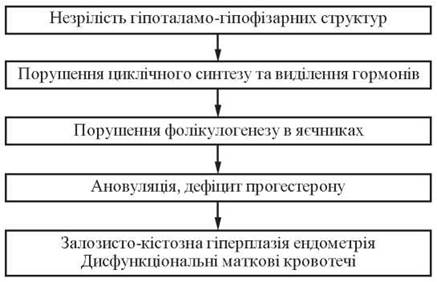
Fig. 10. Pathogenesis of pubertal uterine bleeding
Functional uterine bleeding is fundamentally caused by the prolonged persistence of a single follicle or the atresia of multiple follicles. In both scenarios, the cycle remains monophasic. The endometrium is exposed to an absolute excess of estrogens or to the prolonged action of estrogens unopposed by progesterone. As a result, the endometrium undergoes excessive proliferation without secretory transformation.
Typically, bleeding associated with follicular persistence is heavier—due to higher bodily estrogen levels and more pronounced endometrial proliferation—than that associated with follicular atresia.
Pubertal uterine bleeding can be cyclic (where a timely menstruation transitions into bleeding) or acyclic (characterized by a delayed menstruation followed by heavy bleeding). The duration of bleeding ranges from a few days to several months. The episodes may occur infrequently, but they are typically prolonged and heavy.
Juvenile uterine bleeding is generally accompanied by anemia, which occasionally reaches a severe degree. This is attributed to insufficient uterine contractility, hormonal imbalance during puberty, and secondary Disorders of the Blood Coagulation and anticoagulation systems, which further aggravate blood loss.
The leading symptom of dysfunctional uterine bleeding is genital Hemorrhage. Girls typically complain of general weakness, dizziness, headaches, palpitations, sleep and appetite disturbances, reduced working capacity, irritability, and tearfulness.
Physical examination reveals pallor of the Skin and visible mucous membranes, tachycardia, and low blood pressure. The severity of these symptoms depends on the degree of anemia, the duration of bleeding, and hemodynamic disturbances. The girl's condition must be assessed based on her physical development and somatic status, paying particular attention to The Cardiovascular system and Liver function.
When a patient presents with bleeding, it is essential to ascertain the age at menarche, its pattern and regularity, as well as the characteristics of subsequent menstrual periods (duration, heaviness, and pain).
Gynecological examination involves assessing the stage and sequence of secondary sexual characteristic development.
Examination of the genitalia focuses on the development of the external sex organs and the condition of the vulvar and hymenal mucosa. Recto-abdominal examination helps determine the position, size, and mobility of the uterus, the ratio between the uterine body and cervix, and the condition of the uterine appendages.
In cases of dysfunctional uterine bleeding, functional diagnostic tests are recommended to clarify the underlying causes. These typically show a monophasic basal body temperature, a karyopyknotic index (KPI) of 20–40%, while findings regarding FSH, LH, and estrogen levels remain variable.
For differential diagnosis, blood disorders (such as nosebleeds and other hemorrhages; platelet counts, coagulograms, and thromboelastograms should be performed), cardiovascular diseases, liver pathologies, and thyroid dysfunctions must be ruled out.
Differential diagnosis should also exclude gynecological conditions, including hormone-producing ovarian tumors, Genital Tuberculosis, Cervical Cancer, vaginal Sarcoma, genital trauma, as well as disturbed intrauterine and ectopic pregnancies.
The choice of treatment method depends on the intensity of the bleeding, the degree of anemia, the characteristics of physical and sexual development, the time since menarche, and the patient's chronological age.
First and foremost, the bleeding must be stopped—namely, achieving hemostasis—followed by treatment aimed at correcting hormonal imbalances and regulating ovarian function.
To achieve hemostasis, a therapeutic intravenous mixture is prescribed: 5% glucose solution — 500.0 ml, 4 IU of Insulin, 5 IU of oxytocin, 5% ascorbic acid solution — 1.0 to 5.0 ml. Intravenous drips are prepared ex tempore, and the mixture is administered slowly (25–30 drops per min). Without removing the needle from the vein, 10.0 ml of 10% calcium chloride solution is slowly injected using a syringe. This therapy is designed to enhance uterine contractility (Yu. O. Krupko-Bolshova). It is administered daily for 5–7 days. Simultaneously, hemostatic therapy is prescribed.
To increase capillary resistance, normalize their permeability, improve microcirculation, and achieve hemostasis, dicynone (sodium etamsylate) should be prescribed at 0.25 g orally or 0.25 g parenterally every 6 hours until the bleeding stops. To improve blood coagulation properties, vikasol 1% solution is administered intramuscularly at 1.0 ml for 3 days, or 0.015 g orally three times a day for 3–4 days.
Calcium chloride or a 10% calcium gluconate solution is prescribed orally or intravenously at 7.0–10.0 ml.
To manage increased blood fibrinolytic activity, Fibrinolysis inhibitors are used—such as ε-aminocaproic acid at a dose of 1.0 g per 10 kg of body weight per day, divided into 4–6 doses, or intravenously at 50.0–100.0 ml of a 5% solution at a rate of 40–60 drops per min, and pamba (amben) at a dose of 50–100 mg (5–10.0 ml of a 1% solution) administered intravenously.
In the treatment of AUB, medicinal herbs are widely utilized: stinging nettle, water pepper, yarrow, betony, shepherd's purse, marsh pepper, guelder rose, and others, administered as infusions or decoctions.
The positive hemostatic effect of symptomatic remedies is attributed to their comprehensive impact on most pathogenetic Links of the pathological process.
It is important to note that the Clinical presentation of pubertal uterine bleeding is largely determined by The Nature of the blood loss (intensity, duration) and, consequently, the degree of anemia.
Excessive blood loss in girls with AUB leads to secondary anemia, the primary general laboratory sign of which is a decrease in red blood cell count and Hemoglobin (Hb) per unit volume of blood due to its increased loss. As a result, The amount of iron lost by the body exceeds the intake from diet. Furthermore, an insufficient baseline iron status, commonly observed during puberty, also plays a significant role. Thus, one of the most widespread forms of anemia develops—iron deficiency anemia (IDA).
In patients with AUB, mild anemia (Hb below 70 g/l) occurs in 10% of cases. Anemic symptoms manifest as general weakness, fatigue, lethargy, decreased appetite, headache, pallor of the skin and visible mucous membranes, tachycardia, and in some patients, nausea and vomiting.
One-third of girls exhibit symptoms of vegetative-vascular dystonia and psycho-emotional stress, driven both by the PHYSIOLOGICAL CHARACTERISTICS OF adolescence and by uterine bleeding accompanied by secondary anemia.
When treating anemia in girls with AUB, hematologists recommend prioritizing oral iron supplements over intravenous or intramuscular administration. Parenteral preparations are prescribed only for specific indications, with mandatory determination of serum iron to confirm deficiency, as well as in cases of severe enteritis and other major gastrointestinal pathologies.
The presence of ferrous (divalent) iron salts facilitates better iron absorption. Tot'hema is particularly noteworthy due to its pharmacokinetic and pharmacodynamic properties, pronounced antianemic efficacy, and optimal therapeutic iron dose. In addition to ferrous iron, Tot'hema contains the Trace Elements copper and manganese.
The combination of copper and iron in the treatment of iron deficiency anemia is highly rational, as copper improves iron absorption and its retention by erythroblasts, while manganese exerts a catalytic effect on hemoglobin synthesis. Tot'hema is prescribed at a dosage of 10 mg per 1 kg of body weight twice daily (2–4 ampoules daily), taking into account hemoglobin levels and serum iron concentrations throughout the course of treatment. Tot'hema has been established to have high therapeutic efficacy, good tolerability, and a lack of adverse side effects.
Since the therapeutic effect of iron supplements develops gradually, treatment with these agents must be continued for 2–3 months under the monitoring of complete blood counts, despite an improvement in the girls' general condition.
Blood transfusions for treating anemia in girls should be performed strictly according to vital indications due to the risks of serum hepatitis, Organism sensitization, and other post-transfusion complications.
The Role of serotonin in the processes of sexual maturation and menstrual cycle disorders has been well established. Given that serotonin directly affects the function of the hypothalamic-pituitary system, the non-hormonal antiserotonergic drug peritol is recommended to correct central regulatory disorders of menstrual function. Another important property considered is its ability to normalize body weight in cases of deficiency, which is very common among girls with menstrual disorders. The drug is administered at 4 mg three times a day until bleeding ceases. For the subsequent three menstrual cycles, peritol is prescribed 7–10 days before and during menstruation.
Mefenamic acid directly affects Lipid Peroxidation processes, inhibits cyclooxygenase Enzymes (COX-1 and COX-2) responsible for prostaglandin synthesis, and, compared to other nonsteroidal anti-inflammatory drugs, exhibits the lowest ulcerogenic effect and high membrane-stabilizing activity, thereby preventing hypoxic hemolysis of red Blood Cells. It is prescribed orally after meals at 0.5 g 4 times a day until complete hemostasis is achieved. To prevent AUB recurrences, the medication is prescribed during the next three menstrual cycles 7–10 days before and throughout menstruation.
In cases of AUB, physiotherapeutic Methods are applied: endonasal electrophoresis of a 2% calcium chloride solution, especially if menstrual disorders resulted from an acute respiratory viral infection; and galvanization of the upper cervical sympathetic ganglia. A course typically consists of 10–15 procedures.
When treating AUB, symptomatic therapy is not always effective, necessitating the use of hormone therapy. Therefore, in the presence of anemia with hemoglobin levels below 80 g/l and hematocrit below 25%, hormonal hemostasis should be performed, as it acts rapidly and effectively. Cessation of bleeding generally occurs within 24 hours.
When administering Steroid Hormones to girls to normalize menstrual function, the following principles must be observed:
— purposefully influencing and optimizing The process of puberty;
— striving to normalize folliculogenesis and steroidogenesis in the ovaries;
— promoting secretory transformation of the endometrium and its uniform shedding;
— hormone therapy must not exert direct or potential oncogenic effects on target organs or exacerbate chronic somatic diseases;
— hormone therapy should normalize autonomic and higher nervous activity.
According to WHO recommendations, progestogen preparations are increasingly used to treat menstrual disorders, particularly during the establishment of menstrual function.
Progestogens help normalize gonadotropin-releasing hormone, which in turn normalizes ovarian function in girls with AUB, and promote the transformation of the uterine lining from the proliferative phase (driven by the monotonous action of estrogens) into the secretory phase. This pharmacological property is utilized in treating AUB caused by impairments in the central regulatory mechanisms of the adolescent Reproductive System and corpus luteum hormone insufficiency.
The most common progestin medications used in treating adolescent girls are Primolut-Nor, Norcolut, and Duphaston. Primolut-Nor and Norcolut, which contain norethisterone acetate (a 19-nortestosterone derivative), are prescribed at a dose of 5 mg once or twice daily from the 5th to the 26th day of the expected menstrual cycle (a total of 21 days) for heavy uterine bleeding, or from the 19th to the 26th day of the cycle (7 days in total).
Duphaston is an oral analogue of natural progesterone (dydrogesterone) characterized by high bioavailability and stable pharmacokinetic parameters. It exhibits no androgenic, anabolic, or thrombogenic effects on the body, and most importantly, it does not suppress ovulation. Duphaston is used for hemostasis at a dose of 1 tablet (10 mg) twice daily. The duration of Duphaston administration is the same as that of the aforementioned progestins (Primolut-Nor, Norcolut) and depends on the intensity of the bleeding.
The effectiveness of progestin therapy relies on accurately determining the expected phase of the cycle. Cessation of bleeding (hemostasis) is achieved within 1–2 days, or more rarely within 3–5 days.
This therapy serves as a preventive measure against hyperestrogenic states of the uterine endometrium and mammary glands in young girls. As a result, endometrial hyperplasia does not develop, and menstrual periods remain moderate.
For hemostasis in girls using combined estrogen-progestin preparations—specifically COMBINED ORAL CONTRACEPTIVES (COCs)—only monophasic agents containing 30 to 50 mcg of ethinyl estradiol are employed. Preparations containing less than 30 mcg of ethinyl estradiol do not always ensure reliable hemostasis.
Combined monophasic estrogen-progestin drugs are prescribed at 1 to 3 tablets per day (maximum), depending on the intensity of the bleeding. As a rule, bleeding stops within the first 24 hours. The dosage should not be increased (to 3 tablets per day) even if light spotting persists for a few more days.
Once the bleeding stops, the dosage is gradually reduced to 1 tablet per day. The duration of administration is determined by the duration of bleeding and the severity of anemia, lasting for 15–21 days.
The Mechanism of the therapeutic effect of COCs is as follows:
— the progestins contained in COCs induce secretory transformation of the endometrium;
— the natural mechanism of the menstrual cycle is suppressed, but during the 7-day pill-free interval, a "withdrawal bleeding" occurs, triggered by a drop in estrogen hormone levels in the blood;
— after discontinuing COCs, the cyclic release of gonadotropins normalizes, and ovarian steroidogenesis is restored (the "rebound" effect).
Hormonal hemostasis using estrogens in adolescence is undesirable due to "withdrawal bleeding," which can lead to secondary anemia; furthermore, estrogens suppress the central regulatory mechanisms of menstrual function, an adverse effect during the period when menstrual function is being established.
In cases of profuse uterine bleeding that threatens a girl's life, accompanied by severe secondary anemia (Hb 70 g/L and below, hematocrit below 20%), diagnostic and therapeutic Curettage of the uterus is performed, followed by pathohistological examination of the scrapings. This Procedure is carried out under general anesthesia while preserving the integrity of the hymen, which is achieved by the proper Introduction of appropriate instruments into the patient's Vagina.
Pubertal uterine bleeding (juvenile uterine bleeding) refers to dysfunctional uterine bleeding during puberty (from menarche up to 18 years of age).
The cause of pubertal bleeding is a disruption of the girl's hormonal Homeostasis in the absence of organic disorders of the reproductive system (such as tumors, developmental anomalies, blood clotting disorders, or thrombocytopenia).
The main symptoms of PUB are:
— prolonged genital bleeding lasting more than 7–8 days;
— bleeding episodes with an interval of less than 21 days;
— blood loss exceeding 100–120 mL per day.
The severity of the condition is determined by:
— the pattern of blood loss (intensity, duration);
— the degree of secondary posthemorrhagic anemia (mild, moderate, severe).
There are three degrees of severity of anemia:
— mild — Hb level of 90–119 g/L;
— moderate — Hb level of 70–89 g/L;
— severe — Hb level of 69 g/L and below.
Factors contributing to the onset of PUB:
— adverse course of the antenatal period;
— acute and chronic infectious diseases;
— chronic somatic disorders;
— acute and chronic forms of stress;
— pathological conditions of the Endocrine glands (thyroid gland, adrenal glands, hypothalamic syndrome).
Examination of adolescent girls with PUB is carried out According to the algorithm shown in Fig. 11.
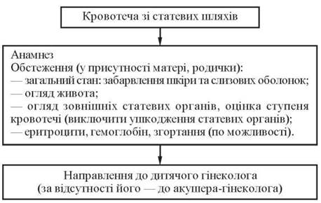
Fig. 11. Algorithm for examining adolescent girls with puberty-related uterine bleeding
The diagnosis of PUB is established based on the medical history and examination data.
Medical history:
— onset of bleeding;
— duration of bleeding and its characteristics;
— menarche, CHARACTERISTICS OF THE course and establishment of menstrual function, previous treatment;
— characteristics of the mother's Pregnancy and Childbirth;
— past somatic illnesses;
— inflammatory Diseases of the reproductive and urinary systems;
— presence of allergies;
— dietary habits, presence of hypovitaminosis;
— socio-living conditions, environmental factors;
— psychological trauma, acute or chronic stress.
Examination:
— general condition (taking into account hemodynamic parameters: pulse, blood pressure);
— coloration of the skin and mucous membranes;
— physical development, height, body weight;
— degree of development of secondary sexual characteristics (mammary glands, axillary and pubic hair distribution), presence of hyperandrogenism;
— abdominal examination (ruling out an abdominal mass).
Gynecological examination (performed in the presence of the patient's mother or close relatives):
— inspection of the external genitalia (degree of development, presence of developmental anomalies, nature of bleeding);
— rectoabdominal examination (to assess the condition of internal reproductive organs);
— examination using vaginal specula and bimanual examination in sexually active girls;
— pelvic ultrasound (to evaluate the Size and Structure of the uterus, the state of the ovarian follicular apparatus, and the presence of endometrial hyperplasia).
Laboratory evaluation:
— complete blood count (CBC), including determination of bleeding onset, cessation, and duration, red blood cell count, hemoglobin, hematocrit, and platelet levels;
— biochemical blood test with serum iron level determination;
— coagulogram;
— when necessary, assessment of blood gonadotropic (LH, FSH, PRL) and sex hormone levels, and colpocytogram.
Consultations with relevant specialists: endocrinologist, hematologist, pediatrician, neurologist.
Differential diagnosis in girls presenting with abnormal genital bleeding is carried out according to the following scheme:
Examination:
— general physical examination, BP, pulse;
— inspection of the external genitalia;
— gynecological examination — rectoabdominal or vaginal examination (in sexually active girls);
— pelvic ultrasound;
— laboratory blood tests (red blood cells, HB, platelets, bleeding time).
Based on the findings obtained from the examination, a diagnosis is established and the management algorithm for the patient is determined (Fig. 12, 13).
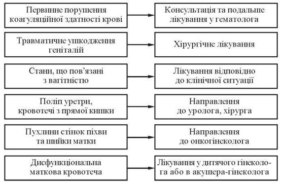
Fig. 12. Management strategy for patients with pubertal uterine bleeding depending on the differential diagnosis
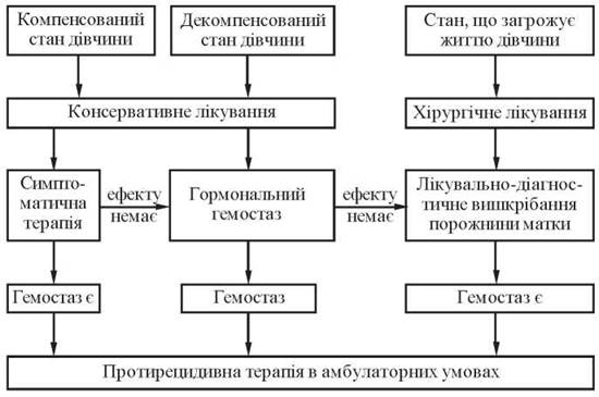
Fig. 13. Treatment algorithm for pubertal uterine bleeding
Management of PUB involves two stages:
— Stage I — direct hemostasis (performed in pediatric gynecology departments or general gynecological wards);
— Stage II — Prevention of disease recurrence (carried out on an outpatient basis).
The individual Selection of therapeutic measures and medications is determined by the specific clinical course of uterine bleeding, the presence or absence of complications, and their severity. It should be kept in mind that before prescribing medical treatment, a thorough medical history must be taken to identify any drug allergies, contraindications to specific medications, and underlying somatic conditions in the adolescent. Hormonal agents are prescribed to adolescent girls with the consent of their parents (Tables 16, 17).
Table 16. Medications used for hemostasis (symptomatic therapy) in girls with pubertal uterine bleeding
Pharmacotherapeutic group of drugs |
Drug name |
Route of administration |
Daily dose |
Duration of administration |
Posterior pituitary hormone |
Oxytocin |
Intravenous drip in 500 ml of 5% glucose solution |
1 ml |
5-7 days |
Hemostatic agents |
Etamgesylate (Sodium etamsylate) |
Intramuscularly, orally |
2 ml 1-4 times, 0.25 g 4 times |
7 days |
Menadione sodium bisulfite (Vikasol) |
Intramuscularly, orally |
1% 1 ml 0.015 g three times |
3 days |
|
Vascular wall-strengthening agents |
Ascorutin |
Orally |
0.25 g three times |
7 days |
Agents that increase tone and myometrial contractility |
Nettle, water pepper, shepherd's purse |
Orally as infusions |
100 ml, three times a day |
7 days |
Antianemic agents |
Iron-containing preparations |
Orally |
Once a day |
Long-term |
Table 17. Medications used for hormonal hemostasis
Drug |
Composition |
Active substance content per tablet, mg |
Daily dose, tab. |
Duration of administration, days |
Progestogen |
Dydrogesterone |
10 |
2 |
15-21 |
Norethisterone acetate |
5 |
1-2 |
15-21 |
|
Combined estrogen-progestogen preparations (COCs) |
Ethinylestradiol + norethisterone acetate |
0.05; 1.0 |
1-3 |
15-21 |
Ethinylestradiol + levonorgestrel |
0.05; 0.15 |
1-3 |
15-21 |
|
Ethinylestradiol + levonorgestrel |
0.03; 0.15 |
1-3 |
15-21 |
Indications for hormonal hemostasis:
— prolonged and heavy bleeding complicated by secondary anemia;
— lack of response to symptomatic therapy in moderate and prolonged bleeding;
— prolonged bleeding accompanied by endometrial hyperplasia (M-echo greater than 10 mm).
Principles of sex steroid hormone therapy in adolescent girls:
— ability to targetedly influence and optimize the puberty process;
— hormone therapy should not disrupt folliculogenesis and must promote the normalization of steroidogenesis and secretory transformation of the endometrium;
— hormone therapy should not exacerbate chronic somatic diseases;
— hormone therapy must not exhibit direct or potential oncogenic effects on target organs.
Indications for surgical treatment (diagnostic and therapeutic curettage of the uterine cavity walls) are as follows:
— profuse uterine bleeding that threatens the patient's life;
— severe secondary anemia (Hb 70 g/l and lower, hematocrit below 25.0%);
— suspected structural changes in the endometrium (endometrial polyp according to pelvic ultrasound).
The conditions required for performing diagnostic and therapeutic uterine curettage must be observed, namely:
— consent from the parents of the minor patient;
— availability of an anesthesiology service for adequate pain management;
— availability of instruments that allow for curettage while preserving the integrity of the hymen, achieved by properly inserting the appropriate instruments into the patient's vagina;
— pathohistological examination of the obtained material.
The treatment of juvenile uterine bleeding (JUB) involves the establishment of a regular menstrual cycle in adolescent girls and plays a crucial role in preventing recurrent menstrual disorders:
— elimination of etiological factors contributing to JUB;
— normalization of the girl's menstrual function through the prescription of medications;
— adherence to A balanced diet and Implementation of general health-promoting measures;
— in the presence of secondary anemia, iron supplements are prescribed.
Depending on the type of hemostasis, anti-relapse therapy is administered (Fig. 14).
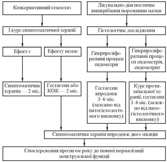
Fig. 14. Algorithm of anti-relapse therapy
Anti-relapse hormone therapy is prescribed for 2-3 menstrual cycles. The duration of hormone therapy depends on the girl's general condition, as well as the condition of the ovaries and endometrium (according to ultrasound data). Furthermore, if hormones are prescribed against the background of endometrial hyperplasia, girls are recommended to be hospitalized at the time of the menstrual-like reaction (Table 18).
Table 18. Medications used to normalize the menstrual cycle (symptomatic therapy)
Pharmacotherapeutic agents |
Drug name |
Route of administration |
Dose |
Course of treatment |
Vitamins |
Oral |
1 mg three times a day |
Days 5–15 of the cycle |
|
Tocopherol acetate |
Oral |
100 mg |
Days 16–28 of the cycle |
|
Ascorbic acid |
Oral |
50 mg three times a day |
Days 16–28 of the cycle |
|
Homeopathic remedies normalizing the menstrual cycle |
Oral |
According to the instructions |
3 months |
|
In the presence of pronounced hyperplastic endometrial processes confirmed by histological examination (adenomatous polyps), progestogens are prescribed from the 5th day after therapeutic and diagnostic curettage to the 25th day of the menstrual cycle for 3 months, with mandatory follow-up pelvic ultrasound monitoring during treatment. At the same time, patients are advised to pay special attention to the necessity of an unscheduled examination with ultrasound in case of a 10-day menstrual delay for the timely detection of endometrial hyperplasia. Based on uterine endometrial ultrasound data after 3 months, the question of further hormone therapy is decided (Table 19).
Table 19. Medications used for hormonal correction (anti-relapse therapy)
Drug |
Composition |
Active substance content per tablet, mg |
Daily dose, tab. |
Course of treatment, days |
Progestogen |
Dydrogesterone |
10 |
2 |
from the 16th to the 25th |
Norethisterone acetate |
5 |
1-2 |
from the 16th to the 25th |
|
Combined estrogen-progestogen preparations (COCs) |
Ethinylestradiol + gestodene |
0,05; 0,075 |
1 |
from the 5th to the 25th |
Ethinylestradiol + desogestrel |
0,03; 0,15 |
1 |
from the 5th to the 25th |
|
Ethinylestradiol + gestodene |
0,02; 0,075 |
1 |
from the 5th to the 25th |
Criteria for the effectiveness of JUB treatment include:
— normalization of the menstrual cycle within a year;
— absence of heavy and prolonged bleeding during menstruation;
— absence of pain syndrome during menstruation;
— absence of pathological changes in the internal genitalia upon rectoabdominal and Ultrasound examination.
In accordance with Order of the Ministry of Health of Ukraine No. 503 dated December 28, 2002, "On the Improvement of Outpatient Obstetric and Gynecological Care in Ukraine," standard therapeutic and preventive measures for menstrual disorders in girls and adolescents have been implemented (App. 1).
Last update: 08/08/2026
Editorial and Educational Adaptation: This material has been compiled based on the primary/original source text. The project team performed an editorial review, corrected technical inaccuracies, structured sections, and adapted the content for an educational format.
What was processed:
- elimination of formatting defects (OCR errors, structural breaks, corrupted characters);
- editorial organization of content;
- standardization of terminology in accordance with academic sources;
- verification of factual statements against the original source text.
All mentions of the author, publication year, and origin of the primary text have been preserved in accordance with the source.