Medical Radiology - Lazar A.P. 2008
Radiation examination of endocrine glands
Adrenal gland disorders
The Base of the Adrenal Glands is medially adjacent to the upper poles of the Kidneys. The mass of each gland is 12-16 g, the vertical dimension is 2-4 cm, the frontal is 2-3 cm, and the sagittal is 5-7 mm. The adrenal glands are located in the retroperitoneal space at the level of the XI-XII thoracic vertebrae. The right Adrenal gland lies slightly lower than the left, is somewhat smaller in size, and has a triangular shape (the left one is semilunar).
The anterior surfaces of the glands are covered by the parietal Peritoneum, the posterior surfaces are adjacent to the Diaphragm, and the lower ones adhere to the upper pole and medial border of the corresponding Kidney. A shallow groove, the hilum, is visible on the anterior surface of each gland, from the depth of which the central vein emerges. Externally, the adrenal gland is covered by a fibrous capsule, from which Connective Tissue trabeculae extend deep into the organ.
The cortex constitutes nearly 90% of the adrenal tissue and consists of three zones: the outer zona glomerulosa (15%), the intermediate zona fasciculata (75%), and the inner zona reticularis (10%). These zones differ in the composition of their constituent Cells and the Chemical Structure of the Hormones they produce. The small Cells of the zona glomerulosa produce the mineralocorticoid hormone aldosterone. Spongiocytes of the zona fasciculata produce glucocorticoid hormones: cortisol, androgens, estrogens, and progesterone. Corticosterone is produced by cells of all three Zones of the adrenal cortex. The adrenal medulla consists of large chromaffin cells—epinephrine and norepinephrine cells (epinephrocytes and norepinephrocytes)—which produce adrenaline and noradrenaline, respectively.
Class="center">
Fig. 232. Cushing's Disease on a computed tomogram. The adrenal glands are enlarged with clear contours (arrows).
Diagnostic imaging Methods for examining the adrenal glands include ultrasound, radionuclide imaging, radiography, CT, and MRI.
Using ultrasound, the adrenal glands are not always visualized, especially in obese individuals. Normal adrenals are echogenic compared to the echogenicity of the kidneys. The glands have a triangular or cone-like shape, a homogenous structure, and fairly distinct contours. Their dimensions are determined sonographically.
On CT and MRI scans, the adrenal glands are homogenous, have smooth, distinct contours, low densitometric density (+10-+20 HU), and yield a low MR signal.
On plain radiographs, the adrenal glands are detected only when calcified, for example, in Addison's disease (hypocortisolism) of tuberculous Etiology. Normal adrenal glands can be visualized on radiographs after performing pneumoretroperitoneum.
Radionuclide imaging of the adrenal glands involves the intravenous administration of 131I-labeled 19-iodocholesterol with an activity of 0.4 MBq/kg. Scintigraphy is best performed a week after administration, when the radiopharmaceutical reaches its maximum accumulation in the organ. In bilateral adrenal hyperplasia (Cushing's disease), an intensified image of the enlarged adrenal glands is revealed. In adenoma and nodular hyperplasia, the image of the affected gland is intensified compared to the healthy one.
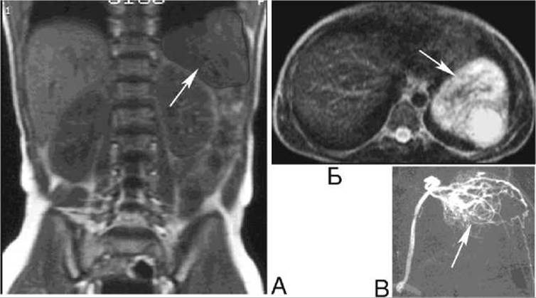
Fig. 233. Left-sided pheochromocytoma on magnetic Resonance tomograms (A - T1-weighted image, coronal section; B - T2-weighted image, transverse section) and on an angiogram (C). The tumor on the MRI and the hypervascular zone on the angiogram are indicated by arrows.
Cushing's disease manifests as enlargement of the adrenal glands, which is detected by ultrasound, CT, and MRI (Fig. 232). This disease mostly results from a pituitary adenoma; therefore, Skull and bone radiography, as well as CT or MRI of the sella turcica region, are also advisable. Due to impaired Mineral METABOLISM, systemic Osteoporosis is observed, and pathological rib and vertebral fractures or aseptic bone necrosis can sometimes be detected.
Pheochromocytoma belongs to the hormonally active tumors of the adrenal medulla, originates from paraganglion chromaffin cells, and produces catecholamines. More than 10% of pheochromocytomas follow a malignant course, accompanied by invasion into adjacent Organs and metastasis to the bones, Lungs, and Brain. Clinically, the disease is characterized by Hypertension with its typical symptoms—headache, cardiac pain, palpitations, etc.
Ultrasound, CT, and MRI reveal enlargement of the affected adrenal gland and its smooth contours. On CT scans, the densitometric density of pheochromocytoma is +12-+32 HU. On MRI, the tumor signal intensity is low on T1-weighted images and high on T2-weighted images, which helps differentiate pheochromocytoma from an adrenocortical adenoma (Fig. 233). Radionuclide imaging reveals a "hot spot" at the site of the adrenal gland in the tumor area (Fig. 234).
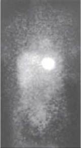
Fig. 234. Left-sided pheochromocytoma on a positron emission tomogram.
Last update: 08/08/2026
Editorial and Educational Adaptation: This material has been compiled based on the primary/original source text. The project team performed an editorial review, corrected technical inaccuracies, structured sections, and adapted the content for an educational format.
What was processed:
- elimination of formatting defects (OCR errors, structural breaks, corrupted characters);
- editorial organization of content;
- standardization of terminology in accordance with academic sources;
- verification of factual statements against the original source text.
All mentions of the author, publication year, and origin of the primary text have been preserved in accordance with the source.