MODERN BOTANY - P. RAVEN - 1990
SECTION IV. DIVERSITY
CHAPTER 11. BACTERIA
Bacteria belong to prokaryotes; they are the simplest, smallest, and most widespread organisms (Fig. 11-1). They were the only form of life on Earth for at least 2 billion years. Bacteria are so distinct from other living organisms that they are placed in a separate kingdom, Monera. They exhibit a diverse array of metabolic types, including some species capable of Photosynthesis. One group of photosynthetic bacteria, the so-called blue-green Algae, or cyanobacteria, was formerly classified with "algae," but today they are recognized as a specific group of bacteria.
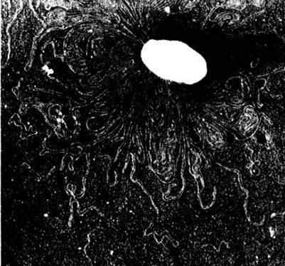
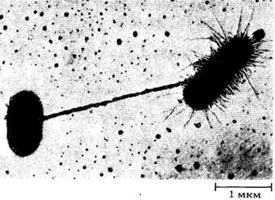
Class="center">Fig. 11-1. A Cell of a common bacterium, Escherichia coli; DNA strands can be seen emerging from The Cell during preparation. The bacterial chromosome consists of a single giant DNA molecule containing approximately 3 billion Base Pairs. The smaller circular DNA molecules visible on the left are Plasmids containing 5,000 base pairs. (The black spots are holes in the photographic film; the scattered white dots are probably dust particles or Cells.)
Bacterial cells lack a nucleus enclosed by a nuclear envelope and do not possess the set of Chromosomes characteristic of eukaryotes. Bacteria also lack true sexual reproduction, although occasional recombination of genetic material may occur. Unlike eukaryotes, bacteria have never been Multicellular Organisms, yet some form filaments or clusters consisting of several cells. Such structures result from incomplete Separation of cell walls following Cell Division; sometimes a group of cells becomes enclosed within a common mucous capsule or sheath. Plasmodesmata between such cells are extremely rare (found only in a few species of cyanobacteria).
Bacteria lack membrane-bound Organelles, but they possess other structures that perform similar functions. The Plasma Membranes of bacteria frequently form intracellular folds (Fig. 11-2) that increase the surface area for enzyme attachment and spatially compartmentalize enzymatic reactions. In some photosynthetic bacteria, pigments are localized on internal membranes, whereas in others they are contained within discrete rounded bodies called chromatophores. The cell walls of most bacteria contain muramic acid, which is absent in eukaryotes.
General Characteristics of Bacteria
The diameter of most bacterial cells is about 1 µm (occasionally 0.1 µm), and some bacteria reach lengths of up to 10 µm (rarely 30 µm). Although each individual bacterial cell is microscopic, the total biomass of bacteria inhabiting Earth exceeds that of all other living organisms combined. Currently, about 2,500 species of bacteria are known.
Fossil bacteria have not been found in the oldest sedimentary rocks of Greenland, which date back 3.9 billion years. However, they have been discovered in Western Australia (see Fig. 1-2) and South Africa in rocks 3.5 billion years old. Organic microstructures resembling cyanobacteria have been found in South African rocks approximately 3.2 billion years old. Chemical analysis of fossil deposits and microfossil impressions indicate that photosynthesis arose about 3.3 billion years ago. In contrast, the first eukaryotes appeared around 1.5 billion years ago. Fossil bacteria were first discovered in ultrathin rock sections using Electron Microscopy only in the 1950s.
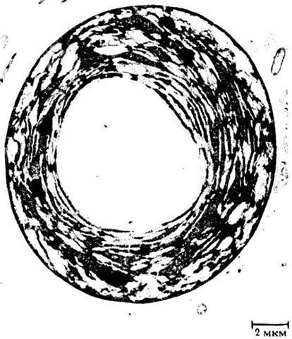
Fig. 11-2. A single cell of the bacterium Prochloron, showing an extensive membrane system. Prochloron is a photosynthetic bacterium containing chlorophylls a and b and carotenoids—pigments otherwise found in green algae and higher plants.
The habitats of bacteria are remarkably diverse; due to their metabolic versatility, some groups can survive where no other Organism can exist. Certain bacteria are obligate anaerobes, meaning they live only in the absence of oxygen. Others are facultative anaerobes, which can survive without oxygen but thrive better in its presence. Cellular Respiration yields significantly more energy than Fermentation (see Chapter 6).
Bacteria are found in the most surprising places and are truly globally distributed. For instance, the bacterium Thermoanaerobacter ethanolicus, isolated from hot springs in Yellowstone National Park (USA), can withstand temperatures exceeding 78°C. Other bacteria inhabit high-pressure environments near deep-sea hydrothermal vents at temperatures above 360°C. Bacteria living in these extreme environments can be cultured in the laboratory only under high pressure; some forms actively grow and double in number at 250°C within 40 minutes! (Such high aqueous temperatures can persist only under the immense hydrostatic pressure of the ocean floor.) Other deep-sea bacteria, such as those inhabiting the guts of marine animals, likewise require high pressure for GROWTH AND DEVELOPMENT.
Diverse bacteria have been discovered in Antarctica, for example, in rock and ice core samples retrieved from depths greater than 430 m. These bacteria are at least 10,000 years old (and possibly a million years old). They remain dormant at temperatures between -7°C and -14°C but become metabolically active when warmed. Such Examples demonstrate that certain bacteria can remain in a suspended state of arrested life processes for exceptionally long periods.
Scientists have recently investigated the physiological limits of bacteria to determine whether life could exist on other planets with different atmospheres. For instance, it was once thought that life would be impossible in Jupiter's alkaline atmosphere, yet certain bacteria from the Livermore Valley in California can grow and reproduce in an alkaline environment at pH 11.5. Other bacteria can tolerate high concentrations of both alkalis and ammonia—conditions reminiscent of Venus's atmosphere. From this perspective, the existence of bacteria on other planets remains plausible. However, most bacteria cannot survive acidic conditions, which is why vinegar (dilute acetic acid) is so effective at protecting food from bacterial spoilage.
Bacteria play a crucial role in the functioning of global ecosystems. Some bacteria are autotrophs and thus make a major contribution to the carbon cycle; certain experimentally cultivated strains can serve as commercial protein sources. The capacity of bacteria for atmospheric Nitrogen Fixation is of paramount ecological significance. Heterotrophic bacteria, much like Fungi, act as decomposers. Thanks to them, nutrients once locked within Living organisms are released and made available to subsequent generations. A single gram of fertile agricultural soil may contain 2.5 billion bacteria, 400,000 fungi, 50,000 algae, and 300,000 Protozoa. At every stage of elemental cycling in Earth's ecosystems, bacteria play an essential part.
Bacteria abound in the marine environment; studies have shown, for example, that 90% of total oceanic biomass consists of organisms smaller than 10 µm, namely bacteria. Depending on geographic Location, between 20 and 60% of primary organic production passes through free-living heterotrophic bacteria.
Bacteria are capable of breaking down A wide variety of Organic compounds. Consequently, their ability to degrade unwanted synthetic pollutants, such as pesticides, Dyes, and crude oil, is under intensive investigation (regardless of how these substances entered the environment). For instance, nylon, first manufactured in 1939, is degraded by a bacterium of the genus Flavobacterium, which evidently evolved two new Enzymes in this remarkably short span of time. Some bacteria break down pesticides so rapidly that they limit their pest-control effectiveness. Other bacteria hold potential for enhanced oil recovery from geological formations.
Beyond their ecological roles, many bacteria are agents of severe Human and Animal diseases, including tuberculosis, cholera, anthrax, Gonorrhea, diphtheria, and tetanus, as well as significant agricultural plant pathogens. In the United States alone, over 200 bacterial species are recognized as plant pathogens. For example, fire blight (Fig. 11-3) has devastated thousands of pear trees, spreading across the USA from the East Coast, where it was introduced in 1880. By the 1930s, the disease had nearly eradicated commercial pear orchards. Today, pear cultivation in the USA is restricted to a few specific regions.
Fig. 11-3. Fire blight, caused by Erwinia amylovora (see Fig. 11-10), is a widespread disease affecting pears, apples, and many other pome fruit trees of the rose family (Rosaceae). The bacterium initially attacks the twigs of these plants and subsequently causes twig and branch cankers (a diseased pear tree is shown). Infected trees die rather rapidly. The bacterial origin of this disease was established in the late 19th century.

Bacteria are utilized as commercial sources of various Antibiotics, such as tyrothricin, bacitracin, subtilin, and polymyxin B. Many bacteria are indispensable for the industrial production of Pharmaceuticals and other chemical compounds, including acetic acid, Amino Acids, and enzymes (see Chapter 30). Furthermore, most cheese-making processes rely on bacterial fermentation of lactose into lactic acid, which precipitates milk Proteins. These same bacteria are used to produce yogurt and cultured buttermilk; additionally, lactic acid plays a preservative role in the fermentation of sauerkraut and pickles.
Fig. 11-4 illustrates Spirulina, a cyanobacterium cultivated in many countries as a protein-rich dietary Supplement for humans and animals.
A group of bacteria known as actinomycetes plays a particularly crucial role in The production of antibiotics, specifically streptomycin, aureomycin, neomycin, and tetracycline. Of the several thousand antibiotics known today, two-thirds are produced by actinomycetes. Other actinomycetes, which live on the roots of certain plants, fix nitrogen and thus play an important ecological role.
Fig. 11-4. A. The spiral filamentous cyanobacterium *Spirulina*, widely cultivated as a protein source. B. Ponds for cultivating Spirulina near Lake Texcoco, close to Mexico City. C. Harvesting moist Spirulina biomass. Spirulina was first used for food by the Spanish conquistadors in Mexico around 1521; its paste contains more protein than soybeans, and this protein has a well-balanced amino acid profile. The daily protein yield is 10 times higher than that of wheat or soy, and it is precisely this high productivity of Spirulina that drives its cultivation.

Bacterial Shape
Bacteria vary remarkably in shape and Cellular Organization (Fig. 11-5). Straight, rod-shaped bacteria are called bacilli, spherical ones are cocci, and spiral-shaped ones are spirilla (Fig. 11-6). Following division, spherical bacteria may form pairs (diplococci), chains (streptococci), or cluster into grape-like bunches (staphylococci). Diplococci cause Pneumonia, whereas Staphylococcal infections lead to Sepsis and suppurations.
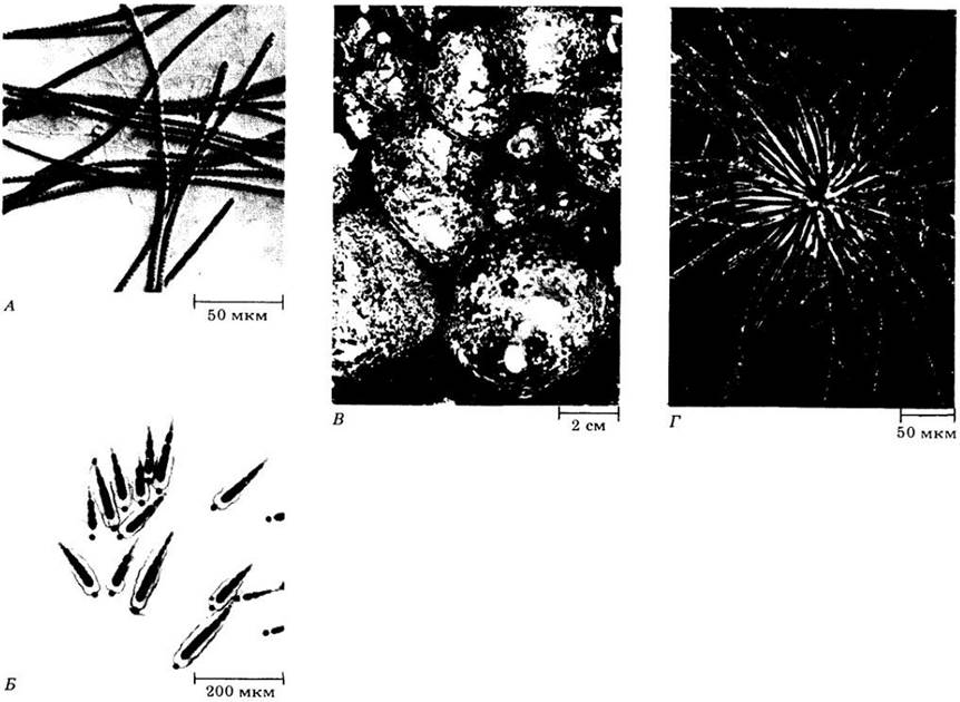
Fig. 11-5. Some types of bacteria. A. Myxobacteria, or slime bacteria, are organized similarly to slime Molds (Ch. 14). Shown here is a scanning electron micrograph of the fruiting bodies of *Chondromyces crocatus*; each consists of approximately 1 million cells. Normally rod-shaped, myxobacteria are capable of locomotion and forming fruiting bodies. B. Spirochetes are spiral-shaped bacteria about 500 µm in length (an impressive size for bacteria). They are capable of undulating movement. *Treponema pallidum*, shown here, is the CAUSATIVE AGENT OF Syphilis. C. Actinomycetes inhabit the soil and decompose organic matter. The characteristic earthy smell of damp soil is due precisely to them. *Streptomyces fradiae*, pictured here, is a commercial source of the antibiotic neomycin. D. Large gliding bacteria are filamentous in shape; they are capable of rhythmic contractions that result in gliding movements and periodic changes in cell shape. In the cells of *Beggiatoa*, shown here, sulfur granules resulting from The oxidation of hydrogen sulfide are clearly visible (a process that stores energy).

Fig. 11-6. Three main bacterial shapes: A — bacilli (rod-shaped); B — cocci (spherical); C — spirilla (curved). Cell Morphology is a relatively constant feature of most bacterial species. Bacilli include, for example, *Clostridium tetani*, the causative agent of tetanus, as well as the common colon bacillus
— *Escherichia coli*. Many bacilli cause plant diseases, such as fire blight of pears and apples (caused by *Erwinia amylovora*), and bacterial wilt of potatoes, tomatoes, and bananas (caused by *Pseudomonas solanacearum*). Cocci include *Diplococcus pneumoniae*, which causes a bacterial form of pneumonia; *Streptococcus lactis*, the bacterium used for souring milk; and *Nitrosococcus nitrosus*, a soil bacterium that oxidizes ammonia to nitrites. Spirilla are less common; they have a spiral shape

Bacilli are generally unicellular forms; however, they sometimes form filaments due to transverse division. These filaments resemble those of fungi, which is why the prefix myco- (from the Greek for "fungus") is often added to the designation of bacilli. For instance, *Mycobacterium tuberculosis* — a rod-shaped bacterium — forms filaments in culture, but not within a host organism; other actinomycetes exhibit a similar morphology (Fig. 11-5, D). Upon division, cyanobacteria are also capable of forming filamentous structures (Fig. 10-4, B, C, 11-7).
Fig. 11-7. Three common genera of cyanobacteria and a related form. A. *Oscillatoria* reproduces solely by fragmentation of the filament. B. *Calothrix* (*Gloeotrichia*), a filamentous form with basal heterocysts. *Gloeotrichia* forms akinetes — large cells with a tough outer coat. C. Gelatinous spheres of *Nostoc commune*; each sphere contains hundreds of filaments and typically inhabits fresh Water. D. *Thiothrix* is a genus of bacteria lacking chlorophyll but closely related to cyanobacteria. *Thiothrix* obtains energy by oxidizing H2S. Chains of cells containing sulfur granules are attached to a substrate (at the center) and form a characteristic rosette.
In the early 1880s, the Danish microbiologist Hans Christian Gram, working in a Berlin morgue, stained infected tissues with a dye similar to modern crystal violet and discovered pneumonia-causing bacteria within them. Many other bacteria also turned purple when stained with this dye; however, Gram was disappointed to find that not all did. Ultimately, Gram's discovery greatly contributed to our understanding of Introduction/37.html">Bacterial cell wall structures, which differ among various groups of bacteria. The current Procedure involves staining the cells with crystal violet, then treating them with a dilute iodine solution (which forms an insoluble complex with the dye), followed by rinsing with alcohol. Alcohol washes the dye out of the walls of Gram-negative bacteria, whereas Gram-positive bacteria retain the dye.
It was later discovered that the matrix of bacterial walls consists of Disaccharides linked by short chains of amino acids (Peptides). The cell wall of Gram-positive bacteria, which ranges in thickness from 15 to 80 nm depending on the species, contains exclusively these peptidoglycan macromolecules. In the walls of Gram-negative bacteria, the peptidoglycan layer is surrounded by lipopolysaccharides (polysaccharide chains with attached Lipids). Gram-positive and Gram-negative cell walls have a thickness of no more than 10 nm.
Gram-positive and Gram-negative bacteria differ in their response to antibiotics. For example, penicillin blocks The formation of peptide bridges in the cell wall of Gram-positive bacteria, causing the cell to lyse as it grows. The action of penicillin on the walls of Gram-negative bacteria is hindered by the lipopolysaccharide layer. The widely used antibiotic actinomycin disrupts Protein Synthesis by binding to the DNA double helix. The actinomycin molecule easily penetrates the cell wall of Gram-positive bacteria, but not that of Gram-negative ones. Therefore, different antibiotics, such as erythromycin, must be used for the latter.
Bacterial cells are capable of adhesion thanks to a specialized layer of intertwined polysaccharide fibers known as the glycocalyx. Although bacteria do not form a glycocalyx in laboratory cultures, it plays a key role in nature during the initial stages and subsequent spread of bacterial infection. It is precisely via the glycocalyx that bacteria attach to various substrates.
Through enzymatic reactions taking place within the glycocalyx, certain bacteria are able to colonize intact tooth enamel or other seemingly resistant surfaces.
Many bacteria form a gelatinous capsule, which is likely secreted by the protoplast (Fig. 11-8). Similar to the cell wall, the capsule is composed of Polysaccharides; however, it is loosely bound to the bacterium and can be washed off.
Fig. 11-8. Micrograph of individual *Bacillus megaterium* cells dispersed in India ink. Capsules are visible as a clear halo.

The cytoplasm of bacteria, like that of other cells, is enclosed by a Plasma Membrane, on the inner surface of which numerous enzymes are localized. The cytoplasm contains A large number of Ribosomes and granular inclusions, as well as one or two regions where DNA is concentrated (typically two, because cell division lags behind the Replication of genetic material). Each of these regions contains a single molecule of circular double-stranded DNA whose length is 700 to 1,000 times greater than that of the cell itself.
Flagella and Fimbriae
Some bacteria possess extremely thin, robust, helical flagella that are several times longer than the cells themselves. These flagella perform rapid rotational movements that propel the bacteria forward. Given their size, bacteria move remarkably fast, covering a distance in one second equal to roughly 20 diameters of the bacterial cell itself. Rotational movement is uncharacteristic of eukaryotes, making the continuous rotation of bacterial flagella a truly unique phenomenon.
Bacterial flagella resemble the (9+2) flagella of eukaryotes only in appearance. They are long (3 — 12 µm), thin, and undulating in shape. Their diameter is typically 10 — 12 nm, which renders them invisible under a Light Microscope. In some bacteria, flagella are distributed uniformly across the entire cell surface, whereas in others they are attached to one or both ends (Fig. 11-9, 11-10). Each bacterial flagelle consists of a single rigid protein molecule of flagellin that emerges from a "collar" in the cell envelope and connects to a complex rotary mechanism. This mechanism comprises a ring rotated by an electrical potential and another, larger stationary ring lying just inside it.
Fig. 11-9. Flagella of Pseudomonas marginalis, a bacterium widely distributed in soil. It causes soft rot in ROOT crops and other vegetables.
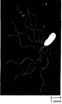
Fig. 11-10. The bacterium Bdellovibrio bacteriovorus parasitizes other bacteria. Frequently found in soil and wastewater, it moves using a single polar flagellum at its rear. The parasitic cell is shown attacking the rod-shaped Erwinia amylovora (right), the causative agent of fire blight in pears and apples.

Fimbriae (pili) are shorter (up to several micrometers) and straighter than flagella, with a diameter of about 7.5 — 10 nm (Fig. 11-11). They are characteristic primarily of Gram-negative bacteria. Fimbriae consist of a protein distinct from flagellin and possess a different Structure. Hollow pili form on bacterial cells during conjugation, although their exact function remains unknown. They may transfer DNA or facilitate Cell-to-Cell adhesion. In most cases, pili help bacteria attach to specific membranes. A better understanding of The Role of fimbriae may aid in combating bacterial diseases.
Fig. 11-11. Fimbriae of Escherichia coli. A hollow fimbria links two cells; numerous fimbriae are visible on the cell to the right.
Specific features of Cyanobacteria
Some photosynthetic bacteria, known as cyanobacteria, form filaments up to a meter or more in length and are capable of accumulating a large biomass. Some cyanobacteria are unicellular, others are joined in chains, and a very few form spherical or irregularly shaped colonies (Fig. 10-4, B, C; 11-7, 11-12, 11-13). Any cell except a heterocyst can divide, and the resulting cells can give rise to new colonies. Like other filamentous and colonial forms of bacteria, cyanobacteria are held together solely by cell walls or gelatinous capsules, meaning each cell essentially leads an independent life; nevertheless, plasmodesmata have been discovered in certain colonial cyanobacteria.
Fig. 11-12. A. A cell of the cyanobacterium Anabaena azollae. An electron micrograph reveals its internal structure. The gelatinous capsule was disrupted during preparation. This microorganism is closely associated with the floating water fern Azolla and fixes nitrogen (see Fig. 10-8, A and ch. 26). B. A Chinese worker harvests Azolla from a body of water to cultivate it in rice paddies.

Cyanobacteria inhabiting the surface waters of fresh and marine bodies of water form communities of microscopic organisms known as plankton; as a rule, these microorganisms feature transparent, irregularly shaped structures called gas vacuoles. These vacuoles regulate buoyancy and allow the organisms to remain suspended in the water Column. When cyanobacteria lose The ability to regulate their density—for example, due to sharp Temperature fluctuations or disruptions in oxygen METABOLISM—they float to the surface and cause water "blooms." In the process, certain cyanobacteria release chemical substances toxic to other organisms, leading to massive die-offs.
Bacterial Motility
Despite their simple organization, bacteria can respond to specific stimuli by moving toward increasing concentrations of nutrients or oxygen. Motile bacteria, such as Escherichia coli, can move toward or away from chemical agents by rotating their flagella; each E. coli cell possesses six to eight flagella. Bacteria exhibit specific sensitivities to various nutrients, such as sugars. Once cells receive a signal regarding "attractants" or "repellents," they can select the appropriate direction of movement.
Julius Adler and his colleagues at the University of Wisconsin determined that E. coli has about 20 different proteinaceous chemoreceptors: 12 for attractants and 8 for repellents. Proteins responsible for binding sugars such as galactose, maltose, and ribose are located in the periplasmic space between the cell wall and The Plasma Membrane. Other chemoreceptors (also proteins) reside in the plasma membrane. The information gathered by chemoreceptors is transmitted in an unknown manner to the flagella, setting them into motion.
Individual E. coli cells move rapidly, executing curvilinear "runs" that each last about a second. A series of runs is frequently interrupted by stationary turns, or "tumbles," lasting a tenth of a second. Following each tumble, the bacterium initiates its next run in a different direction. The frequency of tumbles controls the run length and, consequently, the overall direction of movement. If a bacterium is swimming in the "correct" direction, its tumbles are inhibited; if in the "wrong" direction, the tumble frequency increases.
The ultimate goal of such movements is to migrate toward an attractant source or away from a repellent source. Attractants induce counterclockwise flagellar rotation, whereas repellents induce clockwise rotation. Thus, "runs" are produced by counterclockwise flagellar rotation, and "tumbles" by clockwise rotation. During continuous motion, the flagella work together and gather into a bundle at the rear end of the cell, despite being distributed over virtually the entire surface. When the flagella rotate clockwise, the bundle flies apart, and the cell tumbles. In this case, the flagella rotate independently. The responses of such bacteria to external stimuli are highly complex—considering the prokaryotic level of organization—and foreshadow the much more complex reactions of eukaryotes.
Certain groups of bacteria respond differently to environmental conditions. For instance, some aquatic bacteria contain magnetite crystals (Fе3O2). They can orient themselves within the Earth's magnetic field and steadily swim in a single direction. Microorganisms found in the Northern Hemisphere move northward, following magnetic field lines that angle downward into the depths of the water body. In the Southern Hemisphere, analogous bacteria swim southward. In both cases, the bacteria move deeper into the water column; magnetotaxis may aid them in searching for food.
A completely different mode of locomotion is found in filamentous cyanobacteria and in bacteria lacking flagella (see Fig. 11-5). The movement of these microorganisms is a form of gliding, but it can also involve rotation along the longitudinal axis of the cell. Short segments detached from a cyanobacterial colony can glide at speeds on the order of 10 µm/s. This movement is facilitated by the secretion of slime through Pores in the cell wall and the formation of contractile waves on its outer surface. Some cyanobacteria exhibit convulsive movements.
Bacterial Genetics
Cell Division
The primary mode of bacterial reproduction is asexual; each cell increases in size and divides in two. During division, the plasma membrane and cell wall invaginate, eventually pinching the cell in half (Fig. 11-13). The newly formed cell wall is thicker than the previous one and rapidly splits in the center, providing each daughter cell with a fresh cell wall. Chains of bacteria form when the cell wall does not divide completely; such chains can break apart into multicellular fragments, such as hormogonia in cyanobacteria.
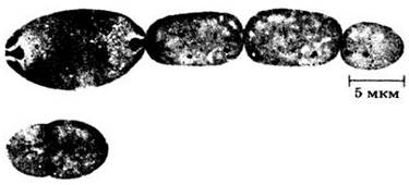
Fig. 11-13. Division of Anabaena. An electron micrograph shows a chain of cells held together by an undivided cell wall; cell division of an individual cell is shown on the left. The leftmost cell in the chain is a heterocyst. This pinching type of cell division is characteristic of many organisms, except plants and certain algae genera, which form a cell plate during division.
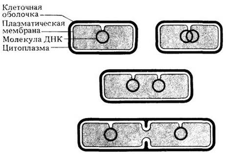
It is believed that the single circular double-stranded DNA molecule carrying Genetic information is attached at a single point to the inner surface of the plasma membrane. Once the DNA replicates, the two identical circular molecules remain connected side by side to the plasma membrane. A new plasma membrane and cell wall form between the two DNA attachment points as the cell divides; eventually, the membrane invaginates inward between the two DNA molecules, and each daughter cell receives an identical DNA molecule (Fig. 11-14).
Fig. 11-14. Schematic diagram illustrating how Bacterial DNA is attached to the plasma membrane, leading to the distribution of DNA between daughter cells. In reality (see Fig. 11-1), the DNA is much longer
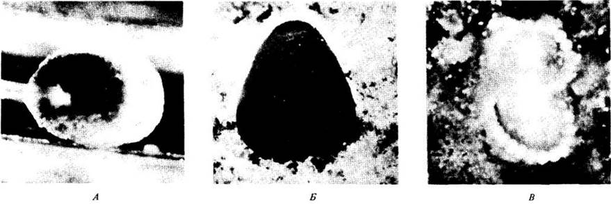
Some bacteria form thick-walled endospores that are resistant to heat and desiccation. Endospores are formed by the division of the bacterial protoplast into two or more parts. A dense spore coat forms around the isolated portion of the protoplast containing the DNA. Endospores can germinate after decades or even hundreds of years. The resistant spores of cyanobacteria are called akinetes. Akinetes differ fundamentally from endospores in that they are enlarged vegetative cells surrounded by a newly formed thick wall (Fig. 11-15). Actinomycetes form chains of endospores at the ends of filaments.
Fig. 11-15. Filaments of Anabaena, a nitrogen-fixing cyanobacterium, consisting of barrel-shaped cells surrounded by a gelatinous matrix. Nitrogen fixation occurs in specialized cells called heterocysts. Like Calothrix (see Fig. 11-7, B), Anabaena forms akinetes. Electron micrographs are shown in Figs. 11-12 and 11-13

Genetic Recombination
Genetic recombination in bacteria occurs As a result of The transfer of a DNA segment from one bacterial cell to another. The introduced DNA fragment may function alongside the recipient cell's DNA and participate in Messenger RNA synthesis. Alternatively, it may integrate into the circular DNA molecule and be passed on to the daughter cell during division along with the rest of the genetic material. Recombination can occur via conjugation (exchange of DNA fragments through direct cell-to-cell contact), Transduction (transfer of DNA from a donor cell to a recipient cell via a virus), or transformation (transfer of free soluble DNA without any intercellular contact or vectors).
Almost all bacteria, In addition to large circular DNA molecules called bacterial chromosomes, possess small circular DNA molecules known as plasmids (see Fig. 11-1). Some plasmids integrate into chromosomes and replicate along with them. In certain bacterial strains, conjugation and plasmid exchange occur quite frequently (see Fig. 11-11), which facilitates the transfer of traits such as Antibiotic Resistance from one bacterial strain to another. The process of recombination is widely used in Genetic Engineering (see Ch. 30).
Genetic material can be transferred from one strain to another through transduction. Bacterial Viruses called Bacteriophages (see Fig. 12-1) can capture small fragments of bacterial chromosomes and transfer them to another bacterium. The viral DNA, along with the bacterial DNA, can integrate into the chromosome of the new host strain.
Fig. 11-16. Replica plating method for screening mutant bacteria. A. Original plate. Colonies were transferred from plate A to plates B and C using a sterile velvet pad pressed against the Agar; some cells adhered to the velvet nap. The velvet was then pressed onto uninoculated agar in other plates, reproducing the entire pattern of colonies from the original plate. Growth of colonies on complete medium is visible on the first two plates, while plate C lacks A number of growth factors. Under these conditions, the colonies indicated by arrows on plate B fail to grow on the third plate C; these are mutants that had already arisen but had not yet been identified on plate A

Alternatively, genetic recombination occurs through transformation. The pioneering experiments on Bacterial Transformation were conducted on Diplococcus pneumoniae, the causative agent of pneumonia. These bacteria form Two Types of colonies: rough (R) and smooth (S). Bacteria forming S-colonies possess capsules and cause pneumonia. In 1928, Frederick Griffith demonstrated that a harmless R strain could be transformed into a virulent S strain by mixing live R bacteria with heat-killed S bacteria (see Fig. 8-2). When bacterial cells are disrupted by chemicals or heat, DNA fragments are released and can enter other cells. By thus demonstrating the existence of a genetically active material involved in transformation, Griffith essentially provided the first proof of the genetic role of DNA. Transformation is now known to occur in many bacteria.
Mutations are an even more important source of bacterial Variability than genetic recombination (Fig. 11-16). It has been calculated that for a given Gene, there is one mutant per 107 cells. An individual Escherichia coli cell has approximately 5,000 genes. Thus, in a culture of these bacteria, there is one cell mutated in any given gene per 2,000 cells; in a culture, 0.05% of the cells will have a mutant phenotype at each cell division. In a culture of 109 cells (the progeny of 30 divisions of a single bacterium), the mutation rate will be 30 × 0.05, i.e., 1.5%. Bacteria reproduce very rapidly. Under optimal conditions, a population of E. coli cells can double in 12.5 min; consequently, the number of mutant cells grows quickly. The high reproduction rate and high mutation frequency of bacteria account for their remarkable ability to adapt.
Bacterial Metabolism
Heterotrophs
Most bacteria are heterotrophs; they are incapable of synthesizing organic compounds from simple inorganic ones and must obtain them in ready-made form. The largest group of heterotrophic bacteria is the saprotrophs. They "feed" on dead organic material. Saprotrophic bacteria and fungi are responsible for the decomposition and recycling of organic matter in soil; many of the compounds produced in this process have a characteristic odor.
Photosynthetic Bacteria
There are at least five groups of photosynthetic bacteria: cyanobacteria, green sulfur bacteria, purple sulfur bacteria, purple non-sulfur bacteria, and Prochloron. Like plants, photosynthetic bacteria contain chlorophyll. Cyanobacteria and Prochloron contain chlorophyll $a$, similar to all photosynthetic eukaryotes. The chlorophylls of other photosynthetic bacteria differ somewhat from chlorophyll $a$, but their structure is fundamentally similar (see Fig. 7-8).
The coloration of these bacterial groups is due to accessory pigments required for photosynthesis. In the two groups of purple bacteria, these are yellow and red carotenoids; in cyanobacteria, the blue pigment phycocyanin and the red pigment phycoerythrin are also found. The photosynthetic processes in cyanobacteria and eukaryotes occur in a similar manner. The cytoplasm of cyanobacteria contains numerous membranes, often arranged parallel to each other, and a large number of ribosomes. These membranes are photosynthetic thylakoids, analogous to the thylakoids of Chloroplasts. However, in cyanobacteria cells, they are not organized into chloroplasts. The main storage carbohydrate of cyanobacteria, as in other bacteria, is Glycogen.
In green and purple sulfur bacteria, sulfur compounds play the same role in photosynthesis as water does in organisms containing chlorophyll $a$, i.e.,

As discussed in Chapter 7, The Study of photosynthesis in purple sulfur bacteria helped C. B. van Niel formulate the general equation for this process:

where H2A is a generalized hydrogen donor.
In purple non-sulfur bacteria, alcohols, Fatty acids, and keto acids serve as hydrogen Donors for photosynthetic reactions.
Photosynthetic sulfur bacteria require hydrogen sulfide or other sulfur compounds, and therefore inhabit environments rich in decomposing organic matter with a distinct sulfur odor. Sulfur can be stored as a reserve in bacterial cells.
Halobacterium halobium — a purple non-sulfur bacterium — is a highly unusual organism. It inhabits concentrated salt solutions in sun-warmed areas near saline water bodies. The plasma membrane of Halobacterium contains inclusions of a purple pigment resembling rhodopsin, the visual pigment of the human eye. These inclusions act as active light-driven "pumps" that extrude protons from the bacterial cell. A similar process occurs during Oxidative Phosphorylation in Mitochondria; among all free-living forms of bacteria, purple non-sulfur bacteria are the closest relatives to mitochondria, which may have evolved from this group of bacteria. This process likely leads to ATP synthesis via the enzyme ATPase located in the pigment-free Regions of the plasma membrane. Thus, Halobacterium is the only currently existing organism that lacks chlorophyll yet is capable of converting sunlight into chemical energy, i.e., performing photosynthesis. Halobacterium is motile and exhibits phototaxis toward violet light, further supporting the concept that its pigment absorbs light via the same mechanism as rhodopsin.
The fifth group of photosynthetic bacteria consists of a single genus, Prochloron, discovered in the early 1970s by Ralph Lewin of the Scripps Institution of Oceanography. Prochloron contains chlorophylls a and b as well as carotenoids—the Photosynthetic Pigments characteristic of green algae and plants (Figs. 11-2 and 11-17). As far as is known, it lives in Symbiosis with ascidians, colonial marine animals inhabiting coastal zones of tropical and subtropical seas. The pigment Complement of Prochloron suggests it could be related to the bacteria from which the chloroplasts of green algae presumably evolved (see ch. 15). On the other hand, based on its ribosomal RNA (rRNA) nucleotide sequences, Prochloron bears a closer resemblance to cyanobacteria than to the chloroplasts of green algae and plants. The potential evolutionary role of Prochloron and its relationships with other organisms are currently under active investigation.
Fig. 11-17. Prochloron is a rather unusual photosynthetic bacterium that lives exclusively in symbiosis with ascidians. A. Larval stage of an ascidian of the genus Didemnum, illustrating the dispersal of Prochloron bacteria. B. Adult Didemnum specimen. C. Inside a dissected Didemnum ascidian, showing a dense mass of Prochloron
Chemoautotrophic Bacteria
Chemoautotrophic bacteria obtain the energy required for synthetic reactions through the oxidation of inorganic substances (such as nitrogen, sulfur, iron compounds, and molecular hydrogen), which serve as Energy Sources analogous to light in photosynthetic organisms. However, their carbon source remains the same: carbon dioxide. Bacteria inhabiting deep-sea hydrothermal vents at temperatures exceeding 360°C are also chemosynthesizers. They generate energy by converting hydrogen sulfide into sulfur and, in the process, sustain an entire community of organisms thriving in the pitch black of the deep ocean.
Archaebacteria
One group of chemoautotrophic bacteria that has attracted significant attention in recent years comprises the methanogenic bacteria. These bacteria are strict anaerobes found in the gastrointestinal tract of ruminants, sewage, swamps, and deep-sea sediments. Most natural gas reserves were formed in the distant past through the METABOLIC ACTIVITY OF methanogenic bacteria. Some of these organisms produce methane from carbon dioxide and hydrogen while harvesting energy; others are capable of reducing elemental sulfur to hydrogen sulfide.
Methanobacteria exhibit high morphological diversity. However, Carl Woese and his colleagues at the University of Illinois recently demonstrated that Various Forms of methanobacteria share homologous rRNA sequences, indicating their close phylogenetic relationship. Surprisingly, these base sequences differ markedly from those of other bacteria and eukaryotes. For example, the rRNA Nucleotide Composition of cyanobacteria is closer to that of Escherichia coli and actinomycetes than to methanogenic bacteria. Unlike all other bacteria, the cell wall of methanobacteria lacks muramic acid, and they feature a distinct type of metabolism. In 1984, a report was published stating that a genus of archaebacteria, Methanosarcina, is capable of fixing atmospheric nitrogen. This is a particularly interesting discovery, given that nitrogen-fixing species are scattered across very few taxa.
Based on these findings, it has been hypothesized that methanobacteria emerged on Earth approximately 3 billion years ago, when the atmosphere was anoxic yet rich in CO2 and H2. Today, they survive only in specific, specialized niches. The distinctions between methanobacteria and other bacterial groups are so profound that it has been proposed to classify them into a separate kingdom: the archaebacteria. As discussed previously, Halobacterium, along with certain other thermophilic and acidophilic bacteria, shares several traits with methanobacteria. For this reason, it may be appropriate to group all these organisms within the kingdom Archaebacteria.
Bacterial Ecology
Soil Bacteria
Various groups of microorganisms participate in specific stages of decomposition and nutrient cycling within the soil; these natural communities possess a highly complex organization (Fig. 11-18). For instance, a penicillin-sensitive strain of Streptococcus is protected by a penicillin-resistant strain of the genus Bacteroides, which degrades penicillin so efficiently that the antibiotic fails to inhibit Streptococcus growth. Many bacteria and fungi break down carbon-containing compounds and release CO2 into the atmosphere. The most important plant-derived organic substances are Cellulose, Lignin, Pectins, starch, and sugars. It has been established that over 90% of the CO2 produced in the biosphere results from the metabolic activity of bacteria and fungi.
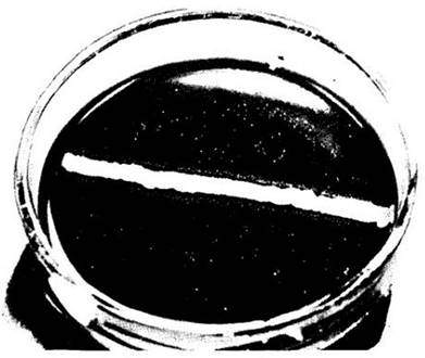
Fig. 11-18. This photograph illustrates the complex interactions among soil microorganisms, showing bacteria growing on penicillin-agar medium. A dense colony of Staphylococcus epidermidis is clearly visible in the center. This strain is penicillin-resistant because it produces the enzyme penicillinase, which breaks down the antibiotic. Flanking the staphylococcal colony are tiny colonies of Neisseria gonorrhoeae, the causative agent of gonorrhea. Although this organism is normally sensitive to penicillin, it can grow in the zone where Staphylococcus has degraded the antibiotic
Some microorganisms hydrolyze proteins into peptides, which are subsequently broken down into their constituent amino acids. Many utilize ammonification—the degradation of amino acids accompanied by the release of ammonium ions (NH+4). Ammonium can be oxidized to nitrite (NO2) by the chemoautotrophic bacterium Nitrosomonas, and nitrite to nitrate (NO-3) by Nitrobacter. The oxidation of ammonium to nitrites and nitrates is termed nitrification. This process releases energy, which chemoautotrophs utilize to reduce carbon dioxide into CARBOHYDRATES. Certain bacteria are also capable of converting nitrates into nitrites and ultimately into ammonium.
Denitrification—The conversion of nitrates into nitrogen gas or nitric oxide—leads to a depletion of soil nitrogen. The reverse process, nitrogen fixation, is of paramount biological significance. Among All living organisms, only bacteria of a few genera are capable of fixing atmospheric nitrogen. The best-known example is the symbiotic bacterium Rhizobium (ch. 26), which forms nodules on the roots of legumes and certain other plants. Actinomycetes are involved in root nodule formation in many woody species, such as alder (Alnus), bayberry (Myrica), and ceanothus (Ceanothus). Nitrogen-fixing actinomycetes contribute to the accumulation of nitrogen in the soil (Fig. 11-19). In addition, certain nitrogen-fixing bacteria associate with leaves, where they utilize carbohydrate-rich plant exudates while supplying the plant with accessible nitrogen in return. Free-living forms, including several cyanobacteria, also play a vital role in nitrogen fixation.
Fig. 11-19. Alders form a symbiosis with nitrogen-fixing actinomycetes of the genus Frankia. This relationship has been thoroughly investigated using red alder at the Harvard Forest in Massachusetts by John Torrey and his colleagues. A. Red alder seedlings grown in various soil mixes following inoculation with Frankia. The seedlings on the left received no Frankia inoculum, whereas those on the right were grown with Frankia and have developed numerous root nodules. B. A root nodule of alder. C. Micrograph of a root section from an alder seedling stained with toluidine blue, showing cortical cells containing Frankia. The hyphae lie in the center of the infected cells, while swollen hyphal tips, known as vesicles, are visible at the periphery. It is believed that The Nitrogenase Enzyme responsible for nitrogen fixation is localized within these vesicles

Chemoautotrophic bacteria, such as Thiobacillus, oxidize sulfur to sulfates, thereby making it available to plants that cannot assimilate elemental sulfur on their own:
2S + 2Н2O + 3O2 —> 4Н+ + 2SO42-.
Sulfates are absorbed by plants, and the sulfur they contain is incorporated into proteins. During protein breakdown (discussed further in ch. 26), Amino acids are released, including sulfur-containing ones (Fig. 11-20). Some bacteria can degrade Sulfur-Containing Amino Acids with the release of hydrogen sulfide (Н2S). Sulfates can also be reduced to Н2S by certain soil microorganisms, such as Desulfovibrio.
Fig. 11-20. The sulfur cycle. Thanks to The activity of bacteria and fungi, many elements—such as carbon, nitrogen, sulfur, and others—do not remain trapped within the molecules into which they were incorporated during metabolic reactions. Soil heterotrophs break down organic matter; the breakdown products can then enter other biological cycles. Without the recycling of organic compounds, all organisms would soon be overwhelmed by their own metabolic wastes. The release of vast amounts of sulfur into the atmosphere through fossil fuel combustion disrupts the natural sulfur cycle.

Parasitic and Symbiotic Bacteria
Some heterotrophic bacteria break down organic matter only when they invade living organisms. This group includes disease-causing forms as well as non-pathogenic microorganisms. The latter may have little effect on the host organism's vital activity or may actually promote its well-being. Specifically, if all bacteria inhabiting the human gastrointestinal tract are eliminated—as often happens, for example, during prolonged antibiotic Treatment—the body becomes much more susceptible to pathogenic bacteria and fungi. In cattle and other ruminants, bacteria hydrolyze cellulose into sucrose, which is essential to the host; otherwise, these animals could not utilize cellulose-rich grass and foliage. Another fundamentally important example is the mutualistic symbiosis between bacteria of the genus Rhizobium and legumes (see ch. 26).
In general, microbes and other parasites interfere with the normal physiological processes of their host organisms, causing severe disease. If death occurs too rapidly, this outcome is unfavorable for the parasite: failing to reach another host, cross-infection does not take place, and the parasite itself perishes.
Human Diseases
Some human diseases are transmitted via airborne droplets. Among the best-known conditions of this kind are bacterial pneumonia (caused by Diplococcus pneumoniae); whooping cough, or pertussis (caused by Bordetella pertussis); and diphtheria (caused by Corynebacterium diphtheriae). The latter microorganism secretes a potent toxin that is rapidly distributed throughout the body, damaging The Heart Muscle, Nervous Tissue, and Kidneys. Today, diphtheria is quite rare because most children are vaccinated against it. A number of other airborne diseases are caused by bacteria of the genus Streptococcus, such as scarlet fever and rheumatic fever. The tuberculosis agent, Mycobacterium tuberculosis, still claims many lives despite improvements in diagnostic and treatment Methods. However, the number of reported deaths in the U.S. in the mid-1980s dropped compared to the 1970s, falling from 30,000 to 20,000 per year.
A series of other bacterial diseases are transmitted through food and water. Examples include typhoid fever, paratyphoid fever (pathogens of the genus Salmonella), and dysentery caused by Shigella dysenteriae. Brucellosis, caused by bacteria of the genus Brucella, is hazardous to both animals and humans, who contract it by drinking milk from infected cows. Milk pasteurization destroys Brucella, making the disease rare in regions where milk is regularly treated. The widespread "traveler's diarrhea" is caused by a specific strain of Escherichia coli that inhabits the Large Intestine.
In 1976, "Legionnaires' disease," which is transmitted via drinking water, was first identified. This mysterious pulmonary illness claimed the lives of 34 members of the American Legion at a conference in Philadelphia. It is now known that the disease is caused by a small, flagellated, rod-shaped bacterium belonging to the genus Legionella. These bacteria enter The Human Body from warm water and multiply rapidly within monocytes—white Blood Cells that play a crucial role in Immunity. It has been established that Legionnaires' disease has affected about 50,000 people in the U.S., with a 15–20% mortality rate. Legionella species are Gram-negative bacteria, and the disease is treatable with erythromycin.
Bacteria cause the spoilage of food and other organic Materials, and some of them are extremely dangerous to humans. Consumption of food contaminated with Clostridium botulinum can be fatal. The endospores of this microorganism survive brief boiling and can germinate even inside sealed tin cans (Fig. 11-21). Food-poisoning Staphylococcus is quite common but, fortunately, less pathogenic. In recent years, however, a number of virulent Staphylococcus strains have emerged that cause dangerous infections. Many of these strains are penicillin-resistant, and some produce the enzyme penicillinase, which destroys the antibiotic. Staphylococcus frequently comes into contact with penicillin-producing fungi or grows alongside them; thus, penicillin resistance provides them with a natural selective advantage (see Fig. 11-18).
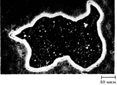
Fig. 11-21. A colony of the bacterium Clostridium botulinum. The food poisoning it causes—botulism—results from consuming spoiled canned goods. Spores of Clostridium botulinum are highly heat-resistant and remain viable in food that has not been thoroughly processed. Clostridium botulinum, which grows only in the absence of oxygen, secretes a potent poison that accumulates in canned foods. The poison is destroyed by boiling for 15 minutes. It is the most potent poison among all known toxic substances: 1 g is enough to poison 14 million people. Foodborne botulism is rare in the U.S.; 114 cases were reported between 1980 and 1983.
Plant Diseases
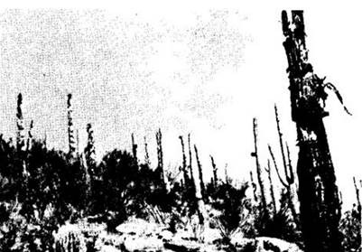
Many plant diseases that cause severe economic damage—effectively resulting in the loss of 1/8 of the world's crop yields—are caused by bacteria. Almost all plants are susceptible to bacterial infection (Figs. 11-3, 11-22).
Fig. 11-22. A population of the saguaro cactus (Carnegiea gigantea), growing at an elevation of 1,000 m on the southern slopes of the Santa Catalina Mountains near Tucson, Arizona, was infected by the bacterium Erwinia carnegieana after the cacti were weakened by freezing temperatures in January 1962.
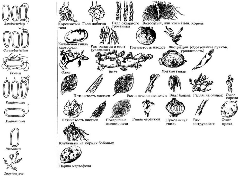
Most plant pathogens are bacilli (rod-shaped forms), and many parasitize their plant hosts. The symptoms of diseases caused by pathogenic bacteria are extremely diverse; in most cases, they manifest as spots on stems, leaves, flowers, and fruits (Fig. 11-23). Almost invariably, these indicate bacterial infection, most frequently caused by closely related organisms of the genera Pseudomonas and Xanthomonas.
Fig. 11-23. Examples of typical bacterial plant pathogens and the various diseases they cause.
Many economically damaging plant diseases, such as blight, soft rot, and wilt, are also caused by bacteria. Blight is characterized by rapidly developing necrosis (dead, discolored patches) on stems, leaves, and flowers. Bacterial blight of apples and pears can destroy young trees within a single growing season. This disease is caused by the bacillus Erwinia amylovora. Bacterial soft rot typically affects the fleshy storage tissues of vegetables, such as tubers (potatoes) and bulbs, as well as juicy fruits like tomatoes and eggplants. The most destructive soft rots are caused by bacteria of the genus Erwinia.
Bacterial vascular wilts affect herbaceous plants exclusively. In these infections, microorganisms invade the xylem vessels and multiply within them, spreading throughout the plant via the flow of water and nutrients, ultimately leading to its death. Initially, the bacteria break down PARTS OF THE vessel cell walls and may even rupture the vessel entirely. They then penetrate the adjacent parenchyma cells, where they continue to proliferate. Sometimes, bacteria form cavities within the plant tissues filled with cellular debris, resinous secretions, and bacterial cells themselves. These cavities communicate with The surface of stems and leaves via cracks. However, as a rule, bacteria do not emerge onto the surface until the plant dies. The most common examples of such diseases are bacterial wilt of alfalfa and beans (each caused by a distinct species of Corynebacterium), bacterial wilt of cucurbits such as pumpkins and watermelons (caused by Erwinia tracheiphila), and black rot of crucifers, such as cabbage (caused by Xanthomonas campestris).
Members of the genus Agrobacterium induce crown gall formation in plants. They are of great importance in Genetic Engineering and will be discussed in ch. 30.
Mycoplasmas are a group of the smallest known bacteria (about 0.1 µm in diameter). A plasma membrane encloses their DNA, RNA, ribosomes, soluble proteins, carbohydrates, and lipids. Mycoplasmas lack a nucleus, organelles, and a cell wall. They likely contain little more than 650 genes, which is five times fewer than the average bacterium.
Classified in a distinct group, mycoplasmas are divided into 6 genera, with 50 or 60 described species belonging to the genus Mycoplasma. By studying rRNA nucleotide sequences, C. R. Woese and his colleagues determined that all mycoplasma genera except one are closely related and probably evolved from a single bacterial Lineage that includes Bacillus and Lactobacillus. At the same time, the genus Thermoplasma evidently acquired its mycoplasma-like features independently. All mycoplasmas can probably be regarded as bacteria that simplified their structure through evolution.
In addition to mycoplasmas found in animal tissues, mycoplasmalike organisms (MLO) have been discovered in 200 plant species, causing about 50 diseases typically characterized by yellowing and stunting. Numerous attempts to cultivate MLO isolated from plants have been unsuccessful.
Some spiroplasmas—long (~ 10 µm), thin (0.2 µm in diameter) helical mycoplasmas—are motile in liquid media and can be cultivated in vitro on artificial media. These include Spiroplasma citri, which causes citrus stubborn disease, and other pathogens. In culture, spiroplasmas exhibit vigorous swirling and wave-like movements. Evidence for the pathogenicity of spiroplasmas isolated from diseased tissues has been obtained for only a small number of species.
Fig. 11-24. Spiroplasmas isolated from a stunted corn plant (Zea mays)

MLO typically invade the sieve tubes of the phloem. It is believed that these microorganisms are carried along with sugar solutions from one sieve tube to another through pores and are subsequently transported throughout the phloem. Some spiroplasmas, however, are capable of actively moving through plant tissues.
Cyanobacteria
Cyanobacteria ("blue-green algae") deserve special Discussion due to their diverse ecological roles. Furthermore, cyanobacteria are frequently treated as plants rather than as a distinct group of bacteria. Although approximately 7,500 species were formerly assigned to cyanobacteria, experimental studies suggest that only slightly more than 200 distinct non-symbiotic species truly belong to this group. These species exhibit a wide range of characteristics depending on their habitat. For instance, Microcoleus vaginatus inhabits moist soil, freshwater, and saltwater across various regions of the globe—from northern Greenland to Antarctica, and from Death Valley to Pikes Peak. When environmental conditions change, the bacterial cells comprising even a single Microcoleus colony can display remarkably diverse morphologies.
Fig. 11-25. A. Mycoplasmalike organisms penetrate through a pore in the sieve plate of a young coconut palm (Cocos nucífera) inflorescence, causing a fatal disease known as lethal yellowing. B. A devastated grove of coconut palms in Jamaica, which now resemble telegraph poles. Lethal yellowing affects many palm species growing in southern Florida and other regions

Like other bacteria, cyanobacteria can thrive in extreme environments—ranging from hot springs to frozen Antarctic lakes, where they form a layer 2—4 cm thick situated in the water 5 m below the permanent ice cover. The green coloration of some polar bears in zoos is caused by cyanobacterial colonies developing within the hollow shafts of their guard hairs.
Cyanobacteria proved to be the pioneer colonizers of the new island of Surtsey near Iceland, which was formed by a volcanic eruption. Conversely, they are absent from acidic waters, where eukaryotic algae, by contrast, are quite numerous.
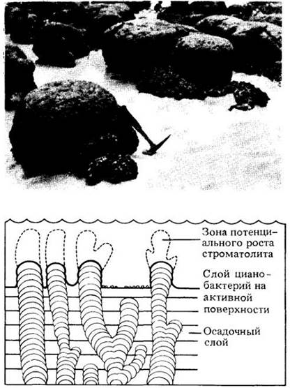
Stratified chalk deposits known as stromatolites (Fig. 11-26) have an immense geological age—2.7 billion years—and were formed through the trapping of calcium by cyanobacteria. Today, stromatolites form only in hot, arid climates in areas with shallow water bodies. The Abundance of stromatolites in the fossil record indicates the existence of similar climatic conditions in distant geological epochs, when cyanobacteria played a decisive role in increasing the level of free oxygen in the early Earth's atmosphere.
Fig. 11-26. Stromatolite formation occurs when extensive colonies of cyanobacteria become calcified and reinforced with calcium carbonate. Stromatolites display a wide variety of shapes, such as the dome-like structures shown in the drawing and photograph. They are frequently found in the fossil record, whereas today they form only under highly specific conditions, such as in the littoral zone of Shark Bay in Western Australia
Many cyanobacteria possess a mucous capsule or sheath, often brightly colored, especially in terrestrial species. This coloration can be light gold, yellow, brown, red, emerald, light blue, violet, or dark blue. Additionally, carotenoids and phycobilins present in cyanobacterial cells modify their hue. Despite their name, only about half of the "blue-green algae" actually exhibit this color. The Red Sea likely derived its name from waterblooms of marine planktonic species of the genus Trichodesmium (vacuolate cyanobacteria), which frequently occur on the sea's surface.
Many marine cyanobacteria inhabit limestones or lime-rich substrates such as mollusk shells or coralline algae. Freshwater species, particularly those living in hot springs, frequently build up thick layers of calcium carbonate within their colonies. In Yellowstone National Park, the filamentous form Mastigocladus thrives in hot water at 55° C, while the unicellular Synechococcus tolerates temperatures up to 73 — 75° C. Cyanobacteria are widely distributed in soils, including deserts, with counts ranging from 20,000 to 50,000 such organisms per gram of soil.
Representatives of many cyanobacterial genera are nitrogen fixers. In warm regions of Asia, rice can grow on the same plot for quite a long time without the application of fertilizers, thanks to nitrogen-fixing cyanobacteria. Cyanobacteria, particularly of the genus Anabaena, inhabit the leaf cavities of the water fern Azolla, which in turn maintains an almost obligate relationship with these cyanobacteria. Due to their ability to fix molecular nitrogen, cyanobacteria can colonize bare rock surfaces and soil alongside Lichens, liverworts, and other organisms with which they form symbiotic associations. Marine Trichodesmium species fix approximately a quarter of all nitrogen assimilated by the oceans, which represents a substantial amount.
Nitrogen fixation takes place in heterocysts—large specialized cells produced by filamentous forms (see Figs. 11-13, 11-15). The heterocyst wall is analogous to the typical cyanobacterial cell wall but features two additional outer layers: an outer polysaccharide layer and an inner glycolipid layer. The internal membranes of the heterocyst form a concentric and reticulate network. Because heterocysts lack Photosystem II (Chapter 7) and perform only cyclic Photophosphorylation, no oxygen is evolved; any oxygen present is immediately reduced by hydrogen—a byproduct of nitrogen fixation—or removed through the heterocyst cell wall. Nitrogenase is sensitive to oxygen, making nitrogen fixation an anaerobic process. Heterocyst differentiation in Nostoc and other genera is inhibited by ammonia and nitrates; however, when the concentration of these nitrogenous compounds drops below a certain threshold, heterocysts begin to appear anew. As already mentioned, some cyanobacteria form resistant spores known as akinetes, which differ structurally and functionally from heterocysts.
Cyanobacteria can establish symbiotic relationships with certain Sponges, amoebas, flagellated Protozoans, colorless diatoms and green algae, other cyanobacteria, mosses, vascular plants, and oomycetes. In these associations, they may lose their cell wall and function analogously to chloroplasts. The symbiotic cyanobacterium divides synchronously with the host cell, much like a chloroplast. The evolutionary relationships between cyanobacteria and certain eukaryotes will be discussed in Chapter 15.
Last update: 07/08/2026
Editorial and Educational Adaptation: This material has been compiled based on the primary/original source text. The project team performed an editorial review, corrected technical inaccuracies, structured sections, and adapted the content for an educational format.
What was processed:
- elimination of formatting defects (OCR errors, structural breaks, corrupted characters);
- editorial organization of content;
- standardization of terminology in accordance with academic sources;
- verification of factual statements against the original source text.
All mentions of the author, publication year, and origin of the primary text have been preserved in accordance with the source.