Fundamentals of Biochemistry - Filippovich, Y. B. 1999
Carbohydrates and Their Metabolism
Structure of Carbohydrates
General characteristics of CARBOHYDRATES. Carbohydrates are defined as Organic compounds containing an aldehyde or ketone group and multiple alcoholic hydroxyls. Their elemental composition is expressed by the general formula CmH2nOn. There are few exceptions to this rule, and although this definition is not entirely precise, it provides the simplest way to characterize this diverse group of Organic compounds as a whole.
Neither the Class name nor the general formula of these compounds gives a clear picture of their chemical nature and Structure. Moreover, the term "carbohydrates", first proposed by K. Schmidt, a professor at the University of Dorpat (1844), suggests that they should be regarded as carbon "hydrates". In reality, this is completely incorrect. Therefore, quite some time ago (1927), another term was proposed to denote this class of compounds—"glucides"—which, unfortunately, has been making its way into chemical nomenclature with great difficulty.
Carbohydrates include compounds with diverse and often entirely different properties. Among them are low-molecular-weight and high-molecular-weight substances, crystalline and amorphous forms, substances soluble in Water and insoluble in it, hydrolyzable and non-hydrolyzable compounds, those easily oxidized and those relatively resistant to oxidizing agents, and so on. This multiplicity of qualities is closely linked to the Chemical Nature of carbohydrates and The structure of their molecules; it determines the specific role that carbohydrates play in vital processes and in The formation of animal and plant Tissues.
In all organisms without exception, carbohydrates serve as a material whose oxidation releases the energy required to drive Chemical Reactions. Such carbohydrates are considered reserve Materials. Along with this, intermediate products of carbohydrate oxidation are utilized for the synthesis of many other organic compounds.
The aforementioned Functions of carbohydrates (structural, energetic, and metabolic) are regarded as canonical. However, it has recently become clear that carbohydrates exhibit many other non-standard, non-canonical functions. Numerous carbohydrates and carbohydrate-containing Biopolymers possess unique structures and Specificity. For instance, Blood group substances, which are Glycoproteins where 80% of the molecule consists of carbohydrates, acquire astonishing interaction specificity precisely due to their asymmetric centers, stereoisomers, tautomers, and conformers. Oligosaccharide fragments of Cell wall glycoproteins and Glycolipids project outward beyond The Cell membranes like antennas, acting as locators with receptor functions. Specifically, they serve as binding sites for protein toxins (e.g., cholera, botulinum, tetanus, diphtheria, and Shiga toxins, etc.), Bacteria (e.g., Escherichia coli interacting with mannose-containing Oligosaccharides), Viruses (e.g., Influenza virus), and the like. The structures of immunoglobulin oligosaccharide fragments are highly reproducible and moderately conserved, ensuring specific carbohydrate-Protein Interactions between the domains of these remarkably refined protective Proteins. Over 250 Enzymes feature oligosaccharide fragments that selectively interact with numerous Lectins—proteins that form conjugates with carbohydrates. Thus, alongside Nucleic Acids and Proteins, carbohydrates are viewed from a modern perspective as informational molecules, i.e., code words in the molecular language of life. Consequently, the contours of a new field in Carbohydrate Biochemistry—glycobiology and glycotechnology—are becoming increasingly clear.
Depending on their composition, structure, and properties—specifically their behavior when heated with dilute aqueous acid solutions (i.e., their susceptibility to Hydrolysis)—Carbohydrates are divided into two groups: simple and complex. Simple carbohydrates do not undergo hydrolysis. Complex carbohydrates break down upon hydrolysis to form simple carbohydrates.
Simple carbohydrates. The overwhelming majority of simple carbohydrates have the composition CnH2nOn. Because simple carbohydrates do not hydrolyze, they are called Monosaccharides. All simple carbohydrates are crystalline solids, readily soluble in water, and typically have a sweet taste. In terms of chemical properties, they are bifunctional and can be characterized as polyhydroxy aldehydes or polyhydroxy ketones. This means their molecules contain an aldehyde or ketone group and several alcoholic (hydroxy) groups. Depending on the number of oxygen atoms in the molecule, monosaccharides are divided into several groups, whose names are formed from Greek numerals with The addition of the suffix -ose, characteristic of carbohydrates. Therefore, the series of simple carbohydrates as a whole is referred to as the series of monoses. Over 200 natural monoses are currently known. Depending on THE POSITION OF the carbonyl group within the molecule, monosaccharides exist as isomers: aldoses and ketoses.
Because asymmetric carbon atoms are present in monosaccharide molecules, and consequently the molecules as a whole are constructed asymmetrically, this group of compounds exhibits optical, or mirror-image, isomerism. As with other optically active compounds, the mirror-image forms of monosaccharides are called optical antipodes. Equimolecular mixtures or compounds of the latter with each other are called racemates. Monosaccharide stereoisomers that differ in the spatial arrangement of the hydrogen and OH group at the carbon atom adjacent to the aldehyde group are epimers. Natural sugars belong predominantly to the D-series, which is apparently related to the specifics of their primary Biosynthesis in plants.
Among aldoses, D-ribose, D-glucose, D-mannose, and D-galactose are especially widespread in nature:

Among ketoses, the best known are D-ribulose and D-fructose; among heptoses, D-sedoheptulose is worth noting:
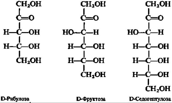
All of these ketoses actively participate in carbohydrate transformations within the Organism.
A characteristic feature of monosaccharides is their pronounced capacity for tautomeric transformations. Two Types of monosaccharide Tautomerism are distinguished: keto-enol and ring-chain.
Keto-enol tautomerism in monosaccharides consists of The conversion of the form with carbonyl oxygen in the aldehyde or ketone group into the enol form (with an OH group attached to a carbon atom linked by a double bond). Due to keto-enol tautomerism, epimeric monosaccharides can convert into one another.
Ring-chain tautomerism in monosaccharides consists of the existence of cyclic (ring) forms and a chain (i.e., open carbon chain) form of the monosaccharide in dynamic equilibrium. METABOLISM/2.html">THE CONCEPT OF the existence of cyclic monosaccharide forms was first proposed (1870) by A. A. Kolli, a professor at the Moscow Higher Technical School. Ring closure occurs when the CO group of the monosaccharide approaches the hydroxyl of a carbon atom located 3–4 links away from it. A hydrogen atom addition reaction from the aforementioned alcoholic group takes place at the carbonyl oxygen, resulting in the formation of a new hydroxyl, termed glycosidic or hemiacetal. Simultaneously, an oxygen bridge closes, giving rise to a five- or six-membered heterocycle (of the furan or pyran type):

The carbon atoms of the six- or five-membered heterocycle in cyclic monosaccharide formulas, and sometimes all other side groups, are omitted, in which case they take the following appearance when written:
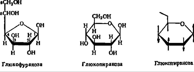
Bonds between the ring carbon atoms pointing toward the observer are depicted by thick lines, while those located behind the plane of the paper are shown with thin lines; this facilitates the spatial perception of the cyclic formula. Hydrogen atoms and OH groups are represented as positioned above and below the plane of the heterocycle, respectively.
Because the carbon atom of the former CO group becomes asymmetric at the moment of ring closure, the appearance of the cyclic monosaccharide form is accompanied by the formation of two new optical isomers. The isomer in which the glycosidic hydroxyl points in the same direction as the alcoholic hydroxyl at the penultimate carbon atom of the monosaccharide (which determines its assignment to the D- or L-series) is called the α-form. The form with the opposite spatial arrangement of the glycosidic radical is called the β-form. Below are corresponding Examples of α- and β-forms, also referred to as anomers (from the Greek ana—up, since glycosidic radicals are usually located at the 1st, top carbon atom):

Ketoses, like aldoses, form cyclic structures. As an example, let us examine the ring-chain tautomerism of fructose. Like other monosaccharides, fructose forms 4 cyclic structures that exist in dynamic equilibrium with the open-chain form (see p. 310).
Typically, the cyclic forms of monosaccharides strongly predominate over the open-chain form in solution; furthermore, as a rule, one of the cyclic monosaccharide forms is present in the equilibrium mixture of all forms in a greater amount than the others. Thus, among the 4 cyclic forms of D-glucose, β-D-glucopyranose heavily predominates in solution (64%); meanwhile, the content of the aldehyde form of glucose in the equilibrium mixture is a mere 0.024%. The concentration of α- and β-glucofuranoses in the mixture is also negligible, and the remaining portion of glucose is represented by the α-anomer. Overall, pyranose forms drastically predominate over furanose forms.

Ring-chain tautomerism in monosaccharides is responsible for a fascinating property of simple carbohydrates. In the crystalline state, monosaccharides exist exclusively in a cyclic form. Depending on crystallization conditions, either the a- or the ß-form is produced. For instance, crystallization from water yields glucose in the a-form, whereas crystallization from pyridine yields the ß-form. Upon dissolving a-D-glucose in water, its specific rotation initially exhibits a Characteristic value of +112.2°. However, upon standing, this value gradually decreases until it reaches a stable equilibrium of +52.5°. Similarly, the initial specific rotation of a ß-D-glucose solution (+17.5°) changes over time, increasing to +52.5°. Because multiple specific rotation values can be observed for some time after dissolving crystalline glucose preparations until equilibrium is established, this phenomenon was named multirotation or mutarotation (from Latin multum — much, rotatio — turning). It is caused by the establishment of an equilibrium in solution among all possible cyclic and acyclic modifications of glucose, each possessing its own specific rotation, with their mixture yielding an average specific rotation value (+52.5°).
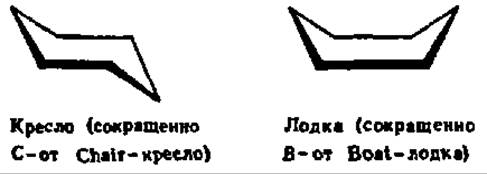
Carbohydrate Conformations are extremely diverse. It is well known that six-membered alicyclic compounds (such as cyclohexane) exist in geometrically distinct shapes that the molecule adopts without altering Bond Lengths or Bond Angles. These shapes are referred to conformational isomers. Conformational isomerism is also characteristic of monosaccharides, which predominantly feature a pyranose structure. However, whereas cyclohexane has only two conformations — the chair and the boat forms:
monosaccharides in the pyranose form exhibit 8 conformations — 2 chair forms and 6 boat forms due to the presence of a heteroatom (oxygen) in the six-membered ring:
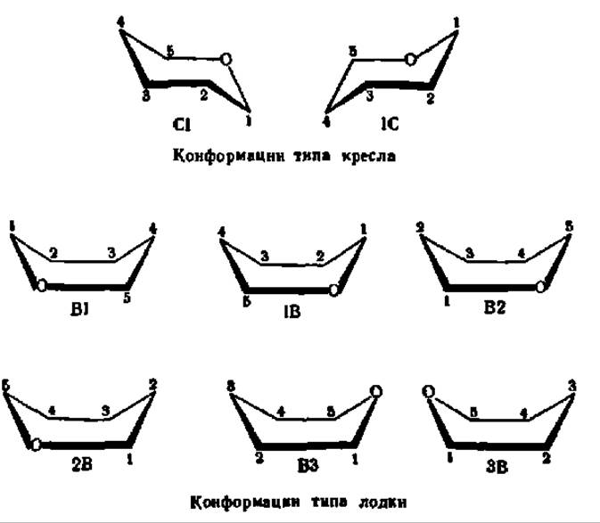
Among the 8 listed conformations of the pyranose ring, the chair conformations are the most stable. In turn, the C1-isomer is preferred, as the vast majority of its substituents are oriented equatorially, in the plane of the ring. It is specifically in the C1-conformation that the majority of monosaccharides exist, for example:
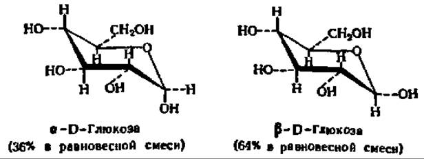
Naturally, the ß-anomer of D-glucose predominates over the a-anomer in the equilibrium mixture because the glycosidic hydroxyl group in the C1 conformation of the former is positioned equatorially:

This holds true not only for glucose anomers but also for the anomers of various other monosaccharides.
Ring-chain tautomerism in monosaccharides is a property dependent on the simultaneous presence of a C=O group and alcohol radicals within their molecules. The behavior of the glycosidic hydroxyl group, formed during the cyclization of a monosaccharide, is unique: it enters into chemical reactions much more actively than the other hydroxyl groups. Derivatives of cyclic monosaccharides obtained by substituting the hydrogen atom of the glycosidic hydroxyl group with a radical are termed Glycosides, while the radical itself is called the aglycone. Examples include a- and ß-methyl glycosides (see p. 109).
Natural glycosides, which typically exhibit potent physiological activity, feature A wide variety of often highly complex radicals acting as aglycones. For instance, plants are rich in glycosides with steroid aglycones; these act as powerful toxins that protect plants from viruses and pathogens. Marine invertebrates are also notable for their high content of glycosides with diverse aglycones. Many of these glycosides are widely used in medicine. Complex carbohydrates are likewise typical glycosides.
Monosaccharides undergo all typical reactions associated with the aldehyde group and alcohol radicals. Specifically, the aldehyde group is characterized by Oxidation and reduction reactions, carbonyl oxygen substitution, polycondensation (resinification), etc., while the alcohol radicals undergo etherification, Esterification, and other interactions well known from organic chemistry. In biochemistry, however, particular importance is attached to the redox reactions of monosaccharides and the formation of their phosphate esters.
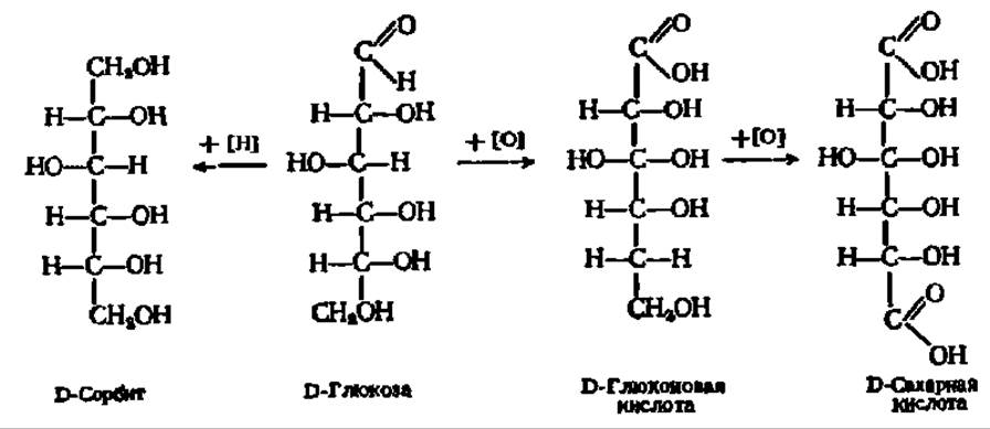
Thus, upon oxidation or reduction of glucose, for instance, which proceeds very readily, the corresponding acid or alcohol is formed:
Among the phosphoric esters, glucose-1-phosphate, glucose-6-phosphate, fructose-1,6-diphosphate, ribulose-5-phosphate, and others are of particular importance:
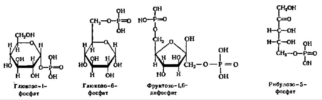
These compounds play a highly active role in biochemical processes occurring in living organisms. Phosphorylation (i.e., the incorporation of a phosphoric acid residue into a monosaccharide molecule) serves as the initial, preparatory stage for both The breakdown of simple carbohydrates and The biosynthesis of more complex carbohydrates from them. According to A. E. Chichibabin (1932), monosaccharide phosphorylation facilitates their transition from a cyclic structure to an open-chain conformation.
The most important representatives of monoses are ribose, glucose, mannose, galactose, and fructose. D-Ribose is a constituent of Selection/9.html">NUCLEIC ACIDS AS a structural element of nucleotide residues, where it occurs in the ß-D-furanose form. Crystalline ribose melts at 87° C, and its specific rotation in an aqueous solution is 23.7°. The reduction of ribose yields a pentahydric alcohol, ribitol, which takes part in the formation of many bioactive compounds:

D-Glucose is one of the most widespread naturally occurring sugars. Crystalline sugar from a syrup obtained by acid Treatment of starch was first isolated by K. Kirchhoff (1811); a quarter of a century later, Jean-Baptiste André Dumas named it glucose. In living organisms, it occurs either in a free state or in a bound form, serving in the latter case as the basis for such crucial natural compounds as cane (beet) sugar, starch, Cellulose, etc. It forms crystals with tпл= 146° С for a-glucopyranose and 148—150° С for ß-glucopyranose. Upon heating in pyridine, the a-form (specific rotation +112.2°) converts into the ß-form (specific rotation +17.5°). Therefore, a-D-glucopyranose crystallizes from aqueous and aqueous-alcoholic solutions, whereas ß-D-glucopyranose crystallizes from solutions in pyridine. The reduction of glucose yields D-sorbitol, whereas its oxidation yields D-gluconic acid and subsequently saccharic acid.
D-Galactose is a component of A number of complex carbohydrates, including lactose (milk sugar). It crystallizes as a monohydrate. Anhydrous crystals melt at 164° C; the specific rotation (final value) is +81°.
D-Mannose forms natural complex carbohydrates known as Mannans, which are frequently carbohydrate components of glycoproteins and mucilages, as well as Plant Cell Walls. Mannopyranose melts at 132° C, and its a- and ß-forms have different angles of rotation (+30° and —17°, respectively); the final angle of rotation established As a result of mutarotation is +14.5°.
D-Fructose occurs both in a free state (e.g., in honey) and in a bound form (e.g., as part of cane sugar and certain high-molecular-weight natural carbohydrates—fructosans). It is 2.5 times sweeter than glucose and 1.7 times sweeter than cane sugar. Anhydrous fructose melts at 102—104° C. The specific angle of rotation in The equilibrium state (after reaching a constant value upon mutarotation) is —92°, which is why fructose is also referred to as levulose.
Recently, data have accumulated indicating the existence of a new group of monosaccharides designated by N. K. Kochetkov et al. (1975) as glycolactic acids. Monosaccharides of this group are composed of conventional monosaccharide residues and lactic acid linked together by an ether bond (the second half of the name for this new group of monosaccharides derives from the Latin acidum lacticum, meaning lactic acid). The most important representative of glycolactic acids is muramic acid:
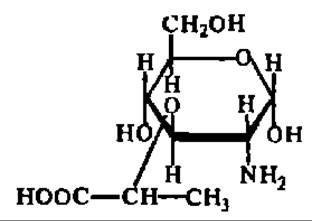
It occurs as crystals with a melting point (tпл) of 150–155 °C; the equilibrium specific rotation of its solutions ranges from 103° to 123°, depending on the pH value. N-acetylmuramic acid is a component of the peptidoglycans in bacterial cell walls; the polysaccharide fragments of peptidoglycans are attacked by Lysozyme (see pp. 110–111).
Complex carbohydrates. Complex carbohydrates are those whose molecules break down upon hydrolysis to yield simple carbohydrates. The composition of complex carbohydrates is expressed by the general formula CmH2nOn, where m > n. Complex carbohydrates are divided into two main groups:
1. Oligosaccharides — sugar-like complex carbohydrates characterized by a relatively low molecular mass (a few hundreds), good solubility in water, easy crystallization, and, as a rule, a sweet taste. Oligosaccharide molecules are composed of a small number (from the Greek *oligos*, meaning few or small) of simple carbohydrate residues.
2. Polysaccharides — high-molecular-weight complex carbohydrates formed from many hundreds of simple carbohydrate residues. Their molecular mass typically reaches hundreds of thousands. They do not form well-defined crystals, and only some of them possess a pseudocrystalline structure. Polysaccharides are either insoluble in water or form solutions whose properties resemble those of colloids, which can be explained by the large molecular mass of the dissolved particles. A sweet taste is not characteristic of polysaccharides.
Oligosaccharides. Depending on the number of monosaccharide residues making up their molecules, oligosaccharides are classified into Disaccharides, trisaccharides, and so on. Of greatest interest among oligosaccharides is the group of disaccharides—compounds that are widespread in nature, many of which are of immense practical importance.
In terms of chemical structure, disaccharides are glycosides of monosaccharides, whose aglycones are also monosaccharide residues. Disaccharides in which the monosaccharide residue serving as the aglycone is attached to the glycosidic radical of the main monosaccharide via its own glycosidic hydroxyl are called glycosido-glycosides. If, however, the bond between the aglycone and the main monosaccharide is formed through the alcoholic hydroxyl of the aglycone, the disaccharides are called glycosido-glucoses. This is illustrated by the formulas of trehalose and maltose:
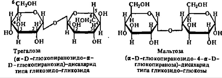
In accordance with their chemical structure, disaccharides of the trehalose type (glycosido-glycosides) and the maltose type (glycosido-glucoses) exhibit substantially different chemical properties: the former do not undergo any reactions characteristic of the aldehyde or ketone group—that is, they are neither oxidized nor reduced, do not form osazones, do not enter into polycondensation reactions (do not resinify), do not exhibit mutarotation, and so forth. For maltose-type disaccharides, conversely, all of the aforementioned reactions are highly characteristic. The reason for this difference is quite clear from what has been said above regarding the two types of disaccharide structures and The properties of their constituent monosaccharide residues. It lies in the fact that ring-chain tautomerism is possible only in maltose-type disaccharides, resulting in the formation of a free aldehyde or ketone group that exhibits its characteristic properties:

In trehalose-type disaccharides, where both glycosidic hydroxyls are utilized to form the glycosidic bond between the monosaccharide residues, ring opening is impossible.
Through their alcoholic hydroxyl groups, both types of disaccharides undergo identical reactions: they form ethers and esters, interact with metal oxide hydrates [for example, dissolving Cu(OH)2], and so on.
A large number of disaccharides exist in nature; among them, the aforementioned trehalose and maltose, as well as sucrose, cellobiose, and lactose, are of primary importance.
Sucrose is a glycosido-glycoside type disaccharide composed of a-D-glucopyranose and ß-D-fructofuranose residues. Upon hydrolysis, sucrose breaks down into two monosaccharides: a-D-glucose and ß-D-fructose (see p. 128). Sucrose is one of the most widespread and practically important disaccharides in nature. It is better known as cane or beet sugar, since virtually all commercial sugar is obtained from the juice of these two plants. Sucrose is also found in the sap of many other plants, though in relatively low concentrations, whereas its content in maple, birch, and palm saps is quite high.
Sucrose readily crystallizes (tпл = 184 °C), is easily soluble in water and sparingly soluble in alcohol. Its solutions do not exhibit mutarotation, which is typical of glycosido-glycosides, but they rotate the plane of polarized light to the right at [a]D1 = +66.5°.
Since the monosaccharides resulting from the hydrolysis of sucrose have opposite specific rotation angles (in the equilibrium state, +52.5° for glucose and –92° for fructose), the total rotation angle after hydrolysis becomes negative. Consequently, hydrolyzed sucrose is referred to as invert sugar, and the hydrolysis process itself as inversion (from Latin *inversio* — overturning, swapping).
1 The symbol [a]D denotes the specific rotation [a] measured in polarized light whose wavelength corresponds to that of the sodium spectral line (D = 588 nm).
Maltose belongs to the glycosido-glucose type of disaccharides and is composed of two a-D-glucopyranose residues joined by a glycosidic bond at the 1,4 positions (see p. 314). This type of bond is called an a-1,4-glycosidic bond. Maltose is obtained during the hydrolytic Cleavage of starch using a specific enzyme (see p. 329) abundant in germinating barley grains. Since sprouted, dried, and ground barley seeds are called malt, the disaccharide formed from starch with the participation of the malt enzyme was named maltose.
Maltose crystallizes from aqueous solutions as a monohydrate that lacks a sharply defined melting point. Aqueous solutions of maltose (it is very soluble in water) undergo mutarotation because ring-chain tautomerism gradually converts the initial form first into a semi-cyclic form and then into other maltose tautomers containing, instead of a-D-glucopyranose, its ß-modification or a- and ß-glucofuranose rings. The equilibrium specific rotation angle of maltose is +136°.
Maltose is formed in PLANT AND ANIMAL organisms as an intermediate product during starch hydrolysis and can be detected in a free state. Upon hydrolysis, maltose breaks down into two glucose molecules.
Cellobiose, like maltose, consists of two D-glucose residues and belongs to the glycosido-glucose type of disaccharides. Its molecular structure differs from that of maltose only in that the glycosidic bond between the monosaccharide residues originates from the glycosidic hydroxyl located in the ß-position. Consequently, the cellobiose molecule is characterized by the presence of a ß-1,4-glycosidic bond:
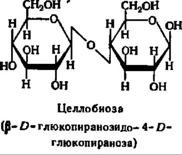
Cellobiose crystallizes exceptionally well, is readily soluble in water and sparingly soluble in alcohol, and undergoes mutarotation: [a]D = +34.6°. It is easily oxidized, acts as a strong reducing agent, forms a characteristic osazone, and yields exclusively D-glucose upon hydrolysis.
Cellobiose is widely distributed in the plant kingdom; it has been found in germinating seeds, apricot pits, and tree sap. Cellobiose is formed during the Enzymatic hydrolysis of cellulose (plant fiber), which constitutes plant cell walls.
Lactose is ß-D-galactopyranosyl-4-D-glucopyranose:
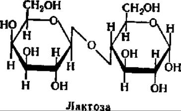
It is contained in significant amounts in milk (5–8%) and is therefore known as milk sugar. However, lactose is not exclusive to mammals (although its role in their Nutrition is undeniable), as it has also been found, for example, in the pollen tubes of certain plants. Lactose is sparingly soluble in water and can be obtained by evaporating whey. It crystallizes as a monohydrate with a melting point (tпл) of 202° C. Because the glucose residue possesses a free glycosidic hydroxyl group, mutarotation is possible, and lactose can therefore exist in two forms. The equilibrium mixture of its a- and ß-forms has a specific rotation of + 52.2°. Unlike other disaccharides, lactose crystals are non-hygroscopic.
Polysaccharides. Polysaccharides are substances composed of a large number of monosaccharide residues or their derivatives. If a polysaccharide contains residues of a single type of monosaccharide, it is called a homopolysaccharide. In cases where a polysaccharide is composed of two or more types of monosaccharides alternating regularly or irregularly within the molecule, it is classified as a heteropolysaccharide.
The most important natural Homopolysaccharides include starch, Glycogen (animal starch), cellulose, dextran, and Chitin. The first four polysaccharides yield only D-glucose upon hydrolysis, while the latter releases a D-glucose derivative, N-acetylglucosamine. In addition, many other homopolysaccharides are known. For instance, the polysaccharide inulin yields only fructose upon hydrolysis, mannan yields mannose, galactan yields galactose, araban yields arabinose, and so forth. These polysaccharides are named after the End products of their cleavage.
The most important natural Heteropolysaccharides include hyaluronic and chondroitinsulfuric acids, heparin, bacterial capsular polysaccharides, agarose, red algal porphyrans and carrageenans, brown algal alginic acids, and the structural polysaccharides of Protozoa.
The Biological Significance of polysaccharides is diverse. Many of them (starch, glycogen, inulin, etc.) serve as nutrient reserves in plant and animal organisms. Some polysaccharides (such as chondroitinsulfuric acid, capsular polysaccharides, and cellulose) perform exclusively supporting and protective functions. A number of polysaccharides (mannans, galactans, etc.) serve as both structural and nutritional materials. Hyaluronic acid, which constitutes the intercellular substance of animal tissues, alongside its structural function, regulates the distribution of vital substances within tissues. Heparin prevents blood clotting in humans and animals. In many cases, polysaccharides form very stable complexes with proteins, producing glycoproteins that perform a number of crucial functions in the organism.
The chemical Introduction/33.html">Structure of Polysaccharides is uniform. All of them are polyglycosides in whose molecules hundreds, thousands, and sometimes tens of thousands of monosaccharide residues and their derivatives are linked by oxygen bridges. The essential partner in the formation of these oxygen bridges is the glycosidic hydroxyl of the monosaccharides, while the other partner is one of the alcoholic hydroxyls of the monosaccharide, most frequently at position 4 (in linear polysaccharides) or positions 4 and 6 (in branched forms).
Polysaccharides are relatively easily hydrolyzed upon boiling with dilute acid solutions or upon incubation with appropriate enzymes. Alkalis do not hydrolyze polysaccharides. For example, glycogen can be heated for several hours with a 30% alkali solution without any decomposition. This serves as the basis for the Quantitative Assay of glycogen. Characteristically, in a number of cases, the enzymatic hydrolysis of such common polysaccharides as starch and cellulose yields disaccharides (maltose and cellobiose, respectively), whereas nonspecific acid hydrolysis yields a monosaccharide (D-glucose).
Starch is one of the most widespread Reserve Polysaccharides in plants. It accumulates intensively as a result of Photosynthesis and is stored in seeds, tubers, and other plant parts. Seeds and tubers contain 40–70% starch, while other plant parts contain from 4 to 25%. Upon acid hydrolysis, starch breaks down to yield D-glucose, which is its structural unit, along with a small amount of glucose-6-phosphate, since all types of starch contain a small amount (0.02–0.16%) of phosphorus. It has been established that glucose within starch exists in the form of a-D-glucopyranose.
Natural starch consists of two distinct fractions that differ in Structure and properties. Approximately 20% of starch is composed of amylose (from the Greek *amylon* meaning starch). The remainder constitutes the second fraction, known as amylopectin (from the Greek *pektos* meaning jelly-like). This terminology reflects certain properties of these two types of starch. Amylopectin is sparingly soluble in hot water, forming a viscous solution (starch paste) that sets into a gel-like mass upon cooling. Amylose, on the other hand, is readily soluble in warm water and does not form a paste. Taking advantage of this circumstance, amylose is separated from amylopectin by repeated extraction with warm water. For the same purpose, the ability of amylose to precipitate under the action of butyl alcohol upon saturating a hot solution containing a mixture of amylose and amylopectin is utilized. Chromatographic Methods are also employed. For example, after passing dispersed starch through a calcium phosphate Column and subsequently washing with a phosphate buffer, amylose is eluted, while amylopectin remains on the sorbent.
The molecular weights of amylose and amylopectin differ: for amylose preparations that have not undergone degradation during isolation, it ranges from 100,000 to 400,000, whereas for amylopectin it generally exceeds 20 ∙ 106. These data were obtained by Gel filtration through Sephadex G-200 and Sepharose 2B. For instance, potato starch has been separated into 9 fractions with molecular weights ranging from 7 ∙ 106 to 73 ∙ 106. Apparently, the molecular weight values of the polysaccharides discussed here and below depend on the isolation method used. When the Native State of the preparation is preserved, the molecular weights are very high. Correspondingly, the polycondensation coefficient of a-D-glucopyranose in amylose molecules is estimated at several hundred, whereas in amylopectin it is estimated at several tens or even hundreds of thousands.
The chemical structures of amylose and amylopectin also differ. The molecules of the former are generally strictly linear. In them, a-D-glucopyranose residues are linked to one another exclusively by a-1,4-glucosidic bonds; that is, oxygen bridges are formed through the glycosidic hydroxyl of the 1st carbon atom of one a-D-glucopyranose molecule and the alcoholic hydroxyl at the 4th carbon atom of another:
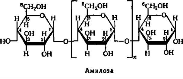
In accordance with this structure, amylose can be characterized as an a-1,4-glucan. Thus, amylose is a linear polysaccharide whose molecules possess a thread-like structure (Figs. 103 and 104). The a-D-glucopyranose residues within amylose have a boat-like conformation. In this case, the structural formula of amylose takes the following form:
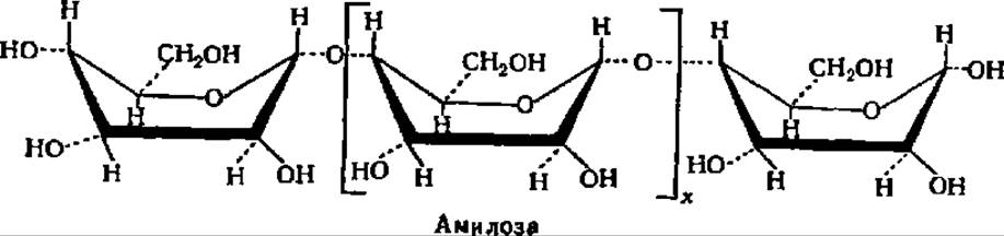
The boat conformation of the a-D-glucopyranose residues in the amylose molecule facilitates the spiralization of the polyglycoside chain. In this process, a single turn of the helix includes 6–7 glucose residues. With the length of each glucose residue being 0.5 nm, a helix with a diameter of about 1 nm is formed (Fig. 103). It is postulated that amylose molecules, like those of other linear polysaccharides, can interact with one another over a certain length, forming secondary structures of a double-helix type with intertwining polysaccharide chains.
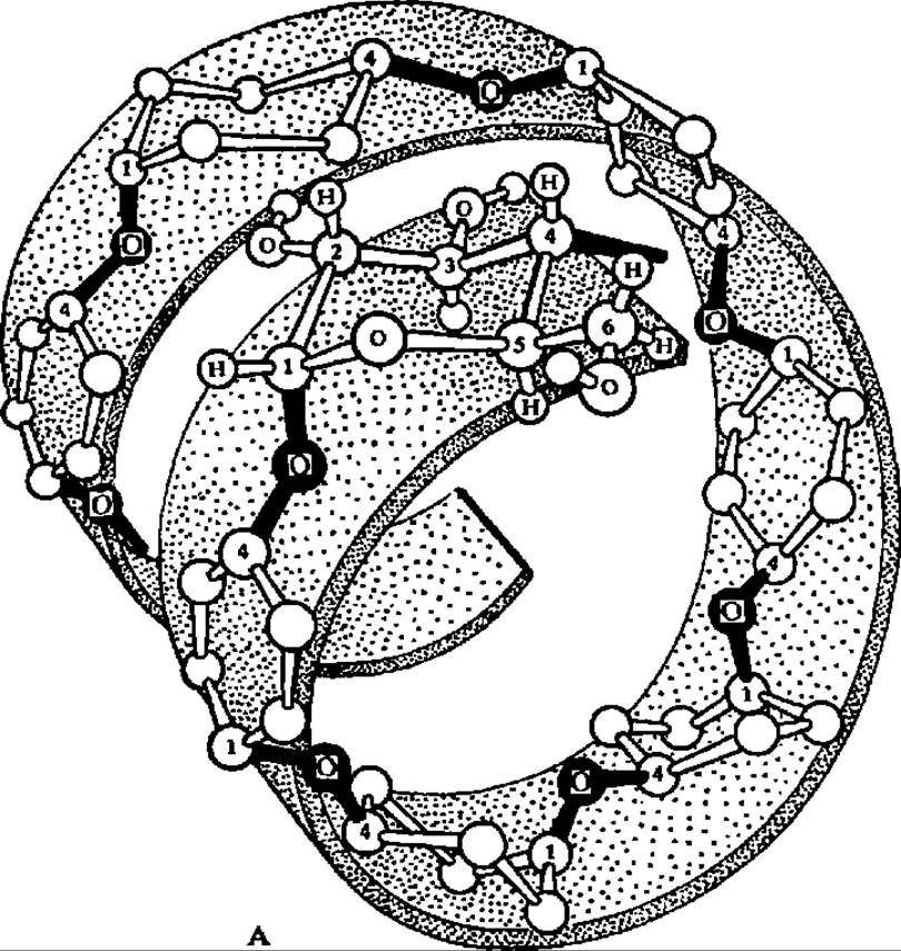
Fig. 103. Spiral conformation of the amylose molecule (A), points of action of a-amylase during hydrolysis (B), and STRUCTURE OF THE adsorption complex between the spiralized starch regions and iodine molecules (C)
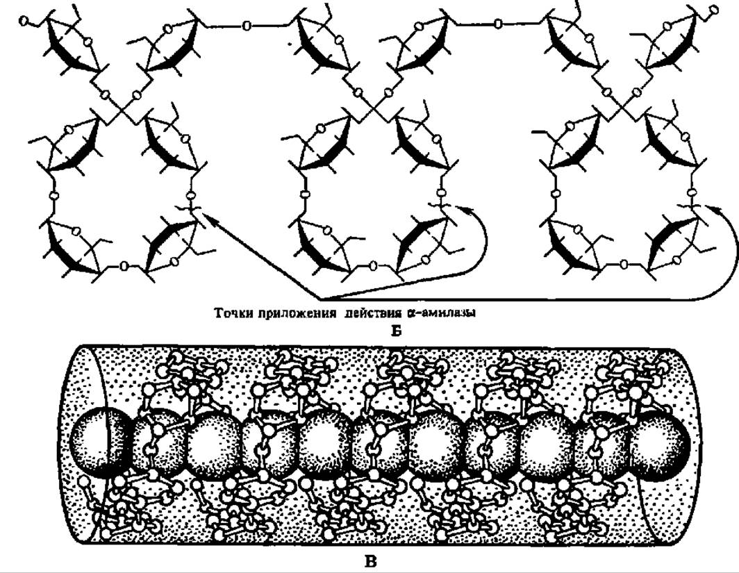
Fig. 103. Continued
Amylopectin has spherical molecules with a radius of gyration ranging from 82 to 255 nm. Their spherical shape is ensured by the fact that the molecule is composed of numerous (several hundred) short polyglycoside chains, each containing an average of 20 a-D-glucopyranose residues. Within each short chain, the glucose residues are linked by a-1,4-glucosidic bonds. The chains are connected to one another through a-1,6-glucosidic bonds. The structure of the branched region of the amylopectin molecule is as follows:
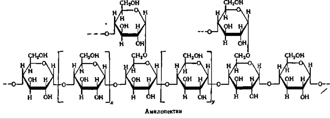

Fig. 104. Structure of starch and glycogen molecules:
A — amylose; B — amylopectin; C — glycogen; each circle denotes a glucose residue; D — modern cluster model of the amylopectin molecule (a — compact, pseudocrystalline region; b — less compact, amorphous region; the dot indicates the reducing group); E — modernized model of the glycogen molecule, demonstrating the presence of concealed polyglycoside chains; F — structure of particulate glycogen; the central line is a polypeptide chain to which ß-particles are attached via hydroxyamino acid radicals, forming an a-particle constructed on THE PRINCIPLE OF a complex drupe; G — three-dimensional model of an amylopectin molecule fragment (R — reducing end; H — non-reducing ends)
The general structure of the amylopectin molecule, according to early data, is shown in Fig. 104, B. A more modern model of the amylopectin molecule (D), derived from a detailed study of enzymatic hydrolysis products and X-ray analysis of this polysaccharide, is presented in the same figure. It is termed cluster-like because the arrangement of its polyglycoside units closely resembles a bunch of grapes. The degree of polymerization of a-D-glucose residues in the units indicated by thick lines reaches 45, and by thin lines, 15. The length of each pseudo-crystalline region is 6 nm, while the amorphous region is three times smaller.
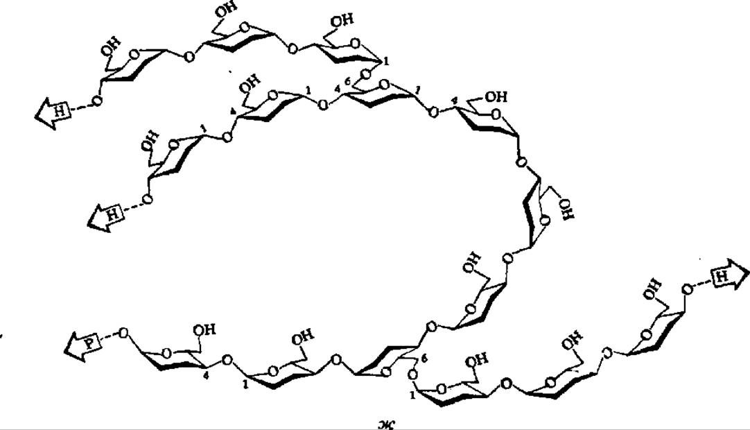
Fig. 104. Continued
It is believed that a-D-glucopyranose in amylopectin also adopts a boat conformation. Consequently, individual segments of the polyglycoside chains that make up the amylopectin molecule are apparently spiraled similar to amylose (Fig. 104, g).
Both starch fractions give a color reaction with iodine in a KI solution; however, amylose turns a pure blue color, while amylopectin turns purple. The starch-iodine reaction is not associated with chemical interaction between them, but rather involves the formation of adsorption-type complexes. Since the helical regions of one or another type of starch molecules play a leading role in the formation of these complexes, the difference in color tone is quite natural. It is hypothesized that iodine molecules are drawn inside the polyglycoside helix, where the corresponding bonds that give rise to the colored complexes are closed (Fig. 103, c).
Upon brief heating of powdered starch, its giant molecules disintegrate, forming a mixture of simpler polysaccharides of lower molecular weight—dextrins. The dextrinization of starch upon heating is accompanied by an increase in its solubility in water. Starch treated in this way is called soluble starch. The breakdown of starch molecules into dextrins proceeds especially intensively when starch paste is heated with a 10% H2SO4 solution. Upon further Processing, the Molecular Weight of the dextrins progressively decreases, and the final breakdown product is ß-glucose. High-molecular-weight dextrins are colored red by iodine, while low-molecular-weight ones do not give a color reaction with iodine. Thus, the Hydrolysis of Starch proceeds stepwise and can be represented by the following scheme:

where x>y>z.
Dextrinization and saccharification of starch are widely used in the alcohol industry, in glue production, etc.
Since starch is composed of optically active a-D-glucopyranose residues, its solutions rotate the plane of polarization of light to the right ([a]D = +195°).
Glycogen serves as a reserve nutrient in the body of humans and animals, which is why it retains the name "animal starch." However, it has also been found in Fungi, Yeast, and corn kernels, which calls its "animal" designation into question. The glycogen content in the Liver of animals reaches 20%, and in Muscles, 4%. Breaking down into simple products via a rather complex pathway called Glycogenolysis (see p. 351), glycogen satisfies the body's need for energy and metabolites. Thus, its biological role is exceptionally significant.
Glycogen is relatively soluble in hot water, although some types of natural glycogen are poorly soluble. Like starch, glycogen gives a color reaction with iodine, and the color tone (red-purple or red-brown) indicates that glycogen is closer to amylopectin than to amylose. Indeed, glycogen and amylopectin are very similar. For instance, the molecular weight of certain fractions of native glycogen is close to that of amylopectin, although overall glycogen exhibits extreme heterogeneity in this regard: animal liver glycogen preparations contain fractions with molecular weights ranging from 10 million to 3 billion, with a predominance of molecules ranging from 200 million to 600 million. Incomplete hydrolysis of glycogen yields dextrins, while complete hydrolysis yields D-glucose. Like amylopectin, glycogen is optically active, and the specific rotation of its solutions (+196°) is very close to that of starch.
The reasons for the similarity in the properties of glycogen and amylopectin lie in the structural resemblance of their molecules: it is essentially the same; the only difference is that in the glycogen molecule, the average length of the short chains linked here by a-1,6-glycosidic bonds is equal to 12 a-D-glucopyranose residues. Thus, the glycogen molecule is somewhat denser and more compact than the amylopectin molecule (see Fig. 104). The glycogen of lower animals is closer to amylopectin, so in many cases it is difficult to draw a sharp boundary between the former and the latter, although accumulating data on the structure of glycogen (see Fig. 104, D and E) clearly indicate a greater Symmetry of its molecule, the presence of a significant number of buried (not exposed on the molecular surface) polyglycoside units in it (these are part of the B units in Fig. 104, D), as well as the existence of a macromolecular structure in glycogen. The latter is characteristic of the so-called particulate glycogen, first isolated by A. Lazarev (1942) under mild conditions using differential ultracentrifugation. It is represented by huge globules called a-particles (up to 200 nm in diameter) that resemble mulberries or raspberries. In turn, a-particles are composed of ß-particles, which in turn consist of y-particles with a diameter of 20–40 nm (see Fig. 104, E).
Cellulose is the primary structural polysaccharide of plants. Leaves contain 15–30% cellulose, wood 50–70%, the stems of fibrous plants (e.g., flax) even more, and cotton fibers (seed hairs) represent almost pure cellulose. The very name of this polysaccharide emphasizes its major role in plant cell wall construction; in this regard, the term cellulose (from Latin cellula – cell) is also widespread. It has been calculated that if all the cellulose contained in plants were burned, The amount of CO2 in the atmosphere would increase by half.
Cellulose is characterized by very low solubility in the vast majority of agents; only an ammoniacal solution of Cu(OH)2 and a concentrated solution of Ca(SCN)2 upon heating dissolve it noticeably. The resistance of cellulose to Solvents is explained by the fact that its long, thread-like molecules interact with each other to form strong micelles, which, in turn, are assembled into fibrils aligned along the fiber axis. Thus, the finest elementary cotton fibers with a diameter of 20 nm consist of numerous cellulose molecules (0.6–0.7 nm in diameter) packed very densely. The detachment of individual cellulose molecules from these stable aggregates is extremely difficult, and only a few substances capable of disrupting intermolecular bonds in micelles can dissolve cellulose.
Upon hydrolysis of cellulose in the presence of a specific enzyme found in a number of bacteria, certain insect species, Molds, and germinating seeds, cellobiose is formed. However, upon acid hydrolysis, ß-D-glucopyranose is produced with an almost quantitative yield; it is the main structural element of the cellulose molecule:
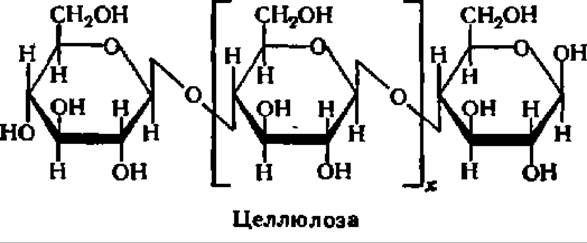
The type of bond between residues is analogous to that in amylose molecules, but unlike the latter, cellulose is a ß-polyglycoside, since the glucose residues are connected to each other by ß-1,4-glycosidic bonds. Like amylose, cellulose molecules have no branches; they are strictly linear in structure, but much longer than amylose molecules. The number of D-glucose residues in a cellulose molecule is very large, reaching several thousand, which corresponds to a native cellulose molecular weight of 10–20 million. These seemingly minor differences in the structure of amylose and cellulose lead to a dramatic difference in their properties.
A deeper difference between amylose and cellulose is revealed when comparing the structures of these polysaccharides taking into account the conformation of their constituent D-glucose residues. It has been established that ß-D-glucopyranose within cellulose exists in a chair conformation:
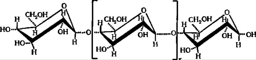
This excludes the possibility of polyglycoside chain spiraling, and the cellulose molecule retains a strictly linear structure.
A solution of cellulose in Schweitzer's reagent exhibits slight optical activity ([a]D = 3.21°).
As with other polysaccharides, a large number of alcoholic hydroxyls remain free in cellulose molecules (at the 2nd, 3rd, and 6th carbon atoms of each ß-D-glucopyranose residue). Corresponding Chemical reactions are possible at these OH groups. Among them, those leading to derivatives widely used in Ion-exchange Chromatography for the Separation of Amino Acids, Peptides, proteins, NUCLEOTIDES, and nucleic acids are particularly important. These include carboxymethylcellulose (CM-cellulose) and diethylaminoethylcellulose (DEAE-cellulose).
CM-cellulose is obtained by treating alkali cellulose with monochloroacetic acid:

As a result, cellulose is enriched with COOH groups, which confer cation-exchanger properties upon it.
DEAE-cellulose is synthesized by treating cellulose with ß-chloroethyldiethylamine hydrochloride in an alkaline medium:
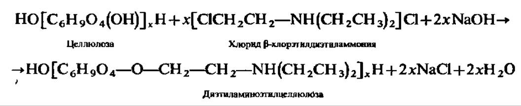
Diethylaminoethyl groups confer anion-exchanger properties on DEAE-cellulose.
Dextran is a polysaccharide produced by certain species of bacteria. Its molecular weight is enormous: various dextran preparations exhibit molecular weight (M) values ranging from 12 million to 1 billion. A dextran molecule consists of relatively short polyglycosidic chains, each containing 10–12 a-D-glucopyranose residues. The a-D-glucose residues within these chains (see p. 31) are linked by a-1,6-glycosidic bonds, whereas the chains themselves are interconnected by additional 1,4-glycosidic bonds. When dextran is treated with epichlorohydrin, Sephadexe materials featuring a cross-linked structure are obtained; these swell remarkably well and are used as molecular sieves.
Chitin is the principal structural component of the integumentary tissues of insects and crustaceans. This polysaccharide is widespread in nature; it has been estimated that crabs alone synthesize 1 million tons of chitin annually. Once freed from proteins or CaCO3, alongside which it participates in building up the integumentary Tissues of the aforementioned animals, chitin appears as a white substance resembling paper pulp. Chitin exhibits very poor solubility, and only formic acid and saturated solutions of certain salts are capable of bringing it into a partially dissolved state. Apparently, this is why precise data on the molecular weight of chitin have not yet been obtained.
The elementary structural unit of chitin is N-acetyl-ß-D-glucosamine, linked by ß-1,4-glycosidic bonds into a linear molecule:
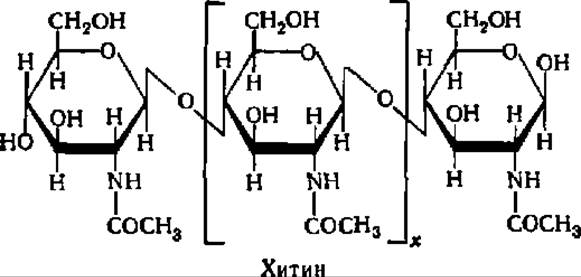
The structure of chitin, as can be seen from the formula given above, strongly resembles that of cellulose. The X-Ray Diffraction patterns of these two polysaccharides are very similar.
Hyaluronic acid is a crucial constituent of the Extracellular matrix of animal tissues. Its content is particularly high in the Skin, vitreous body of the eye, tendons, and the like. Hyaluronic acid can be obtained from these tissues by extraction with dilute alkalis, trichloroacetic acid, or phenol. Following precipitation from the extract with alcohol, hyaluronic acid is freed from tightly bound protein by enzymatic Digestion of the latter.
The molecular weight of the hyaluronate-protein complex reaches several million. However, hyaluronic acid preparations display relatively low molecular weight values (270,000–500,000). Apparently, degradation of hyaluronic acid molecules occurs during the isolation process.
Being a heteropolysaccharide, hyaluronic acid contains two distinct structural units—N-acetyl-ß-D-glucosamine and ß-D-glucuronic acid—in a 1:1 ratio. They are linked to each other in an alternating fashion by ß-1,3- and ß-1,4-glycosidic bonds:
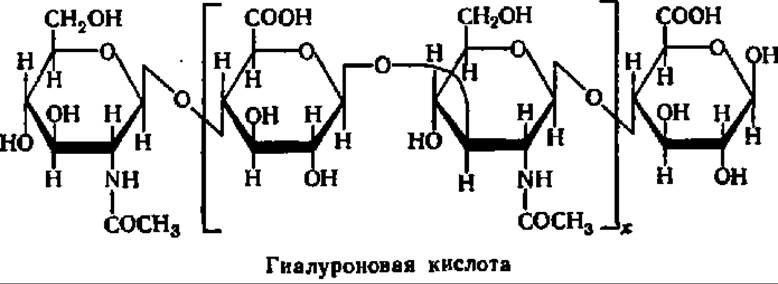
In animal tissues, hyaluronic acid performs functions that extend far beyond structural ones. Permeates tissues as an extracellular component, hyaluronic acid regulates the entry into Cells of compounds that are either required for cellular activity or are metabolic products thereof. This function of hyaluronic acid is largely carried out with the participation of an enzyme, hyaluronidase, the properties of which are being intensively studied.
Chondroitin sulfate is an indispensable component of Cartilage, Bone tissue, tendons, Heart Valves, and other similar animal tissues. Its content in nasal septum cartilage, for example, is 20–40%.
Chondroitin sulfate is difficult to isolate in pure form because it is firmly bound to protein, namely Collagen. It is likely that significant degradation of chondroitin sulfate molecules occurs during isolation, given that the molecular weight of its preparations does not exceed 50,000, whereas the M of its protein complex ranges from 4 to 50 million.
In the form of a purified preparation, chondroitin sulfate is a white substance that undergoes hydrolysis to yield glucuronic acid and N-acetylgalactosamine sulfate, which are connected to each other by ß-1,3- and ß-1,4-glycosidic bonds analogous to those found in hyaluronic acid:
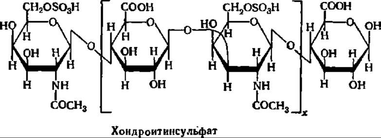
The sulfo group attached to the N-acetylgalactosamine residue in chondroitin sulfate can also occupy the 4-position. This variant of chondroitin sulfate is designated as chondroitin sulfate A. There are Other types of chondroitin sulfates that differ from the forms mentioned above in certain structural details.
Heparin is a specific heteropolysaccharide that prevents blood clotting in animals and humans. It also exhibits antilipemic, antimitotic, and regulatory activities toward a number of enzymes. Heparin is found in the liver (up to 100 mg/kg of tissue), Lungs, Spleen, Thyroid Gland, blood, and presumably in other tissues and Organs; it has been obtained in crystalline form. The molecular weight of heparin isolated from various sources ranges from 4,000 to 20,000. Determinations of the molecular weight of heparin from various sources using gravimetric, viscosimetric, and gel-filtration methods have yielded a narrower range: 11,000–12,900. The heparin molecule consists of glucuronic acid and a-glucosamine residues in the form of a double sulfuric acid derivative:

Under the action of various enzymes (heparinase, disaccharide sulfoesterase, sulfamidase, sulfoesterase, etc.), heparin breaks down into its constituent structural elements and degradation products, i.e., ultimately into glucuronic acid and glucosamine. The half-life of heparin in the body is 17.5 ± 6.5 min in rabbits and 34 ± 13.5 min in dogs.
Currently, research on the isolation, purification, and Study of the composition, structure, and functions of a number of other homo- and heteropolysaccharides is expanding widely. These include hemicelluloses, pectic substances, glucomannans and galactomannans of higher plants, algal polysaccharides (Agar, carrageenans, alginic acids, galactans, mannans, laminarians, etc.), extracellular (xanthan, pullulan, etc.) and capsular bacterial polysaccharides, and, finally, protozoan polysaccharides (paramylon, etc.). Both the chemistry and, in particular, the biochemistry of many of these compounds are of great interest.
Last update: 06/08/2026
Editorial and Educational Adaptation: This material has been compiled based on the primary/original source text. The project team performed an editorial review, corrected technical inaccuracies, structured sections, and adapted the content for an educational format.
What was processed:
- elimination of formatting defects (OCR errors, structural breaks, corrupted characters);
- editorial organization of content;
- standardization of terminology in accordance with academic sources;
- verification of factual statements against the original source text.
All mentions of the author, publication year, and origin of the primary text have been preserved in accordance with the source.