Human Biochemistry Volume 1 - Murray R. 1993
Structure and Functions of Proteins and Enzymes
Enzymes: General Properties
Quantitative Assay of Enzymatic Activity
Unlike conventional organic or inorganic substances, Enzymes are present in Cells in extremely small amounts, making the determination of their concentration in tissue extracts or biological fluids a distinct challenge. Fortunately, highly sensitive and specific assay Methods have been successfully developed based on the measurement of enzymatic catalytic activity.
To estimate The amount of an enzyme in a sample of a tissue extract or biological fluid, The rate of the reaction catalyzed by that enzyme is measured. Under defined conditions, the measured rate is directly proportional to the amount of enzyme present. Because it is generally difficult to determine the exact number of enzyme molecules or their total mass in a sample, results are expressed in arbitrary units of enzyme activity, allowing for the relative comparison of enzyme levels across different extracts. Units of activity are most conveniently expressed as the number of micromoles (µmol, 10-6 mol), nanomoles (nmol, 10-9 mol), or picomoles (pmol, 10-12 mol) of substrate consumed or product formed per unit time (per minute). The corresponding international units of enzyme activity are designated as μU, nU, or pU.
Example of Quantitative Assay of Enzymatic Activity: Determination of Dehydrogenase Content
When measuring reaction rates involving NAD+ or NADP+ (Reactions Catalyzed by dehydrogenases), advantage can be taken of the fact that NADH and NADPH (though not NAD+ and NADP+) strongly absorb light at a wavelength of 340 nm (Fig. 7.4).
The oxidation of NADH to NAD+ (or the reverse process) is accompanied by A change in the optical density (D) of the solutions at 340 nm, and under certain conditions, the rate of change in D is proportional to the enzyme activity (Fig. 7.5).
To obtain a calibration curve (Fig. 7.6), a plot is constructed showing the dependence of the rate of optical density change (the slope of the lines in Fig. 7.5) on the volume of the added enzyme preparation. The amount of enzyme present in the test solution can be determined from the observed rate of change in D at 340 nm.
Class="center">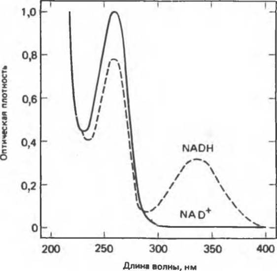
Fig. 7.4. Absorption spectra of NAD+ and NADH. Solution concentration 44 mg/L, optical path length 1 cm. NADP+ and NADPH exhibit analogous spectra, respectively.
Coupled Enzyme Assay
In the previous example, the enzymatic activity was assessed by measuring the rate of product formation (NADH). The rate of product formation (or, less frequently, the rate of substrate consumption) can be used to determine the activity not only of dehydrogenases, but also of Other Enzymes. The specific method for quantitative assessment is dictated by the PHYSICOCHEMICAL PROPERTIES OF the product or substrate. In many cases, it is convenient to subject the resulting reaction product to the action of a dehydrogenase for which this product serves as a substrate (Fig. 7.7).

Fig. 7.5. Principle of measuring The activity of an NADH- or NADPH-dependent dehydrogenase. The rate of change in optical density at 340 nm, caused by The conversion of the reduced coenzyme to its oxidized form, is measured. The oxidized substrate (S), reduced coenzyme, and buffer are added to the cuvette, and absorbance is recorded at 340 nm. Initially, a high optical density is observed due to the strong absorption of NADH (or NADPH). Upon adding 0.025-0.2 mL of the standard enzyme solution, the optical density decreases.

Fig. 7.6. Calibration curve for determining the amount of enzyme. The ordinate represents the slope of the lines shown in Fig. 7.5, and the abscissa represents the amount of enzyme.

Fig. 7.7. Determination of hexokinase activity in a system involving a coupled enzymatic reaction catalyzed by glucose-6-phosphate dehydrogenase. Glucose-6-phosphate dehydrogenase, glucose, ATP, Mg2+, and NADP+ are added in excess. Under these conditions, the rate of the overall coupled reaction depends on the amount of added hexokinase. This rate is determined by The formation of NADPH, which absorbs light at 340 nm.
Last update: 06/08/2026
Editorial and Educational Adaptation: This material has been compiled based on the primary/original source text. The project team performed an editorial review, corrected technical inaccuracies, structured sections, and adapted the content for an educational format.
What was processed:
- elimination of formatting defects (OCR errors, structural breaks, corrupted characters);
- editorial organization of content;
- standardization of terminology in accordance with academic sources;
- verification of factual statements against the original source text.
All mentions of the author, publication year, and origin of the primary text have been preserved in accordance with the source.