Human Anatomy - H. I. Koliadenko 2009
The Study of Viscera (Splanchnology)
Digestive System (Digestive Apparatus)
The Digestive System (Fig. 89) begins with the Oral Cavity, which consists of the oral cavity proper and the oral vestibule. The oral cavity proper communicates via the fauces, which connect with the Pharynx. These are followed by the Esophagus, Stomach, Small Intestine, and Large Intestine, and the digestive system terminates in the rectum and anus. The digestive Organs include the Salivary Glands (parotid, submandibular, and sublingual), the Liver, and the Pancreas.
The oral cavity (cavitas oris) consists of the oral vestibule (vestibulum oris) and the oral cavity proper (Fig. 90).
The oral vestibule communicates with the external environment through the oral fissure, which is bounded anteriorly by the Lips, laterally by the Cheeks, and by the alveolar processes and Teeth. These structures divide the oral cavity into two parts. The lips are two muscular folds formed by the orbicularis oris Muscle. Their outer surface is covered with Skin, while the inner surface is lined with mucous membrane that continues into the mucosal lining of the alveolar processes. Along the midline between the lips and Gums, the mucous membrane thickens to form two folds: the frenula of the upper and lower lips. The outer margin of the lips has a reddish hue due to the underlying Muscles shining through the epithelial layer. The submucosa of the lips contains mucous glands, the excretory ducts of which open onto The surface of the mucous membrane. The lips form the angles of the Mouth.
Class="center">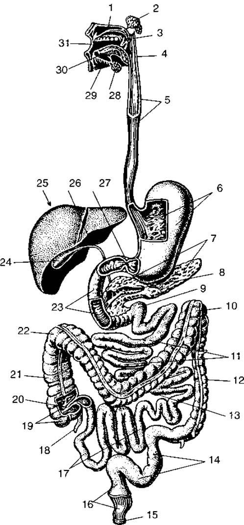
Fig. 89. Digestive system:
1 — hard palate; 2 — parotid gland; 3 — soft palate; 4 — pharynx; 5 — esophagus; 6 — stomach; 7 — pancreas; 8 — pancreatic duct; 9 — intestinal loop; 10 — left colic flexure; 11 — jejunum; 12 — descending colon; 13 — transverse colon; 14 — sigmoid colon; 15 — external anal sphincter; 16 — rectum; 17 — ileum; 18 — Appendix; 19 — cecum; 20 — ileocecal valve; 21 — ascending colon; 22 — right colic flexure; 23 — duodenum; 24 — Gallbladder; 25 — liver; 26 — Bile duct; 27 — pyloric sphincter; 28 — submandibular gland; 29 — sublingual gland; 30 — Tongue; 31 — oral cavity
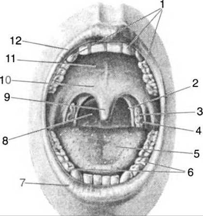
Fig. 90. Oral Cavity and fauces:
1 — upper dental arch; 2 — posterior palatine arch; 3 — palatine tonsil; 4 — anterior palatine arch; 5 — dorsum of tongue; 6 — lower dental arch; 7 — lower lip; 8 — fauces; 9 — uvula; 10 — soft palate; 11 — hard palate; 12 — upper lip
Vascularization of the lips is provided by the superior and inferior labial Arteries, which are Branches of the facial artery.
The lips are innervated by the Facial Nerve.
The cheeks are covered externally with skin and internally with mucous membrane. Between them lies the buccinator muscle, which is particularly well-developed in infants. The ducts of small mucous glands, whose bodies lie in the submucosa of the cheeks, open onto the buccal mucosa.
The oral cavity proper (cavum oris) is bounded anteriorly and laterally by the alveolar processes and teeth, and superiorly by the hard palate and part of the soft palate. The second part of the soft palate bounds the oral cavity posteriorly. Inferiorly, the oral cavity proper is formed by the Muscles of the oral Diaphragm.
The hard palate is formed by the palatine processes of the maxillae and the palatine bones, covered by a mucous membrane that fuses with the periosteum of these bones, making it immobile here. The mucous membrane contains numerous small mucous glands. Along the midline, the hard palate features a narrow white line — the palatine raphe — with 3–4 transverse palatine folds in its anterior part. The hard palate continues into the soft palate, which separates the oral cavity from the nasopharynx. The free, dangling portion of the soft palate is called the palatine velum. It is a muscular fold covered with mucous membrane that hangs freely downward. In the middle of the soft palate is the uvula, which is a PROJECTION OF THE central part of the velum. On each side, the palatine velum extends into two arches: one runs toward the ROOT of the tongue and is called the palatoglossal arch, while the other continues into the mucous membrane of the lateral wall of the pharynx and is called the palatopharyngeal arch. Between the palatine arches are small depressions — tonsillar fossae — which house the palatine Tonsils. Inside the tonsil, there are numerous lymphatic follicles where lymphocytes are produced, contributing to the body's unified immune system.
The Medial surface of the tonsils features numerous tonsillar pits that extend into tonsillar crypts. These structures increase the surface area of contact between the tonsil and the external environment.
At the rear of the oral cavity is a small opening — the fauces — which leads into the pharynx.
Salivary glands. The ducts of three pairs of major salivary glands and numerous minor ones open into the oral cavity, secreting saliva.
Saliva is a serous fluid containing mucus, organic substances, and minerals. The organic substances are represented by Enzymes: ptyalin and maltase, which facilitate The breakdown of complex CARBOHYDRATES — Polysaccharides — into simpler fractions — mono- and Disaccharides.
According to their Structure, salivary glands are classified into tubular, alveolar, and tubulo-alveolar.
The parotid gland (glandula parotis, Fig. 91) has an irregular triangular shape. It is located in the parotid-masseteric region of the face and is surrounded by the parotid-masseteric fascia. Anteriorly, the gland is partially covered by the masseter muscle. The parotid duct emerges from its anterior margin, passes across the masseter muscle and adipose tissue to reach the buccinator muscle, pierces it, and opens onto the mucosa of the oral vestibule opposite the second upper molar. The saliva of this gland is watery, serous (proteinaceous), and mucus-free. Structurally, this gland is a compound alveolar gland, with a mass ranging from 11 to 30 g.
The submandibular gland (glandula submandibularis) is half the size of the parotid gland and is located near the angle of the Mandible beneath the mylohyoid muscle. The gland is tubulo-alveolar and secretes a seromucous fluid. Its mass is 10–15 g. The duct of the submandibular gland wraps around the posterior border of the mylohyoid muscle and opens onto the sublingual caruncle.

Fig. 91. Glands of the oral cavity:
1 — parotid gland; 2, 3 — parotid duct; 4 — buccinator muscle; 5 — buccal glands; 6 — labial glands; 7 — upper lip; 8 — lingual glands; 9 — lower lip; 10 — sublingual caruncle; 11 — major sublingual duct; 12 — minor sublingual ducts; 13 — mandible; 14 — muscles of the tongue; 15 — mylohyoid muscle; 16 — sublingual gland; 17 — submandibular duct; 18 — submandibular gland; 19 — masseter muscle
The sublingual gland (glandula sublingualis) is located directly beneath the mucous membrane of the floor of the oral cavity. The gland is tubulo-alveolar, mixed, and secretes serous fluid and mucus. Its anterior border adjoins the inner surface of the body of the mandible, while its posterior border adjoins the submandibular gland. Numerous small ducts (10–12) of this gland open through tiny orifices along the sublingual fold. The largest of these, the major sublingual duct, merges with the submandibular duct and opens through a common orifice on the sublingual caruncle.
Minor salivary glands secrete mucus. Depending on their Location, they are referred to as labial, buccal, or lingual glands.

Fig. 92. Permanent teeth of the right half of the oral cavity:
1, 2 — incisors; 3 — canine; 4, 5 — premolars; 6, 7, 8 — molars
Salivary glands consist of individual epithelial lobules separated by Connective Tissue. Secretions are produced within the epithelial lobules and released via a duct system—initially small and subsequently larger ducts—into the oral cavity, where they help moisten and coat the oral mucosa with mucus, impregnate food with enzymes, and facilitate partial Digestion.
Teeth (dentes) (Figs. 92, 93) are located in the alveolar sockets of the upper and lower jaws. A tooth is an extremely durable organ composed of organic matter permeated with inorganic salts, bearing a macroscopic resemblance to bone. The tooth comprises a crown, a neck, and a root. The entire mass of the tooth is made up of dentin. The crown is covered with enamel, which is even harder than dentin (its hardness approaches that of quartz). The neck and root of the tooth are covered with a grayish substance called cementum, which is histologically similar to bone. The pulp cavity inside the tooth contains the dental pulp, which consists of Blood Vessels, nerves, and loose connective tissue.
At the root apex, there is an opening that continues into the root canal, through which blood Vessels and nerves enter the tooth. The tooth crown projects above the gingiva (gums), which surrounds the neck of the tooth, whereas the root is lodged within the dental alveolus and surrounded by the periosteum, or periodontium. Based on the shape of their crowns and their Functions, teeth are classified into incisors, canines, premolars, and molars.

Fig. 93. Cytology/practical/54.html">Longitudinal section of a tooth:
1 — enamel; 2 — dentin; 3 — cementum; 4 — root canal; 5 — pulp cavity
The dental formula conventionally represents the number of teeth, with the numerator indicating the teeth in one half of the upper jaw and the denominator representing those in the lower jaw:
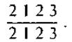
The dental formula should be read as follows: 2 incisors, 1 canine, 2 premolars, and 3 molars in each upper and lower half of the jaws.
The crown of each tooth has several surfaces. The surface facing the tooth of the opposite jaw is called the occlusal surface; the surface facing the vestibule is the vestibular surface; the one facing the tongue is the lingual surface; and the surface facing the adjacent tooth is the contact (proximal) surface.
The incisors have a sharp, cutting, chisel-like edge, and a cone-shaped root compressed laterally. The cutting edge of a canine resembles a truncated cone, and it has a single root. Upper canines are larger and more massive than lower ones.
Premolars feature two cusps on their masticatory surface: an inner lingual cusp and an outer vestibular cusp. Lower premolars have a single slightly flattened root, whereas upper premolars may have two roots or a single root bifurcated into two.
Molars have a cubic crown. Their masticatory surface features four cusps: two lingual and two vestibular. The first lower molar sometimes has 5 cusps—two lingual and three vestibular. Upper molars possess three roots: two lateral vestibular and one inner lingual. Lower molars have two roots: an anterior and a posterior. The third molars, or wisdom teeth, erupt between the ages of 17 and 25 or later, hence their designation as wisdom teeth; they may also fail to develop altogether. Wisdom teeth are vestigial structures. In 15% of people, wisdom teeth are congenitally absent in the upper jaw.
Development of teeth in ontogeny and phylogeny. In children, teeth erupt at 6–8 months of age. These are temporary, so-called deciduous teeth; numbering 20, they fully erupt by the age of two. Their formula is

that is, 2 incisors, 1 canine, no premolars at this age, and 2 molars in each half of the upper and lower jaws, respectively.
The central lower incisors erupt first, followed by all upper incisors, and by 10–12 months all incisors have appeared. Premolars erupt at 12–15 months, followed by canines at 18–20 months, and all deciduous teeth are present by 20–24 months. At the age of six, deciduous teeth begin to shed, being replaced by permanent ones. The tooth-replacement process is gradual and concludes by age 12–15.
The contact between the upper and lower incisors is termed the bite (occlusion). Normal occlusion is characterized by the opposing teeth of both jaws coming into contact, with each upper jaw tooth meeting the corresponding lower jaw tooth positioned lateral to the identically named contacting tooth.
Teeth perform the functions of biting off, holding, and masticating food, and also contribute to clear articulation in speech.
Tooth development begins during the second month of embryonic development. Following the fusion of the two maxillary and mandibular prominences in the embryo, two grooves form, separating the gums from the labial folds. The dental lamina develops from the gingival epithelium, upon which enamel organs appear as thickenings corresponding in number to the deciduous teeth. These thickenings take the form of buds connected to the lamina by a narrow neck. Mesenchyme grows toward the buds, forming the dental papilla. The enamel organ gives rise to the tooth enamel, whereas the dentin and dental pulp originate from the dental papilla. Cementum develops from embryonic connective tissue.
During development, teeth such as incisors, canines, and premolars possess two sets of primordia: one gives rise to deciduous teeth, and the other to permanent teeth. The primordia of permanent molars appear only by the 6th month of fetal development.
During phylogeny, teeth evolved from specialized dermal scales of primitive fish, appearing as hard structures long before the Skeletal System. In fish, amphibians, and reptiles, teeth are replaced multiple times throughout their life cycle.
The tongue (lingua) is a muscular organ formed of striated muscle and covered by a mucous membrane (Fig. 94). Its muscles are divided into two groups: extrinsic and intrinsic. The extrinsic muscles include the genioglossus, hyoglossus, and styloglossus.
The genioglossus muscle protrudes the tongue forward and downward.
The hyoglossus muscle retracts the tongue backward and downward.
The styloglossus muscle pulls the tongue backward and upward.
The intrinsic muscles of the tongue run in longitudinal, transverse, and oblique directions. The latter extend obliquely downward from the dorsal surface of the tongue, functioning as suspended muscle fibers.
The tongue has an elongated, oval shape. Its upper surface is called the dorsum, the anterior part is the apex (tip), and the posterior part is the root. The body of the tongue lies between the tip and the root. Beneath the tongue, the oral mucosa forms the frenulum of the tongue.
The mucous membrane of the tongue is covered with stratified squamous epithelium lacking a submucosa, rendering it directly fused with the underlying muscles. The margins and dorsum of the tongue are studded with lingual papillae, which are classified by shape into filiform, fungiform, foliate, and vallate (circumvallate) papillae.
Filiform papillae cover the entire dorsum of the tongue and perform mechanical and tactile functions.
Fungiform papillae are scattered among the filiform papillae and are responsible for taste sensation. Circumvallate papillae, surrounded by a trench or wall, are located between the dorsum and the root of the tongue, arranged in a V-shaped pattern (sulcus terminalis), numbering from 7 to 12.
Foliate papillae are located on the posterolateral margins of the tongue and also participate in taste perception.
The lingual tonsil is situated on the upper surface of the root of the tongue.
The tongue performs several vital functions: it is an organ of articulate speech, as the pronunciation of most sounds requires its movement; it shifts food from one part of the oral cavity to another; and it serves as the primary ORGAN OF TASTE.
The pharynx is located on the anterior surface of the cervical spine, extending from the base of The Skull and continuing into the esophagus at the level of the VI cervical vertebra. It is 12–15 cm long. The pharynx is divided into nasal, oral, and laryngeal parts.
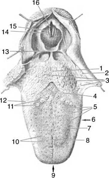
Fig. 94. Dorsal surface of the tongue:
1 — root of the tongue; 2 — palatine tonsil; 3 — lingual tonsil; 4 — terminal sulcus; 5 — fungiform papillae; 6 — body of the tongue; 7 — dorsum of the tongue; 8 — median sulcus of the tongue; 9 — apex (tip) of the tongue; 10 — filiform papillae; 11 — vallate papillae; 12 — foliate papillae; 13 — epiglottis; 14 — vestibular fold (false vocal cord); 15 — vocal fold (true vocal cord); 16 — rima glottidis
The nasal part of the pharynx (nasopharynx) communicates with the Nasal cavity via the choanae. On the lateral walls of the nasopharynx, the pharyngeal openings of the auditory (Eustachian) tubes connect it to the tympanic cavity. The nasopharynx lies at the level of the soft palate and extends up to the Base of the skull.
The oral part of the pharynx (oropharynx) has a single opening, the fauces. The anterior wall of the lower pharynx contains an opening leading into the Larynx. The pharynx has no distinct lower floor; rather, it narrows and transitions directly into the esophagus.

Fig. 95. Pharyngeal cavity:
1 — nasal cavity; 2 — hard palate; 3 — soft palate; 4 — sphenoid sinus; 5 — pharyngeal opening of the auditory tube; 6 — pharyngeal tonsil; 7 — anterior arch of the atlas; 8 — uvula (palatine); 9 — palatine tonsil; 10 — epiglottis; 11 — cricoid Cartilage; 12 — esophagus; 13 — Trachea; 14 — thyroid cartilage; 15 — Hyoid bone; 16 — genioglossus muscle; 17 — oral vestibule; 18 — oral cavity proper
The pharyngeal wall consists of three layers: an inner mucous membrane with a submucosal layer, a middle muscular layer, and an outer connective tissue or adventitial layer.
The muscular layer of the pharynx is formed by five pairs of striated muscles. Three of these pairs are circular constrictors whose contractions propel food from the pharynx into the esophagus. Two pairs of muscles run longitudinally and serve to elevate the pharynx. The pharynx is surrounded by loose connective tissue, allowing it to move freely during swallowing.
The upper wall of the nasopharynx houses the unpaired pharyngeal tonsil, two paired tonsils lie near the auditory tubes connecting the nasopharynx to the Middle ear, another unpaired tonsil is located in the region of the tongue root, and two palatine tonsils are also present. These tonsils form the Pirogov-Waldeyer lymphatic ring, named after the scientist who first investigated and detailed its structure.
The esophagus is a muscular tube that begins at the level of the VI–VII cervical vertebrae and transitions into The Stomach at the level of the XI thoracic vertebra. In an adult, the esophagus is approximately 25 cm long. From the thoracic cavity, it passes through the diaphragm into the Abdominal cavity, where it connects to the cardiac part of the stomach.
The esophagus is conventionally divided into three parts: cervical, thoracic, and abdominal. It lies anterior to THE Vertebral Column and features four curvatures along its course: two in the sagittal plane and two in the frontal plane. The esophageal wall consists of mucosal (inner), muscular (middle), and external (adventitial) layers. Additionally, the abdominal part of the esophagus is covered by a serous membrane.
The mucous membrane forms longitudinal folds that fill the lumen of the esophagus. Beneath the mucosa lies the submucosal layer, which contains mucous glands whose ducts open onto the mucosal surface. The mucus secreted by these glands lubricates the mucosa and facilitates the passage of food. The folds of the mucous membrane flatten out during swallowing.
The muscular coat of the esophagus consists of two layers: an inner layer with transverse or circular muscle fibers, and an outer layer with longitudinal fibers. Numerous fibrous extensions originate from the muscular coat, anchoring the esophagus like cords to the surrounding organs. At the site where the esophagus passes through the diaphragm, a circular muscle forms a sphincter-like structure around it.
The adventitial coat, composed of loose connective tissue, contains a small number of elastic fibers that anchor the esophagus to other organs in the posterior Mediastinum. Blood vessels and Lymphatic vessels—which drain Lymph from the esophagus—run within the adventitia, along with the esophageal nerve plexus.
Along its course, the esophagus has three anatomical constrictions: the first at its origin, the second where it is crossed by the left main bronchus and trachea, and the third at the diaphragmatic hiatus.
The stomach (ventriculus s. gaster, Fig. 96) is the most dilated portion of the digestive tract. Its capacity ranges from 1.5 to 3 liters, but this is not a fixed limit, as the stomach can stretch significantly in response to large volumes of food and liquid—sometimes up to 10 liters.
The stomach lies beneath the diaphragm, with 5/6 of its body situated to the left and 1/6 to the right of the median plane of the body. At the level of the X–XI thoracic vertebrae, the stomach has its entrance, or cardia, which is the junction with the esophagus. At the level of the XII thoracic and I lumbar vertebrae, the stomach has its exit, the pylorus, marking the transition into the small intestine. Between the entrance and the exit lies the body of the stomach, which features an upper elevated portion known as the fundus, or vault of the stomach. The upper, concave surface forms the lesser curvature of the stomach, while the lower, convex surface forms the greater curvature. The anterior surface of the fundus is in contact with the left dome of the diaphragm, while its posterior surface faces the Spleen. The lesser curvature is directed toward the Inferior surface of the left lobe of the liver, whereas the greater curvature abuts the transverse colon and partially the spleen.
The wall of the stomach consists of three layers, or tunics: the mucous membrane, the muscular coat, and the serous coat (Fig. 97).
The mucous membrane (tunica mucosa) is a continuation of the esophageal mucosa and extends to the entrance of the duodenum. At the transition point, it forms a circular fold known as the pyloric valve. Supported by a smooth muscle membrane and the submucosa, the mucosa gathers into numerous folds of varying sizes. Long folds run longitudinally, interconnected by shorter transverse folds, creating a reticular pattern on the gastric mucosa. The ducts of the gastric glands open onto the mucosal surface, with approximately 100 glands present per square millimeter of mucosa. These glands are classified into main (zymogenic) glands, which secrete gastric juice, and parietal glands (located in the fundus), which secrete Hydrochloric acid. Accessory glands secrete mucus. Solitary lymph nodules are located within the thickness of the gastric mucosa.

Fig. 96. STRUCTURE OF THE stomach wall:
1 — folds of the mucous membrane; 2 — muscular coat; 3 — serous coat; 4 — pyloric opening; 5 — folds of the esophageal mucosa
In a stomach filled with food, the mucous membrane smooths out, and the folds disappear.
The muscular coat (tunica muscularis) is formed by smooth muscle fibers arranged in three directions: an outer (longitudinal) layer, a middle (circular) layer, and an inner (oblique) layer. At the pyloric opening, the muscle forms a thickening known as the pyloric sphincter.
The serous coat of the stomach (tunica serosa) is part of the Peritoneum that covers the anterior and posterior surfaces of the stomach, with the exception of a small area on the posterior wall. In the Regions of the lesser and greater curvatures, the folds of the serous membrane form the peritoneal ligaments of the stomach.
The gastrophrenic ligament extends to the left from the lesser curvature to the diaphragm. To the right lies the hepatogastric ligament, which continues into the ligament connecting the superior part of the duodenum to the porta hepatis. These three ligaments form the lesser omentum. The gastrocolic ligament originates from the greater curvature of the stomach, giving rise to a fold that descends as a double layer into the lesser pelvis, forming the two anterior layers of the greater omentum. The length of the intestine is 5–7 m. The duodenum is short, measuring 25–30 cm in length; 2/5 of the small intestine's length belongs to the jejunum, and 3/5 to the ileum.

Fig. 97. Mucous membrane of the fundus
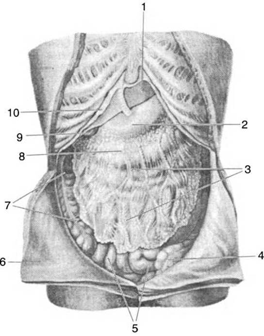
Fig. 98. Greater omentum:
1 — liver; 2 — stomach; 3 — greater omentum; 4 — sigmoid colon; 5 — small intestine; 6 — parietal peritoneum; 7 — ascending colon; 8 — transverse colon; 9 — gallbladder; 10 — diaphragm
The wall of the small intestine consists of the following layers: mucous, submucous, double-layered muscular, and serous.
The mucous membrane forms numerous open circular folds that increase its absorptive surface area. These folds are supported by the underlying submucosa. The mucosa is densely covered with intestinal villi (22–40 per 1 mm2). Intestinal villi are mucosal projections composed of loose connective tissue and covered by a single-layered columnar epithelium. Each villus contains a network of branching blood capillaries and lymphatic vessels. Digested food is absorbed through the intestinal villi: venous capillaries absorb Proteins and carbohydrates, while lymphatic vessels absorb fats. Each villus is covered with microvilli, numbering about 1,500–3,000 per Cell, which further expand the absorptive surface. Digestive enzymes have been identified within the intestinal villi, facilitating membrane digestion and nutrient absorption. Throughout its length, the intestinal mucosal epithelium is interspersed with tubular intestinal glands that produce intestinal juice, as well as goblet Cells that secrete mucus. The mucosa contains defensive structures in the form of Solitary lymphoid nodules that perform an immune function. Additionally, the small intestine contains elongated aggregations of lymphoid tissue known as Peyer's patches (about 30–40 in total, measuring 1–3 cm in length), with the majority located in the ileum and fewer in the jejunum.
The muscular coat of the small intestine consists of an outer layer of longitudinal smooth muscle and an inner circular layer. The muscular coat is best developed in the duodenum and progressively thinner in the ileum. Contractions of the muscular coat drive peristaltic movements, food mixing, absorption, and the evacuation of chyme into the large intestine.
The serous membrane is the outermost layer. It covers all sections of the small intestine, except for the duodenum, which is covered by the serous membrane only on its anterior surface.
The duodenum begins at the pylorus of the stomach, is horseshoe-shaped, and fixed (fused with the posterior abdominal wall) (Fig. 99), thereby remaining immobile. Its superior (descending) part lies at the level of the first lumbar vertebra, extends to the right along the vertebral bodies, and reaches the third lumbar vertebra. Then, forming a leftward bend, it transitions into the ascending part, which joins the jejunum at the level of the second lumbar vertebra. Along the posterior wall of the duodenum lies the duodenal fold, which terminates in the major duodenal papilla—the opening site for the common bile duct and the pancreatic duct, both running along the fold. Often, superior to the major papilla, There is a minor duodenal papilla, which serves as the opening for the accessory pancreatic duct, while the HEAD of the pancreas is embraced by the middle portion of the duodenum.
The primary function of the duodenum is digestion. Trypsinogen, the main protein-digesting enzyme, is converted into Trypsin within the intestine. Bile, secreted by the liver through the common bile duct, facilitates the emulsification and digestion of fats.
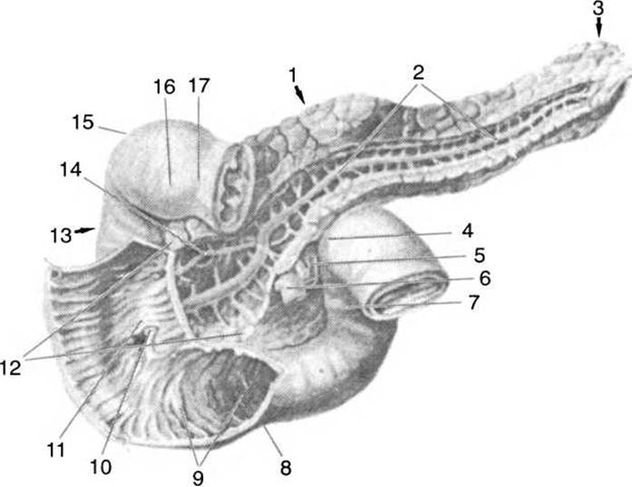
Fig. 99. The pancreas and duodenum:
1 — body of the pancreas; 2 — pancreatic duct; 3 — tail of the pancreas; 4 — duodenojejunal flexure; 5 — superior mesenteric artery; 6 — superior mesenteric vein; 7 — ascending part of the duodenum; 8 — horizontal (inferior) part; 9 — circular folds; 10 — duodenal papillae; 11 — longitudinal fold; 12 — head of the pancreas; 13 — descending part of the duodenum; 14 — accessory pancreatic duct; 15 — superior duodenal flexure; 16 — superior horizontal part; 17 — pylorus
The jejunum and ileum are covered by the peritoneum on all sides, suspended from the posterior abdominal wall by the mesentery, and form numerous intestinal loops.
Throughout their entire length, their mucous membrane forms 600–650 folds covered with intestinal villi and microvilli located on the apical surface of the columnar epithelial cells.
Due to its muscular coat, the small intestine is in constant motion. These movements are of two types: pendular movements—resulting from the alternating contraction of longitudinal and circular muscles—and peristaltic movements—resulting from the simultaneous contraction of both muscle layers. These movements ensure the mixing of digested food within the small intestine and its propulsion toward the large intestine. Furthermore, the intestinal walls are permanently in a state of tonic contraction. This is why, upon loss of muscle tone in a deceased person, the length of the intestines increases by 1.5–2 meters compared to a living human.
The large intestine (intestinum crassum) begins in the right iliac fossa, where the ileum terminates. The opening between the ileum and the large intestine is surrounded by a mucous membrane that forms the ileocecal valve (or valve of Bauhin). It automatically closes the entrance to the small intestine after food residues are propelled into the large intestine.
The large intestine is 1.5–2 meters long and is divided into three sections: the cecum, the colon, and the rectum (Fig. 100).
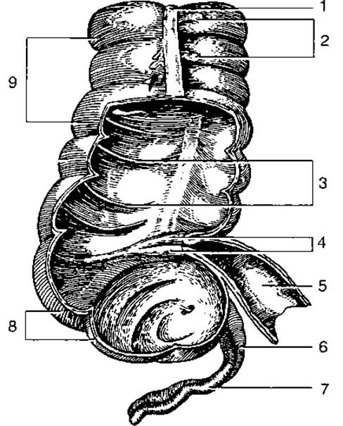
Fig. 100. Cecum, vermiform appendix, and ascending colon:
1 — taenia coli; 2 — omental Appendices; 3 — semilunar folds of the colon; 4 — ileocecal valve; 5 — ileum; 6 — mesoappendix; 7 — vermiform appendix; 8 — cecum; 9 — colic haustra
The structure of the large intestine wall resembles that of the small intestine, albeit with certain differences. Specifically, the longitudinal layer of the muscular coat in the large intestine is not continuous but forms three bands (taeniae): the mesenteric, omental, and free taeniae. The mesentery is attached to the mesenteric taenia, the greater omentum to the omental taenia, and the free taenia is associated with neither. Because these bands are shorter than the large intestine itself, they gather it into folds or sacculations, also known as haustra (haustra coli). Between these haustra lie sulci that form semilunar folds on the mucous membrane. Externally, the serous coat of the intestine features fat-filled outgrowths (epipploic appendages). The mucous membrane is lined with a single-layered columnar epithelium. Intestinal villi and Peyer's patches are absent in the large intestine; instead, it contains solitary lymphoid nodules, numerous goblet cells, and tubular glands.
The cecum (intestinum caecum) is 7–8 cm long, and the vermiform appendix (appendix vermiformis), measuring 8–9 cm (sometimes 18–20 cm), extends from it. The cecum lacks a mesentery, whereas the appendix has its own mesoappendix.
The colon (intestinum colon) is a continuation of the cecum. It is divided into four sections: the ascending, transverse, descending, and sigmoid colons.
The ascending colon (colon ascendens) is 14–18 cm long, located in the right region of the abdominal cavity, adjacent to the posterior abdominal wall and the right Kidney; forming the right colic flexure, it transitions into the transverse colon (colon transversum), which is 25–30 cm long and attached to the posterior abdominal wall by the mesocolon. This section lies horizontally between The Liver and the spleen, with its middle part curving anteriorly in an arch. Anteriorly, the colon is covered by the greater omentum. Below the inferior border of the spleen, anterior to the left kidney, the transverse colon forms the left colic flexure and transitions into the descending colon (colon descendens).
The descending colon is 10–12 cm long, lies on the left side of the abdominal cavity, and is adjacent to the posterior abdominal wall. In the region of the left iliac fossa, the descending colon transitions into the sigmoid colon (colon sigmoideum), which has its own mesentery. In the sigmoid colon, the muscular bands broaden slightly. At the level of the third sacral vertebra, the sigmoid colon transitions into the rectum, which is 15–20 cm long. This segment marks the end of the digestive tract (Fig. 101).
The rectum forms two flexures: the sacral flexure, convex posteriorly, which conforms to the curvature of the pelvic surface of the sacrum, and the perineal flexure, located after the intestine passes through the pelvic diaphragm. The mucous membrane of the rectum features 5–10 longitudinal folds known as rectal columns, between which lie anal sinuses—small recesses where foreign bodies may become lodged. The lower third of the rectum is dilated and called the ampulla. The rectum terminates in the anus, which is provided with two sphincters: the internal anal sphincter, composed of smooth muscle and operating involuntarily, and the external anal sphincter, composed of striated muscle and subject to voluntary control. The upper part of the rectum possesses a mesentery.
Upon contraction, the longitudinal fibers of the muscular coat of the large intestine widen the bowel, whereas the circular fibers narrow it. Contractions of the large intestine are wave-like in character.
Fecal masses are formed and Water is absorbed in the large intestine. In children, the large intestine is somewhat shorter than in adults, averaging 40–45 cm in length. During the first year of life, it doubles in size. The appendix in children has a conical shape. In newborns, the muscular bands are poorly developed, and the serous coat lacks haustra and sacculations, which appear by the 6th month of life. During the first year of life, children lack an omentum. The formation of the large intestine takes 3.5–4 years. Its mass increases up to the age of 40, after which it gradually decreases due to the thinning of the muscular coat. The appendix undergoes involution and appears as a small outgrowth.
The serous membrane lining the walls of the abdominal cavity is called the peritoneum. It consists of a parietal layer, which lines the cavity walls, and a visceral layer, which covers the Internal Organs. The cavities containing the internal organs are formed between these layers.
The pancreas is located posterior to the stomach at the level of the first lumbar vertebra. Its mass is 70–100 g, its length is 20–22 cm, and its width is about 4 cm. Superiorly, the gland is covered by a thin connective tissue capsule. It consists of a head, body, and tail. The head lies within the horseshoe-shaped concavity of the duodenum, the body lies at the level of the first lumbar vertebra and is adjacent to the posterior abdominal wall, while the tail reaches the spleen and the left kidney.

Fig. 101. Rectum:
1 — end of the strip on the sigmoid colon; 2 — sigmoid colon; 3 — longitudinal muscle fibers; 4 — levator ani muscle; 5, 7 — retractors; 6 — external anal sphincter; 8 — ampulla of the rectum; 9 — constriction at the site of the transverse fold
The pancreas consists of a single-layered epithelium and belongs to complex tubulo-acinar glands, which form separate lobules connected by connective tissue septa extending from the capsule.
The pancreatic cells secrete pancreatic juice, which contains enzymes such as trypsin, Chymotrypsin, amylase, lipase, and others that digest proteins, fats, and carbohydrates. Daily secretion ranges from 500 ml to 2 L of alkaline pancreatic juice.
Pancreatic cells are conical in shape. Each cell is divided by The Nucleus into a broad basal part and a tapered apical part. The apical part appears granular and represents the secretory portion of The Cell.
Pancreatic juice flows from the glandular cells into intralobular ducts, which in turn drain into interlobular ducts, and finally into the pancreatic duct. This main duct runs the entire length of the gland from the tail to the head and opens via a common orifice, after merging with the common bile duct, at the major duodenal papilla. At the junction with the duodenum, there is the sphincter of the pancreatic duct.
The pancreas is a gland of mixed secretion. By producing digestive pancreatic juice, it functions as an exocrine gland. However, it also contains cell clusters 0.1–0.3 mm in diameter located within the glandular lobules, known as pancreatic islets, or islets of Langerhans. The number of these islets ranges from 200,000 to 1,800,000. The secretion, or incretion, of these cells—Insulin—enters the bloodstream directly and regulates Carbohydrate METABOLISM.
Thus, the pancreas functions simultaneously as both an exocrine and an endocrine gland.
The liver (hepar; Fig. 102) is located in the upper right region of the abdominal cavity, beneath the diaphragm. It is the largest gland in The Human Body, with a reddish-brown color, and is externally covered by a serous membrane that forms the sagittal falciform ligament, dividing the liver into two lobes: a large right lobe and a much smaller left lobe. These lobes occupy respective positions: the right lobe lies under the right dome of the diaphragm, and the left under the left dome. Inferiorly, the falciform ligament continues into the round ligament of the liver.
The diaphragmatic surface of the liver is convex, while the visceral surface is uneven, slightly concave, and features several impressions corresponding to the adjacent organs: the kidney, the colon, and the duodenum.
On the visceral surface of the liver, There are two longitudinal grooves running anteroposteriorly. The left groove divides the liver into the left and right lobes.

Fig. 102. Liver, pancreas, and duodenum:
1 — left coronary ligament of the liver; 2 — left lobe of the liver; 3 — falciform ligament of the liver; 4 — hepatic duct; 5 — common bile duct; 6 — pancreatic duct; 7 — pancreas; 8 — Origin of the jejunum; 9 — duodenum; 10 — gallbladder; 11 — cystic duct; 12 — quadrate lobe of the liver; 13 — right lobe of the liver; 14 — right coronary ligament of the liver
Between the two longitudinal grooves lies a transverse groove, which forms two additional small lobes in the liver: the quadrate lobe anteriorly and the caudate lobe posteriorly. The transverse groove is called the porta hepatis (hepatic hilum).
The left longitudinal groove contains the ligamentum venosum (the obliterated fetal venous duct) and the round ligament (the obliterated fetal umbilical vein) (Fig. 103).
The right longitudinal groove lodges the gallbladder and the INFERIOR VENA CAVA, into which the hepatic Veins empty.
The porta hepatis transmits the portal vein (vena porta), proper hepatic artery, and nerves, while the common hepatic duct and lymphatic vessels emerge from it. Bile is drained via the common hepatic duct. This duct opens onto the duodenal papilla together with the pancreatic duct.
The liver is almost entirely covered by peritoneum, except for the area where its posterior surface attaches to the diaphragm (via the coronary ligament) and the abdominal cavity. On the visceral surface of the liver, there are two ligaments: the hepatoduodenal and hepatogastric ligaments, which together form the lesser omentum.
Beneath the serous membrane of the liver lies a connective tissue sheath known as Glisson's capsule. It penetrates the glandular tissue of the liver and forms a network (stroma), within the compartments of which hepatic lobules, each 1–1.5 mm in size, are located (Fig. 104). In shape, they resemble polyhedral prisms. Hepatic lobules are composed of cells called hepatocytes, which store Glycogen. Hepatocytes are arranged in cords (plates), with each cord consisting of a double row of cells. These cords radiate toward the interlobular vein located at the center of the lobule. Bile canaliculi originate from each cell, draining into the interlobular bile ducts that form the left and right hepatic ducts (corresponding to the liver lobes). Emerging from the porta hepatis, the right and left hepatic ducts unite to form the common hepatic duct, which then joins the cystic duct.

Fig. 103. Liver (visceral surface):
1 — inferior vena cava; 2 — posterior surface of the liver; 3 — renal impression; 4 — right lobe of the liver; 5 — colic impression; 6 — duodenal impression; 7 — gallbladder; 8 — quadrate lobe; 9 — cystic duct; 10 — round ligament of the liver; 11 — bile duct; 12 — hepatic duct; 13 — portal vein; 14 — proper hepatic artery; 15 — gastric impression; 16 — left lobe of the liver; 17 — esophageal impression; 18 — caudate lobe

Fig. 104. Structure of a hepatic lobule:
1 — central vein of the lobule; 2 — branch of the hepatic artery; 3 — branch of the portal vein; 4 — interlobular artery; 5 — interlobular branch of the portal vein; 6 — bile ducts; 7 — interlobular vein; 8 — branches of the hepatic vein; 9 — interlobular septa; 10 — hepatic cords; 11 — capillaries of the portal vein
Inside the liver, the hepatic artery and portal vein branch into smaller vessels whose branches surround each lobule, which is why they are called interlobular vessels. The interlobular arteries and veins break up into capillaries that enter the interior of the lobule. The venous capillaries radiate through the lobule, from which blood flows into the central vein that runs longitudinally through the lobule and empties into one of the collecting interlobular veins, which drain into 3—4 hepatic veins, which in turn flow into the inferior vena cava.
Within an hour, all of a person's blood passes through the liver lobular capillaries several times. The capillary walls are formed by endothelial cells, interspersed with stellate cells—reticuloendothelial cells, or Kupffer cells—which perform a phagocytic function.
It is believed that the liver synthesizes glycogen at night and bile during the day. Over the course of a day, the liver produces 1000—1200 ml of bile. Bile enters the duodenum, where it emulsifies fats, thereby facilitating the action of lipolytic enzymes in pancreatic juice, activates lipase, and stimulates the absorption of fat breakdown products. Bile contains no enzymes.
The liver performs a barrier function by detoxifying harmful substances generated in the body during metabolism. Glycogen, known as animal starch, is stored in the liver. In addition, it acts as a unique blood reservoir, breaks down erythrocytes, and produces bile pigments—bilirubin and biliverdin—as well as bile acids: glycocholic and taurocholic acids.
The gallbladder (vesica fellea) is pear-shaped and located in the right longitudinal sulcus of the liver—in the fossa for the gallbladder. It consists of a fundus, body, and neck, which continues into the cystic duct that connects with the common hepatic duct. The gallbladder has a capacity of 60 cm3 and a length of 8—12 cm. It serves as a temporary reservoir for bile, which flows here during intervals between digestion. In the gallbladder, bile becomes more concentrated due to the absorption of water by its walls.
The mucous membrane of the gallbladder features a network of fine folds and is lined with a single-layered columnar epithelium. The cystic duct has a spiral valve in the region of the neck. Reflex contractions of the gallbladder muscles can accelerate or slow down the flow of bile. The walls of the gallbladder also contain a muscular layer that facilitates its contraction and the expulsion of bile.
Last update: 08/08/2026
Editorial and Educational Adaptation: This material has been compiled based on the primary/original source text. The project team performed an editorial review, corrected technical inaccuracies, structured sections, and adapted the content for an educational format.
What was processed:
- elimination of formatting defects (OCR errors, structural breaks, corrupted characters);
- editorial organization of content;
- standardization of terminology in accordance with academic sources;
- verification of factual statements against the original source text.
All mentions of the author, publication year, and origin of the primary text have been preserved in accordance with the source.