Human Anatomy - Kotsan I. Y. 2009
History of Human Anatomy
Development of National Anatomy
Knowledge of anatomy and medicine reached Kievan Rus from Byzantium following the adoption of Christianity.
In feudal Rus, there were no medical schools, and medicine developed mainly in monasteries where the clergy established hospitals (monastic medicine). Later on, the renal corpuscles, splenic lymphoid follicles, and the germinative layer of the epidermis were discovered.
In the 17th century (specifically in 1620), the Pharmacy Department («Apteczny Prikaz») was established as a medical administration body, and in 1654, the first medical school was founded under its auspices to teach medical sciences, including human anatomy. Anatomy was taught using skeletons and illustrations. The primary textbook on anatomy was Andreas Vesalius's work De humani corporis fabrica, which was translated into Russian in 1658 by the monk Epiphanius Slavinetsky. In other languages, this work was translated 125 years later.
Peter the Great played a major role in The Development of medicine and anatomy, showing a keen personal interest in the field. During his travels abroad, Peter the Great attended lectures and witnessed autopsies in Amsterdam conducted by the renowned Dutch anatomist Frederik Ruysch (1638–1731), visited the anatomical theater in Leiden, and met with Antonie van Leeuwenhoek, who demonstrated The Structure of Organs under a Microscope. In 1717, Frederik Ruysch, who possessed a brilliant mastery of anatomical preparation techniques for museums, sold his collection to the Russian government. This collection of anatomical specimens, along with specimens of Congenital Malformations (monstrosities) gathered by order of Peter the Great, served as the foundation for Russia's first anatomical museum in St. Petersburg—the Kunstkamera—which played a significant role in natural science education. Some of these specimens have been preserved to this day.
In 1724, the Russian Academy of Sciences was established, and in 1775, at the initiative of Mikhail Lomonosov, Moscow University was founded. The course in anatomy at the university was taught by Lomonosov’s student A.V. Protasov.
The 18th century marked the beginnings of microscopic anatomy. The first in our country to study microscopic anatomy was O.M. Shumlyansky (1748–1795). He defended his doctoral dissertation in Germany on the STRUCTURE OF THE Kidneys, in which he described the capsule surrounding the renal glomeruli and receiving urine from them. This capsule is now known as the Bowman's capsule, named after the foreign scientist who independently described it around the same time as Shumlyansky.
O.M. Shumlyansky attached great importance to The Study of anatomy for physiology and pathology. He fought against the dominance of foreign scholars and advocated for education in the native language.
In 1798, the Medico-Surgical Academy was established in St. Petersburg. New universities with medical faculties were subsequently founded in Dorpat (1802), Vilnius (1803), Kharkiv (1805), Kyiv (1834), and Odesa (1865).
The first HEAD of the Department of Anatomy at the Medico-Surgical Academy in St. Petersburg was P.A. Zagorsky.
P.A. Zagorsky (1764–1846) was an eminent scientist who founded the first Russian anatomical school. In teaching anatomy, he placed special emphasis on cadaver dissection. He published the first original Russian textbook on anatomy, which went through five editions and played a crucial role in the development of Russian Anatomical Terminology.
P.A. Zagorsky's numerous scientific papers were primarily devoted to teratology (the study of human congenital anomalies). He reorganized the museum housing specimens of famous foreign anatomists, created new exhibits of his own, and assembled a collection of teratological specimens. Under his leadership, the museum became a true research institution where various Selection/31.html">Methods of Studying anatomy were developed.
Class="center">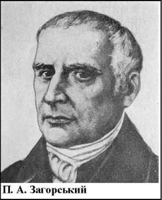
I.V. Buyalsky was a student of P.A. Zagorsky and succeeded him as head of the department in 1833.
I.V. Buyalsky (1789–1866) was an outstanding Russian surgeon, anatomist, and artist, and one of the pioneers of Topographical Anatomy. Endowed with exceptional surgical talent, a brilliant command of anatomical techniques, remarkable oratory skills, and an iron will, he was able to withstand the attacks of German scientists working in Russia who detested him. I.V. Buyalsky created a magnificent anatomical museum, featuring his own corrosion casts. He was the first in Russia to apply the corrosion technique and published «Photographic Drawings of Corroded Arteries and Veins OF THE Human Kidney Taken from Specimens, with a Detailed Description of the Method of Preparing Such Specimens». Buyalsky developed and proposed a method for embalming corpses as well as techniques for «ice» and «sculptural» anatomy. At his own expense, he published anatomical drawings for students, along with «A Short General Human Anatomy» and «Anatomical and Surgical Tables» illustrating vascular ligation techniques. This was the first Russian atlas of operative surgery. Translated into German, it caused a sensation abroad.
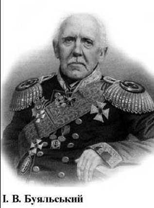
Driven by the growing demands of surgery, surgical or topographical anatomy emerged as an independent science, largely thanks to I.V. Buyalsky and Nikolai Pirogov—a genius anatomist and surgeon. Through the work of Nikolai Pirogov, medicine in general and anatomy in particular made a giant leap forward in their development.
Nikolai Pirogov (1810–1881) graduated from the medical faculty of Moscow University at the age of 18, defended his dissertation at 22, and at 25 became head of the Department of Surgery at the University of Dorpat. The works he published during this period earned him international renown. At the age of 29, Pirogov took over the Department of Surgery at the Medico-Surgical Academy in St. Petersburg, where he initiated educational reforms centered around the rigorous study of anatomy as the foundation for future physicians.
Nikolai Pirogov possessed brilliant surgical skills and established the first hospital surgical clinic that matched the standards of the finest European clinics. World acclaim came to Pirogov with his seminal work, «Surgical anatomy of Arterial Trunks and Fasciae». He introduced a groundbreaking research method into anatomy—serial cross-sectional slicing of frozen cadavers (ice anatomy)—and based on this method, authored «A Complete Course of Applied Anatomy of the Human Body» and the atlas «Topographical Anatomy Illustrated by Sections Made Through the Frozen Human Body in Three Directions». These were the pioneering manuals of topographical anatomy.

A monumental contribution of Nikolai Pirogov was the development of ether anesthesia, which he tested on himself and volunteers before applying it in combat conditions in the Caucasus. Pirogov traveled twice to Sevastopol during the Crimean War, where his administrative and organizational talents fully unfolded. Unfortunately, his work was overshadowed by slander and press persecution that denied him peace of mind, prompting him to resign upon returning from Sevastopol. For a time, Pirogov served as the trustee of the Odesa and Kyiv educational districts, but he could not remain long due to his progressive views, affection for students, and defense of their rights, which greatly displeased the ruling circles. The final years of his life were spent on his small estate in the Vinnytsia region, where he opened a pharmacy, cultivated medicinal herbs, maintained a magnificent garden, and founded a small hospital where he operated on and treated patients as long as his health permitted. He died of oral mucosal Cancer in November 1881, having stopped writing his unfinished «Diary of an Old Physician» just a month before his death. In the village of Vyshneve (now Pirohove) in the Vinnytsia region of Ukraine, his embalmed body rests and a memorial museum has been established. Re-embalming Procedures of Pirogov’s body were carried out in 1945, 1956, 1979, 1981, and 1994.
Nikolai Pirogov’s name is commemorated in Pirogov's triangle (hyoid triangle), Pirogov's tonsillar ring (pharyngeal lymphoid ring), and Pirogov’s node (a Lymph node located in the deep ring of the femoral canal). In his honor, the Society of Russian Physicians was founded, and a named prize of the USSR Academy of Medical Sciences was established. Pirogov's scholarly works have lost none of their significance today. His follower, Professor of Anatomy at Kyiv University O.P. Walter (1817–1888), introduced and substantiated METABOLISM/2.html">THE CONCEPT OF the «physiological anatomy of the living body» and authored the finest anatomy textbook of his time in Russian.
P.F. Lesgaft (1837–1909), a contemporary of Nikolai Pirogov, embodied the talents of a scientist, educator, physician, administrator, and public figure. He was the founder of domestic theoretical anatomy and the science of physical education, and published the first works addressing the Organization of monitoring the physical development of children and adolescents. He stood at the origins of domestic biomechanics, and is recognized as one of the founders of national anthropology and Functional Anatomy. He insisted on the necessity of experimental and X-ray methods in studying organs and systems, and pioneered the Practical Application of X-rays in anatomy. Having earned doctorates in both medicine and surgery, he taught at the medical faculties of the universities of Kazan and St. Petersburg, as well as at the Medico-Surgical Academy in St. Petersburg, and established a remarkable biological laboratory featuring superb imported collections of comparative anatomical specimens.
He founded higher academic courses for female educators and physical culture instructors, and developed a progressive system of physical education successfully utilized to promote fitness and sports both nationally and internationally.
P.F. Lesgaft established the GENERAL PATTERNS OF Blood vessel branching, identified the factors influencing bone shape, and described the architecture of Bones and joints, as well as the Structure of Muscles, the rectum, and the Perineum. He maintained that the living human being should be the primary subject of anatomical research. Lesgaft presented his research findings in the two-part work «Fundamentals of Theoretical Anatomy». He authored such works as «The Tasks of Anthropology and the Methods of Its Study», «On the Relation of Anatomy to Physical Education», and «Anthropology and Pedagogy», as well as the two-volume textbook «Human Anatomy». All of Lesgaft's works were grounded in materialist philosophy and the principles of The Unity of the Organism and its environment, as well as the unity of form and function.
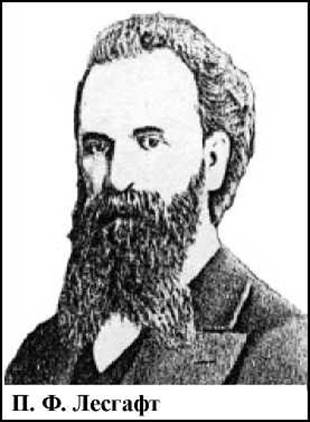
P. F. Lesgaft was an extraordinary educator and teacher. His progressive activities caused discontent among the ruling circles, making his life full of hardship and persecution by the tsarist government. P. F. Lesgaft died in November 1909 in a foreign land, in Cairo, where he had been sent for medical Treatment.
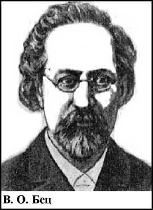
Many discoveries in the field of anatomy were made in Kyiv by the prominent scientist V. O. Betz.
V. O. Betz (1834–1894) was a renowned Ukrainian anatomist, physician, educator, and tutor. He graduated from the medical faculty of Kyiv University and became a professor of anatomy at the age of 34. He was a brilliant lecturer and a talented teacher. V. O. Betz gained immense fame through his scientific works, which primarily focused on the structure of the Cerebral Cortex in adults, children, embryos, and various animals. He discovered giant pyramidal Cells in the precentral gyrus of the CEREBRAL CORTEX AND, by comparing his morphological data with the physiological experiments of other scientists, established the presence of the motor center in the cerebral cortex. This brought him worldwide acclaim. In other works, V. O. Betz established the lamination of the cerebral cortex and identified eleven distinct areas in the cortex based on their microscopic structure. In doing so, he laid the foundations for the science known as the "cytoarchitectonics of the cerebral cortex."
V. O. Betz also studied The Mechanism of hepatic Blood Circulation and discovered the chromaffin reaction of The adrenal medulla. He investigated the Skeletal System, conducting the first systematic studies on Bone Development and growth, and summarized the results of this work in his monograph Morphology of Osteogenesis. He extensively promoted museum curation, assembling a large collection of plaster casts and Human and Animal Brain specimens, which was awarded a medal at the All-Russian Exhibition in St. Petersburg and honored at the World Exhibition in Vienna.
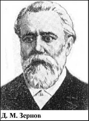
Another anatomist who made significant contributions to brain anatomy was D. M. Zernov (1843–1917), who provided a more refined Classification of the brain's sulci and gyri. D. M. Zernov's primary work focused on The Nervous System and Sensory Organs. He wrote an exceptional textbook on anatomy that went through 14 editions. D. M. Zernov opposed the reactionary racist theory of Lombroso, who believed that a distinct criminal type existed, characterized by specific anatomical Skull features. By demonstrating the absence of racial and ethnic differences in brain structure, Zernov established an anatomical foundation for combating racism. He founded a prominent school of native anatomists renowned for their scholarly contributions.
The prominent Ukrainian scientist M. A. Tikhomirov conducted extensive research on The Vascular System of the brain.
M. A. Tikhomirov (1848–1902) was a professor of anatomy at Kyiv University, an outstanding educator, and one of the founders of anthropology in Ukraine. He studied the CEREBRAL ARTERIES AND variations in the development of arteries and veins. He authored the work Variations of Arteries and Veins of The Human Body in Relation to the Morphology of the Circulatory system and developed a methodology for teaching Normal Anatomy.
A major contribution to the Development of the anatomy of The Lymphatic system was made by the Ukrainian scientist F. A. Stefanis.
F. A. Stefanis (1865–1917) was a professor of anatomy at Kyiv University and head of the Department of Anatomy. He prepared numerous anatomical specimens and compiled a systematic catalog of the anatomical museum specimens at the Kyiv Medical Institute. He studied the lymphatic system and congenital kidney defects, and invented a device for injecting the Lymphatic vessels of the kidneys, Liver, and Stomach. His disciples formed the foundation of the Kyiv school of lymphology.
The Ukrainian anatomist M. K. Lysenkov made a substantial contribution to the development of domestic anatomy by engaging in all major anatomical disciplines dedicated to the normal Structure of the human body: normal, topographical, and plastic anatomy.
M. K. Lysenkov (1865–1941) was an anatomist, artist, and professor at the medical faculty of Odesa University, as well as a co-author of the well-known Lectures on Topographical Anatomy and Operative Surgery. He proposed a valuable "method for preserving anatomical specimens without immersion in fluids while maintaining their natural volume." He authored Plastic Anatomy, Anatomical and physiological Foundations of Physical Culture of the Human Body, and the textbook Normal Anatomy (co-authored with V. I. Bushkovych).
Among the domestic morphologists who worked at the turn of the 20th century, V. P. Vorobyov, V. M. Tonkov, and V. N. Shevkunenko deserve special mention.
V. P. Vorobyov (1876–1938) was an outstanding Ukrainian anatomist, founder of a major anatomical school, a talented lecturer and educator, a prominent public figure, and one of the pioneers of the functional approach in anatomy; he discovered new laws governing the Structural organization of the nervous system.
V. P. Vorobyov made a significant contribution to the development of educational anatomical museums. He published the paper On the Issue of Establishing Educational Anatomical Museums and organized the world's sole "Museum of Human Evolution." He participated in organizing higher medical education and developing curricula and syllabi in human normal anatomy for medical universities. He created the five-volume Atlas of Human Anatomy and served as the editor and co-author of the textbook for dentists Anatomy, Histology, and Embryology of the Oral Cavity and Teeth.
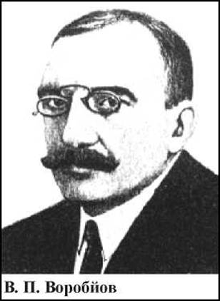
The macro- and microscopic Research Methods developed by V. P. Vorobyov, along with his refined techniques of corpse embalming, gained widespread recognition and adoption.
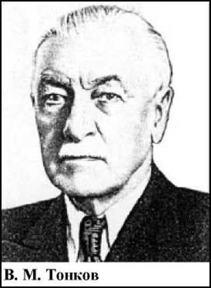
V. M. Tonkov (1872–1954) was a prominent Soviet-era anatomist, talented educator, and administrator. He studied at the Military Medical Academy in St. Petersburg. After defending his doctoral dissertation, he was sent abroad. Upon his return, he headed a department at the Women's Medical Institute, and from 1905 onward, he led the Department of Normal Anatomy at Kazan University, where he engaged in extensive academic and public activities. In 1915, V. M. Tonkov returned to the Military Medical Academy's department, maintaining his association with it for the rest of his Life as a professor, president, and in his final years, as a consultant.
V. M. Tonkov established an anatomical school focused on functional anatomy. His works address the normal and comparative anatomy of humans and animals. As one of the first Russian researchers to apply X-rays in studying skeletal GROWTH AND DEVELOPMENT, he laid the groundwork for domestic X-ray anatomy.
Academician V. M. Tonkov, who headed the Department of Anatomy at the Military Medical Academy in St. Petersburg for over 50 years, together with his disciples (B. A. Dolgo-Saburov, G. F. Ivanov, N. V. Kolesnikov, and others), developed The Theory of collateral circulation and demonstrated the Influence of the nervous system on the development of collaterals. His work in the field of collateral circulation gained recognition both domestically and internationally. V. M. Tonkov contributed greatly to the restructuring of the educational process in anatomy. He was one of the initiators of establishing educational anatomical museums and organized the educational anatomical museum at the Women's Medical Institute in St. Petersburg in 1901.
V. M. Tonkov's scientific legacy is vast. His doctoral dissertation was recognized as a classic, and all his experiments enriched science with abundant novel data and original insights. He authored numerous papers dedicated to teaching anatomy and educational anatomical museums, as well as one of the finest anatomy textbooks, which was reprinted six times. He founded a school of anatomists, whose prominent representative and V. M. Tonkov's successor as head of the department was Professor B. A. Dolgo-Saburov, who successfully carried forward his mentor's work alongside his colleagues.
V. N. Shevkunenko (1872–1952) was a renowned Soviet anatomist and the founder of the modern school of topographical anatomists. He developed The Doctrine of individual Variability in the shape, structure, and topography of organs, which is of great significance for surgery. V. N. Shevkunenko made a substantial contribution to the organization of educational anatomical museums, participating in the creation of the Military Medical Museum in St. Petersburg. He published the first comprehensive domestic manuals on operative surgery and topographical anatomy, and was awarded the USSR State Prize for his unique work, Atlas of the Peripheral Nervous and Venous Systems.
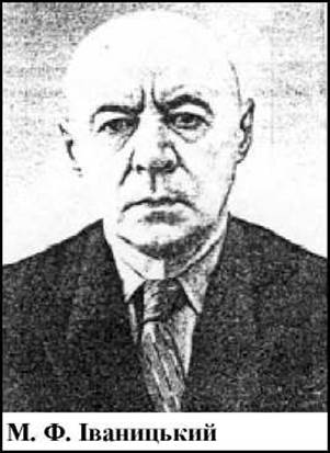
A major role in the development of domestic functional anatomy belongs to the renowned anatomist M. F. Ivanytsky.

M.F. Ivanytskyi (1895–1969) was a Doctor of Medical Sciences, professor, and an exceptional lecturer and educator with an exquisite mastery of anatomical drawing. He laid the scientific foundations of sports anatomy and advanced functional anatomy to meet the demands of athletic practice. His fundamental research centered on the question of "THE POSITION OF the general center of gravity of the human body." He established the course in dynamic anatomy, published Notes on Dynamic Anatomy, and authored the book Movements of the Human Body.
He conducted extensive research in sports anthropology, focusing on constitutional types, body proportions, and the physical development of athletes.
M.F. Ivanytskyi authored numerous works on projection anatomy and the functional Anatomy of The Musculoskeletal System. He wrote the original textbook Human Anatomy for students of physical culture institutes, which went through many editions and played a pivotal role in advancing physical education in our country.
A significant contribution to the development of macro-microscopic anatomy was made by the prominent Ukrainian anatomist and representative of the Kharkiv anatomical school, R.D. Synelnykov.
R.D. Synelnykov (1896–1982) pioneered and extensively applied macro-microscopic research methods to study the musculoskeletal system, peripheral and autonomic nervous systems, Skin glands, mucous membranes, and vascular system from age-specific and comparative-anatomical Perspectives. He headed the Department of Normal Anatomy at the Kharkiv Medical Institute, served as a professor, and worked in the laboratory at V.I. Lenin's Mausoleum. He also supervised the project on the restoration and preservation of N.I. Pirogov’s body. He developed an original method for preparing anatomical museum specimens and authored numerous works on The history of medicine and anatomical research methodologies. He participated in the preparation and publication of the 5-volume Atlas of Human Anatomy, created in collaboration with V.P. Vorobyov. Later, he prepared and published a new Atlas of Human Anatomy, which underwent multiple editions and gained widespread acclaim both domestically and internationally. His son, Ya.R. Synelnykov, continued his father’s legacy, and the latest edition of the 4-volume atlas has been republished in many European languages.
The development of national anatomy was also greatly advanced by Ukrainian anatomists from the Kyiv Medical Institute, M.S. Spirov and I.Ye. Kefeli.
M.S. Spirov (1892–1973) was an Honored Scientist, professor, and head of the Department of Anatomy at the Kyiv Medical University. He investigated the lymphatic vessels of serous membranes, authored a manual on the dissection of human muscles, ligaments, vessels, and nerves, reorganized the department's anatomical museum, and established a scientific school dedicated to the Study of the lymphatic system.

I.Ye. Kefeli (1920–1980) was a professor and head of the Department of Anatomy at the Kyiv Medical University. He specialized in the Peripheral Nervous System and vascular vasculature and authored the student textbook Atlas of Diagrams in Human Anatomy (circulatory and peripheral nervous systems) in Ukrainian.
A notable contribution to national and world morphology was also made by such eminent scientists as A.M. Severtsov (1866–1936), O.O. Zavarzin (1886–1945), O.H. Knore (1914–1981), and others.
Modern morphologists continue to fruitfully build upon the finest traditions of domestic anatomists, histologists, and embryologists.
Contemporary science no longer views the human body as something static and immutable once and for all. Instead, it examines the organism dynamically, in continuous development, striving not only to identify the Structural Features of a given organ but also to investigate the external and internal Factors affecting the body. At the same time, the study of human bodily structure is pursued with a view toward its practical application in clinical medicine.
One of the leading contemporary anatomists in Ukraine is the State Prize laureate of Ukraine, Professor V.H. Koveschnikov.
V.H. Koveschnikov was born in 1931 into a large working-class family. In 1955, he graduated with honors from the Saratov Medical Institute. In 1959, he defended his Candidate dissertation on pressing issues in craniology, and in 1970, he defended his Doctoral dissertation dedicated to the clinically and theoretically significant topic of the planar orientation of the semicircular canals of the Inner ear labyrinth, alongside the ontogenetic and phylogenetic Features of the vestibular apparatus.
V.H. Koveschnikov is the author of over 200 scientific papers and 5 inventions (including a proprietary craniograph model), as well as a co-author of the textbook Human Anatomy and the Anatomical Dictionary for medical universities. His monograph Clinical Anthropology has received widespread acclaim. For over 30 years, he has conducted intensive, comprehensive research into the growth, structure, morphogenesis, and regeneration of Bone tissue under the Influence of Environmental factors of varying Etiology and intensity. Under his guidance, a major series of experimental studies was carried out to uncover the patterns of adaptive and re-adaptive skeletal transformations across different regimes of physical activity, as well as dosed static and dynamic loads during various age periods. These findings have earned widespread recognition among sports physicians, traumatologists, orthopedists, gerontologists, physiologists, and pediatricians. Among morphologists, the universal morphofunctional classification of bone growth plate zones—the epiphyseal Cartilage according to Koveschnikov—is well-known and widely used.
A hallmark of the research conducted under V.H. Koveschnikov’s supervision is its applied nature, acute socio-scientific relevance, and pressing urgency. These studies represent systematic steps in investigating exogenous factors, their impact on the organs and systems of the human body, and the pathways for correcting and preventing adverse consequences. He is entirely deserving of his reputation as one of the founders of national functional Osteology.
Professor V.H. Koveschnikov also devotes significant attention to the organization and optimization of the educational process. He is a person consumed by scientific inquiry, deeply devoted to his work and his students.
Last update: 08/08/2026
Editorial and Educational Adaptation: This material has been compiled based on the primary/original source text. The project team performed an editorial review, corrected technical inaccuracies, structured sections, and adapted the content for an educational format.
What was processed:
- elimination of formatting defects (OCR errors, structural breaks, corrupted characters);
- editorial organization of content;
- standardization of terminology in accordance with academic sources;
- verification of factual statements against the original source text.
All mentions of the author, publication year, and origin of the primary text have been preserved in accordance with the source.