LEHNINGER PRINCIPLES OF BIOCHEMISTRY - VOLUME 2. BIOENERGETICS AND METABOLISM - 2014
PART II. BIOENERGETICS AND METABOLISM
14. GLYCOLYSIS, GLUCONEOGENESIS, AND THE PENTOSE PHOSPHATE PATHWAY
14.4. Gluconeogenesis
The Central Role of glucose in METABOLISM arose early in the evolutionary history of our biosphere, and for modern organisms—from microbes to humans—this sugar remains a nearly universal fuel and building block. Some mammalian Tissues depend almost completely on glucose for their metabolic energy. For the human Brain and Nervous system, as well as erythrocytes, Testes, renal medulla, and embryonic tissues, glucose is the sole or primary source of energy. The brain alone requires about 120 g of glucose each day—more than half of all the glucose stored as Glycogen in Muscle and Liver. However, these glucose reserves are not always sufficient; glycogen is depleted between meals, during longer periods of fasting, or after vigorous exercise. During these times, organisms must synthesize glucose from noncarbohydrate precursors. This pathway is called Gluconeogenesis (the "new" synthesis of sugar), which converts Pyruvate and related three- and four-carbon compounds to glucose.
Gluconeogenesis occurs in animals, plants, and microorganisms. The pathway is highly conserved in all Cells and organisms. In animals, the important precursors of glucose are three-carbon compounds such as lactate, pyruvate, and glycerol, as well as Certain Amino Acids (Fig. 14-15). In mammals, gluconeogenesis takes place mainly in the liver, and to a lesser extent in the renal cortex. The synthesized glucose is released into the Blood to supply other tissues. Lactate produced in Skeletal Muscle during anaerobic Glycolysis under heavy physical exertion is returned to The Liver and converted to glucose, which is carried back to the muscle to form glycogen; this is the Cori cycle (Box 14-2; see also Fig. 23-20). In plant seeds, stored fats and Proteins are converted, via several pathways including gluconeogenesis, to the disaccharide sucrose for transport throughout the growing plant. Glucose and its derivatives serve as the building blocks for Plant Cell Walls, NUCLEOTIDES, Coenzymes, and many other essential metabolites. In many microorganisms, gluconeogenesis starts from simple two- or three-carbon organic molecules, such as acetate, lactate, and propionate, present in the growth medium.
Class="center">Fig. 14-15. Carbohydrate synthesis from simple precursors. The pathway from phosphoenolpyruvate to glucose 6-phosphate is common to the biosynthetic conversion of many carbohydrate precursors in animal and plant cells. However, only plants and photosynthetic Bacteria can use CO2 to synthesize CARBOHYDRATES. The conversion of pyruvate to phosphoenolpyruvate involves oxaloacetate, an intermediate in The Citric Acid Cycle, which is discussed in Chapter 16. Thus, any compound that can be converted to pyruvate or oxaloacetate can serve as a Starting Material for gluconeogenesis. These include Alanine and aspartate, which are converted to pyruvate and oxaloacetate, respectively, and Other Amino Acids that yield three- or four-carbon fragments (the glucogenic amino acids; see Table 14-4 and Fig. 18-5). Plants and photosynthetic bacteria have The unique ability to convert CO2 to carbohydrates using The Glyoxylate cycle (p. 212).

Although the reactions of gluconeogenesis are the same in all organisms, the metabolic context and the Regulation of the pathway vary from species to species and from tissue to tissue. In this section, we focus on gluconeogenesis in the mammalian liver. In Chapter 20, we will see how photosynthetic organisms use this pathway to convert the primary products of Photosynthesis into glucose, to be stored as sucrose or starch.
The Metabolic pathways of glycolysis and gluconeogenesis are not identical; they run in opposite directions, although they do share several steps (Fig. 14-16). Seven of the ten ENZYMATIC REACTIONS OF gluconeogenesis are the reverse of glycolytic reactions. However, Three Reactions of glycolysis are essentially irreversible in vivo and cannot be used in gluconeogenesis: the conversion of glucose to glucose 6-phosphate by hexokinase, the phosphorylation of fructose 6-phosphate to fructose 1,6-bisphosphate by Phosphofructokinase-1, and the conversion of phosphoenolpyruvate to pyruvate by pyruvate kinase (Fig. 14-16). In cells, these three reactions are characterized by a large, negative free-energy change, whereas the other glycolytic reactions have a ∆G near zero (Table 14-2). In gluconeogenesis, the three irreversible steps are bypassed by a separate set of Enzymes, catalyzing reactions that are sufficiently exergonic to be effectively irreversible in the direction of glucose synthesis. Thus, both glycolysis and gluconeogenesis are irreversible processes in cells. In animals, both pathways occur largely in the Cytosol, necessitating their coordinated regulation. Independent regulation of the two pathways is achieved through those enzymatic steps that are not shared.
Table 14-2. Free-Energy Changes of Glycolytic Reactions in Erythrocytes
Glycolytic reaction |
∆G′° (kJ/mol) |
∆G (kJ/mol) |
(1) Glucose + ATP —> glucose 6-phosphate + ADP |
-16,7 |
33,4 |
(2) Glucose 6-phosphate ⇄ fructose 6-phosphate |
1,7 |
0 to 25 |
(3) Fructose 6-phosphate + ATP —> fructose 1,6-bisphosphate + ADP |
-14,2 |
-22,2 |
(4) Fructose 1,6-bisphosphate ⇄- dihydroxyacetone phosphate + glyceraldehyde 3-phosphate |
23,8 |
0 to 6 |
(5) Dihydroxyacetone phosphate ⇄ glyceraldehyde 3-phosphate |
7,5 |
0 to 4 |
(6) Glyceraldehyde 3-phosphate + Pi + NAD+ ⇄ 1,3-bisphosphoglycerate + NADH + H+ |
6.3 |
-2 to 2 |
(7) 1,3-Bisphosphoglycerate + ADP ⇄ 3-phosphoglycerate + ATP |
-18,8 |
0 to 2 |
(8) 3-Phosphoglycerate ⇄ 2-phosphoglycerate |
4,4 |
0 to 0,8 |
(9) 2-Phosphoglycerate ⇄ phosphoenolpyruvate + H2O |
7,5 |
0 to 3,3 |
(10) Phosphoenolpyruvate + ADP —> pyruvate + ATP |
-31,4 |
-16.7 |
Note. As defined in Chapter 13 (pp. 14–15), ∆G′° represents the standard free-energy change; ∆G is the free-energy change calculated from the actual concentrations of glycolytic intermediates in erythrocytes under physiological conditions at pH 7.0. The bypass reactions of gluconeogenesis are highlighted in orange. The equations shown are not balanced for hydrogen atoms and charge (p. 28).
We begin our study of gluconeogenesis by examining the three bypass reactions (recall that "bypass" reactions are those that circumvent the irreversible steps of glycolysis).
The conversion of pyruvate to phosphoenolpyruvate proceeds in two exergonic steps
The first bypass reaction of gluconeogenesis is the conversion of pyruvate to phosphoenolpyruvate (PEP). This process cannot occur by a simple Reversal of the pyruvate kinase reaction of glycolysis (p. 79), which has a large negative standard free-energy change and is therefore irreversible in the intact cell (Table 14-2, reaction (10)). Instead, the phosphorylation of pyruvate is achieved through A bypass pathway consisting of a sequence of reactions that, in Eukaryotic cells, require both cytosolic and mitochondrial enzymes. As we will see, the metabolic pathway shown in Fig. 14-16 and described in detail here is one of two possible pathways for the conversion of pyruvate to PEP. This pathway is the primary one when pyruvate or alanine serves as the precursor for glucose synthesis. The second pathway, described below, predominates when lactate is the glucose precursor.
Fig. 14-16. Opposing pathways of glycolysis and gluconeogenesis in rat liver. Glycolytic reactions are on the left, in red; gluconeogenic reactions are on the right, in blue. The major regulatory sites of gluconeogenesis shown in the diagram are discussed later in this chapter and in more detail in Chapter 15. Figure 14-19 shows an alternative pathway for oxaloacetate conversion occurring in Mitochondria.

Fig. 14-17. Synthesis of phosphoenolpyruvate from pyruvate. (a) In mitochondria, pyruvate is converted to oxaloacetate in a biotin-dependent reaction catalyzed by pyruvate carboxylase. (b) In the cytosol, oxaloacetate is converted to phosphoenolpyruvate by PEP carboxykinase, and the CO2 incorporated into oxaloacetate is released again. Decarboxylation leads to a redistribution of electrons, facilitating the attack of the carbonyl oxygen on the γ-phosphate group of GTP.

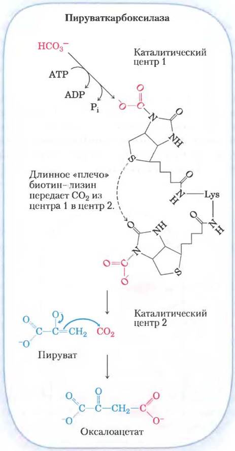
First, pyruvate is transported from the cytosol into mitochondria or is generated in mitochondria from alanine by Transamination, in which the α-amino group of alanine is removed (yielding pyruvate) and transferred to an α-keto acid (transamination reactions are discussed in Chapter 18). Then, the mitochondrial enzyme pyruvate carboxylase, in the presence of the coenzyme biotin, converts pyruvate to oxaloacetate (Fig. 14-17):
Pyruvate + HCO3- + ATP —> oxaloacetate + ADP + Pi (14-4)
Biotin plays The Role of a carrier of activated bicarbonate in the carboxylation reaction (Fig. 14-18). The reaction mechanism is shown in Fig. 16-16. (Note that HCO3 arises from the dissociation of carbonic acid, formed from CO2 + H2O.) Phosphorylation of HCO3 by ATP yields a mixed anhydride (carboxyphosphate); biotin then displaces the phosphate, forming carboxybiotin.
Fig. 14-18. Role of biotin in the pyruvate carboxylase reaction. The biotin cofactor is covalently bound to the enzyme by an amide linkage to the ε-amino group of a Lysine residue, forming a biotinylated enzyme. The pyruvate carboxylase reaction occurs in two phases, which take place at two different catalytic sites on the enzyme molecule. At catalytic site 1, bicarbonate is converted to CO2 at the expense of one ATP molecule. The CO2 then reacts with biotin to form a carboxybiotin-enzyme complex. A long "arm," consisting of biotin and the side chain of the lysine residue to which it is attached, transfers CO2 from the carboxybiotin-enzyme complex to catalytic site 2 on the enzyme surface, where CO2 is released and reacts with pyruvate to form oxaloacetate and regenerate the biotinylated enzyme. The central role of such "flexible arms" in transferring intermediates between active sites of enzymes is illustrated in Fig. 16-17, and the detailed mechanism of the pyruvate carboxylase reaction is shown in Fig. 16-16. A similar mechanism operates in other biotin-dependent carboxylation reactions, such as those catalyzed by propionyl-CoA carboxylase (see Fig. 17-11) and acetyl-CoA carboxylase (see Fig. 21-1).
Pyruvate carboxylase is the first regulatory enzyme of gluconeogenesis, requiring the presence of acetyl-CoA as an activator (acetyl-CoA is formed during Fatty acid oxidation (Chap. 17), and its accumulation signals that Fatty acids can be utilized as an energy source). As we will see in Chap. 16 (see Fig. 16-15), the reaction catalyzed by pyruvate carboxylase can supply intermediates for another vital metabolic pathway—The Citric Acid cycle.
The mitochondrial membrane lacks transporters for oxaloacetate; therefore, before being transported into the cytosol, the oxaloacetate formed from pyruvate is converted into its reduced form (malate, the salt of malic acid) through the action of the mitochondrial enzyme malate dehydrogenase, utilizing NADH:
Oxaloacetate + NADH + H+ ⇄ L-malate + NAD+ (14-5)
In this reaction, The change in Standard Free energy is quite significant, but under physiological conditions (including a very low oxaloacetate concentration), ∆G ≈ 0, and the reaction proceeds readily in the reverse direction. Mitochondrial malate dehydrogenase participates in both gluconeogenesis and the citric acid cycle, although the net fluxes of metabolites in these two processes run in opposite directions.
Malate leaves the mitochondria via a specific transport system located in The inner mitochondrial membrane (see Fig. 19-30). In the cytosol, malate is re-oxidized to oxaloacetate with the generation of NADH:
Malate + NAD+ —> oxaloacetate + NADH + H+ (14-6)
Oxaloacetate is then converted into PEP by the action of phosphoenolpyruvate carboxykinase (Fig. 14-17). This Mg2+-dependent reaction requires the presence of GTP as a phosphoryl group donor:
Oxaloacetate + GTP ⇄ PEP + CO2 + GDP (14-7)
Under intracellular conditions, the reaction is reversible; The formation of one energy-rich phosphate bond (in PEP) is counterbalanced by the Hydrolysis of another phosphate bond (in GTP).
The equation for this bypass metabolic pathway is obtained by summing equations (14-4) through (14-7):
Pyruvate + ATP + GTP + HCO3- —> PEP + ADP + GDP + Pi + CO2 (14-8)
∆G'° = 0.9 kJ/mol
Thus, for the conversion of a single pyruvate molecule into PEP, two high-energy phosphate groups must be expended (one from ATP and one from GTP), each possessing an energy of approximately -50 kJ/mol under intracellular conditions. Conversely, during the conversion of PEP to pyruvate in glycolysis, only a single ATP molecule is synthesized from ADP. Although the standard free energy change ∆G′° for the two-step conversion of pyruvate to PEP is 0.9 kJ/mol, the actual free energy change, accounting for intracellular intermediate concentrations, has a substantial negative value (-25 kJ/mol). This occurs because the resulting PEP is rapidly consumed in subsequent reactions, keeping its steady-state concentration at a sufficiently low level. Consequently, within The Cell, this reaction is effectively irreversible.
Note that the reaction catalyzed by PEP carboxykinase releases the exact same CO2 molecule that is added to pyruvate in the step catalyzed by pyruvate carboxylase (Fig. 14-17, b). This carboxylation-decarboxylation sequence serves to "activate" pyruvate, since the decarboxylation of oxaloacetate facilitates the formation of PEP. In Chapter 21, we will discuss how a similar carboxylation-decarboxylation sequence is utilized to activate acetyl-CoA in FATTY ACID Biosynthesis (see Fig. 21-1).
To clarify why these reactions take place within the mitochondria: the [NADH]/[NAD+] ratio in the cytosol is 8 • 10-4, which is roughly 105 times lower than in the mitochondria. Because cytosolic NADH is consumed during gluconeogenesis for the conversion of 1,3-bisphosphoglycerate into glyceraldehyde-3-phosphate (Fig. 14-16), glucose biosynthesis cannot proceed without a sufficient supply of NADH. The transport of malate from the mitochondria into the cytosol, followed by its conversion to oxaloacetate, facilitates The transfer of reducing equivalents into the cytosol, where they are deficient. Thus, the conversion of pyruvate to PEP maintains an essential balance in gluconeogenesis between the cytosolic NADH produced and consumed.
The second alternative bypass reaction from pyruvate to PEP predominates when lactate serves as the primary starting material for glucose synthesis (Fig. 14-19). This metabolic pathway utilizes lactate generated via glycolysis—for instance, in erythrocytes or under anaerobic conditions in muscle—and plays a particularly critical role in large vertebrates following intense physical exertion (Box 14-2). The conversion of lactate to pyruvate in the hepatocyte cytosol is accompanied by the generation of NADH, eliminating the need to export reducing equivalents (in the form of malate) from the mitochondria in this scenario.
Fig. 14-19. Alternative pathways for the formation of phosphoenolpyruvate (PEP) from pyruvate. THE CONTRIBUTION OF each process is determined by the availability of lactate or pyruvate and the cytosolic demand for NADH in gluconeogenesis. On the right is the pathway that predominates when lactate is the starting substrate, because the Lactate dehydrogenase reaction generates NADH directly in the cytosol, obviating the need to export it from the mitochondria (see text).

Pyruvate formed in the lactate dehydrogenase reaction is transported into the mitochondria and converted to oxaloacetate by pyruvate carboxylase, as described above. However, this oxaloacetate is converted directly to PEP by the mitochondrial isoenzyme of PEP carboxykinase, and the PEP is then exported from the mitochondria to continue gluconeogenesis. The mitochondrial and cytosolic Isoenzymes of PEP carboxykinase are encoded by distinct chromosomal genes, providing yet another example of how two different enzymes catalyzing the same reaction can be localized in different cellular compartments or serve different metabolic Functions (recall hexokinase isoenzymes).
Second Bypass Pathway: Conversion of Fructose 1,6-Bisphosphate to Fructose 6-Phosphate
Another glycolytic reaction that cannot be directly utilized in gluconeogenesis is the phosphorylation of fructose 6-phosphate by phosphofructokinase-1 (PFK-1; Table 14-2, reaction (D)). This reaction is characterized by a large negative standard free energy change and is consequently irreversible under cellular conditions. Therefore, the Formation of fructose 6-phosphate from fructose 1,6-bisphosphate (Fig. 14-16) is carried out by a different enzyme: Mg2+-dependent fructose 1,6-bisphosphatase (FBPase-1). This process involves the essentially irreversible hydrolysis of the phosphate group at the C-1 position (rather than a phosphoryl group transfer to ADP):
Fructose 1,6-bisphosphate + H2O —> fructose 6-phosphate + Pi
∆G'° = -16.3 kJ/mol
FBPase-1 should be distinguished from the structurally and functionally related enzyme FBPase-2, which plays a regulatory role (see Chap. 15).
Third Bypass Pathway: Formation of Glucose from Glucose 6-Phosphate
This bypass pathway occurs at The final stage of gluconeogenesis and involves the dephosphorylation of glucose-6-phosphate to yield glucose (Fig. 14-16). Reversing the hexokinase reaction (p. 71) would require transferring a phosphate group from glucose-6-phosphate to ADP to form ATP, which is energetically unfavorable (Table 14-2, reaction (1)). The reaction catalyzed by glucose-6-phosphatase is not coupled to ATP synthesis; it is simply the hydrolysis of a phosphoric acid ester:
Glucose-6-phosphate + H2O —> glucose + Pi
∆G′° = -13.8 kJ/mol
This Mg2+-dependent enzyme is found on the luminal surface of The Endoplasmic reticulum in hepatocytes, Kidney cells, and epithelial Cells of the Small Intestine (see Fig. 15-28), but not in other tissues, which are consequently unable to supply glucose to the blood. If other tissues contained glucose-6-phosphatase, this enzymatic activity would hydrolyze the glucose-6-phosphate that those tissues require for glycolysis. The glucose produced via gluconeogenesis in the liver or Kidneys, as well as that obtained from diet, is delivered to the brain and Muscle Tissues via the bloodstream.
Gluconeogenesis is essential, yet energetically costly
The biosynthetic reactions leading from pyruvate to glucose (Table 14-3) can be summarized by the overall equation:
2 Pyruvate + 4 ATP + 2 GTP + 2 NADH + 2 H+ + 4 H2O —> glucose + 4 ADP + 2 GDP + 6 Pi + 2 NAD+ (14-9)
Thus, the synthesis of a single glucose molecule from pyruvate consumes six high-energy phosphate groups—four from ATP and two from GTP. In addition, two molecules of NADH are required to reduce two molecules of 1,3-bisphosphoglycerate. Therefore, Equation 14-9 clearly represents more than just the reverse of the glycolysis pathway from glucose to pyruvate; it demonstrates that only two molecules of ATP are consumed in the reverse direction:
Glucose + 2 ADP + 2 Pi + 2 NAD+ —> 2 pyruvate + 2 ATP + 2 NADH + 2 H+ + 2 H2O
Table 14-3. Sequence of Reactions in Gluconeogenesis Starting from Pyruvate
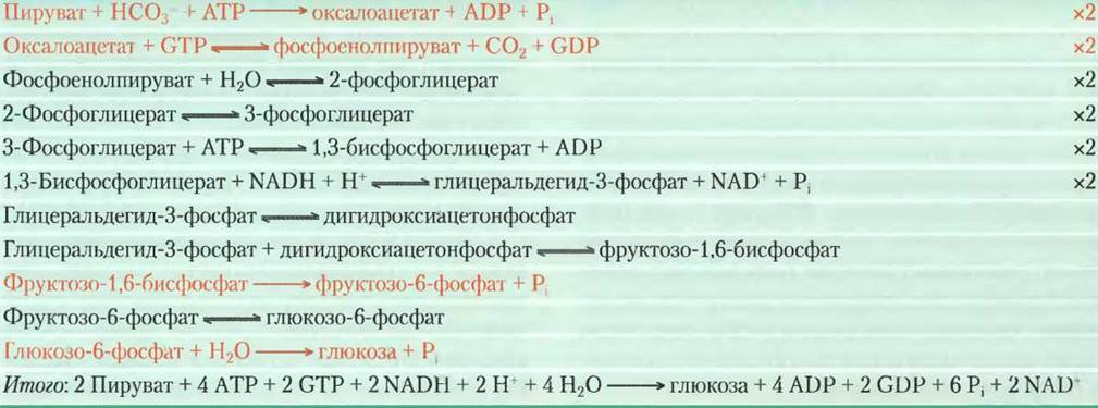
Note. Bypass reactions are highlighted in orange; all other reactions are the reverse steps of glycolysis. Numbers on the right indicate that the given reaction must be counted twice because two three-carbon precursors are required to form one glucose molecule. Reactions required to replenish cytosolic NADH consumed in the glyceraldehyde-3-phosphate dehydrogenase reaction (such as the cytosolic conversion of lactate to pyruvate or the transfer of reducing equivalents from mitochondria to the cytosol in the form of malate) are not considered here. The reaction schemes shown are not balanced for hydrogen atoms and charge (p. 28).
The Synthesis of glucose from pyruvate is an energy-requiring process. Most of this energy cost is necessary to ensure the irreversibility of gluconeogenesis. Under intracellular conditions, the overall standard free-energy change for glycolysis is approximately -63 kJ/mol, whereas for gluconeogenesis it is -16 kJ/mol (under the same conditions). Consequently, both glycolysis and gluconeogenesis proceed irreversibly within the cell.
Citric acid cycle intermediates and certain Amino acids are glucogenic
The glucose biosynthesis pathway described above operates not only when Glucose is formed from pyruvate, but also from other four-, five-, and six-carbon compounds generated in the citric acid cycle (Chap. 16). Citrate, isocitrate, α-ketoglutarate, succinyl-CoA, succinate, fumarate, and malate are citric acid cycle intermediates; they can be oxidized to oxaloacetate (see Fig. 16-7). Some or even all of the carbon atoms from Most amino acids derived from Protein Catabolism are ultimately incorporated into pyruvate or citric acid cycle intermediates. In other words, these amino acids are ultimately converted into glucose and are therefore termed glucogenic (Table 14-4).
Table 14-4. Glucogenic Amino Acids (grouped by their point of entry into the citric acid cycle)

Note. These amino acids serve as precursors for blood glucose or liver glycogen because they can be converted into pyruvate or citric acid cycle intermediates. Of the 20 standard amino acids, only leucine and lysine cannot supply carbon for glucose synthesis.
* These amino acids are also ketogenic (see Fig. 18-21).
In mammals, the most important glucogenic amino acids are alanine and glutamine, which transport amino groups to the liver from other tissues (see Fig. 18-9). Following the removal of amino groups in the mitochondria of liver cells, the carbon skeletons of these amino acids (pyruvate and α-ketoglutarate) readily enter gluconeogenesis.
Mammals cannot convert fatty acids into glucose
The complete conversion of fatty acids into glucose is impossible in mammalian organisms. As we will see in Chapter 17, the catabolism of most fatty acids yields exclusively acetyl-CoA. Mammals cannot convert acetyl-CoA into glucose because the reaction catalyzed by pyruvate dehydrogenase is irreversible, and cells lack any alternative pathway for converting acetyl-CoA into pyruvate.
Plants, Yeasts, and many bacteria possess The ability to convert acetyl-CoA into oxaloacetate (via the glyoxylate cycle; see Fig. 16-20), allowing these organisms to utilize fatty acids as starting Materials for gluconeogenesis. This process is critically important, for example, during seed germination before leaves develop sufficiently to produce the required energy and carbohydrates through photosynthesis; during this stage, stored fats are utilized to generate energy and synthesize cell walls.
Although mammals cannot convert fatty acids into carbohydrates, they can utilize the small amount of glycerol generated from The breakdown of fats (triacylglycerols) for gluconeogenesis. Phosphorylation of glycerol by glycerokinase, followed by oxidation of the central carbon atom, yields dihydroxyacetone phosphate in the liver—an intermediate of gluconeogenesis.
As we will discuss in Chapter 21, glycerophosphate is an essential intermediate in the synthesis of triacylglycerols in adipocytes; however, these cells lack glycerokinase and are therefore unable to phosphorylate glycerol. Instead, adipocytes carry out a truncated version of gluconeogenesis known as glyceroneogenesis, a process in which pyruvate is converted to dihydroxyacetone phosphate via the initial steps of gluconeogenesis, followed by the reduction of dihydroxyacetone phosphate to glycerophosphate (see Fig. 21-21).
Glycolysis and gluconeogenesis are reciprocally regulated
If the pathways of glycolysis (the conversion of glucose to pyruvate) and gluconeogenesis (the conversion of pyruvate to glucose) occurred simultaneously at high rates, the result would be the consumption of ATP and the dissipation of heat. For example, phosphofructokinase-1 and fructose bisphosphatase-1 catalyze reactions operating in opposite directions:

The overall equation for these two reactions is:
АТР + Н2О —> АDР + Pi + heat
These two enzymatic reactions, like several other steps in glycolysis and gluconeogenesis, are regulated allosterically and by covalent modification (phosphorylation).
We will examine The regulatory mechanisms in more detail in Chapter 15. For now, suffice it to say that regulation is coordinated such that when the conversion of glucose to pyruvate is stimulated, the conversion of pyruvate to glucose is inhibited, and vice versa.
Box 14–4. MEDICINE. Why the Pythagoreans Didn't Eat Falafel: Glucose-6-Phosphate Dehydrogenase Deficiency
Fava beans are a key ingredient in the Arab dish falafel (used as food by Mediterranean and Middle Eastern populations since ancient times). The Greek philosopher and mathematician Pythagoras forbade his disciples from eating these beans, likely because they could trigger favism—a severe and sometimes fatal hemolytic condition. In individuals with favism, ingestion of fava beans causes red Blood Cells to lyse within 24 to 48 hours, releasing free Hemoglobin into the blood and leading to anemia, jaundice, and enlargement of the Spleen and liver. Similar symptoms can be provoked by the antimalarial drug primaquine, sulfonamide Antibiotics, and certain herbicides. These symptoms have a genetic basis: a deficiency in the enzyme glucose-6-phosphate dehydrogenase (G6PDH), which affects approximately 400 million people worldwide. In most cases, the enzyme deficiency is completely asymptomatic, and clinical manifestations only appear when the genetic defect is combined with specific environmental triggers.
The first reaction of the Pentose Phosphate Pathway, which generates NADPH, is catalyzed by glucose-6-phosphate dehydrogenase (see Fig. 14–21). This reducing agent, essential for many biosynthetic processes, also protects cells against oxidative Damage caused by hydrogen peroxide (H2O2) and superoxide radicals—highly reactive oxidants generated as metabolic by-products, by certain drugs (such as primaquine), and by divicine, a toxic component of fava beans. Normally, H2O2 is converted to Water through the action of reduced Glutathione and glutathione peroxidase, while oxidized glutathione is recycled back to its reduced form by glutathione reductase and NADPH (Fig. 1). Additionally, H2O2 is split into oxygen and water by the enzyme catalase, an activity that also requires NADPH. When G6PDH is deficient, NADPH production drops, thereby inhibiting the breakdown of H2O2. Consequently, the cell may suffer from Lipid Peroxidation leading to erythrocyte membrane destruction, protein oxidation, and DNA damage.
Fig. 1. The role of NADPH and glutathione in protecting cells against reactive oxygen species. Reduced glutathione (GSH) protects the cell by destroying hydrogen peroxide and hydroxyl radicals. Converting the oxidized form of glutathione (GSSG) back to its reduced form requires NADPH produced via glucose-6-phosphate dehydrogenase.

It is striking how the G6PDH deficiency is distributed geographically. The defect occurs in up to 25% of the population in tropical Africa, PARTS OF THE Middle East, and Southeast Asia—regions with a high prevalence of malaria. Beyond epidemiological observations, in vitro studies have shown that G6PDH-deficient erythrocytes retard the growth of the malaria parasite Plasmodium falciparum. This parasite is highly sensitive to oxidative damage and is eradicated at levels of oxidative stress that do not harm the host Organism. The trade-off between resistance to malaria and susceptibility to oxidative damage maintains the G6PDH-deficient genotype within populations inhabiting malaria-endemic regions. G6PDH deficiency causes severe clinical consequences only when compounded by excessive oxidative stress triggered by drugs, herbicides, or divicine.
The therapeutic action of the antimalarial drug primaquine is thought to rely on inducing oxidative stress in the pathogen. There is a certain irony in that an antimalarial drug can cause disease through the exact same biochemical mechanism. Divicine acts similarly as an antimalarial agent, and consuming fava beans may confer protection against malaria. By shunning falafel, Pythagoreans with normal G6PDH activity may have inadvertently exposed themselves to an increased risk of malaria!
Summary of Section 14.4 Gluconeogenesis
■ Gluconeogenesis is ubiquitous; it is a multi-step process in which pyruvate or related three-carbon compounds (lactate, alanine) are converted into glucose. Seven (reversible) steps of gluconeogenesis are catalyzed by the same enzymes used in glycolysis.
■ Three irreversible steps of gluconeogenesis are bypassed using specialized gluconeogenic enzymes: (1) pyruvate carboxylase and PEPCK catalyze the conversion of pyruvate to PEP via oxaloacetate; (2) fructose bisphosphatase-1 catalyzes the dephosphorylation of fructose-1,6-bisphosphate; (3) glucose-6-phosphatase catalyzes the dephosphorylation of glucose-6-phosphate.
■ The synthesis of glucose from pyruvate is energetically costly: forming a single molecule of glucose requires 4 ATP, 2 GTP, and 2 NADH.
■ In mammals, gluconeogenesis takes place in the liver and kidneys, supplying glucose to the brain, Muscles, and erythrocytes.
■ Pyruvate carboxylase is allosterically activated by acetyl-CoA, which accelerates gluconeogenesis when the cell already has an adequate supply of alternative energy substrates (such as fatty acids).
■ Animals cannot convert the acetyl-CoA derived from fatty acid catabolism into net glucose, whereas plants and microorganisms possess this ability.
■ Glycolysis and gluconeogenesis are reciprocally regulated, preventing the futile cycling of both pathways simultaneously.
Last update: 06/08/2026
Editorial and Educational Adaptation: This material has been compiled based on the primary/original source text. The project team performed an editorial review, corrected technical inaccuracies, structured sections, and adapted the content for an educational format.
What was processed:
- elimination of formatting defects (OCR errors, structural breaks, corrupted characters);
- editorial organization of content;
- standardization of terminology in accordance with academic sources;
- verification of factual statements against the original source text.
All mentions of the author, publication year, and origin of the primary text have been preserved in accordance with the source.