Diagnosis and treatment of patients with recurrent gastroduodenal bleeding - Shaprynskyi V.O. 2009
Anatomical, topographical, and clinical features of the stomach and duodenum
The Stomach is located in the upper compartment of the Abdominal cavity. Most of it lies in the left hypochondrium, and a smaller part is in the epigastric region.
The stomach has anterior and posterior walls that merge along the lesser and greater curvatures. The junction where the Esophagus enters the stomach is called the cardiac orifice, ostium cardiacum, and the initial part of the stomach adjacent to this entrance is the cardia or cardiac part, pars cardiaca.
To the left of the cardiac orifice lies the Cytology/practical/108.html">Fundus of the stomach, fundus ventriculi, separated from the cardiac part by the cardiac notch, incisura cardiaca. In some cases, the fundus of the stomach projects upward so that the cardiac notch is clearly visible between the esophagus and the fundus. In other cases, the esophagus gradually dilates as it transitions into the cardiac part, making the cardiac notch poorly defined. To the right of the entrance lie the body and the Pyloric part of the stomach; the latter is divided into the pyloric antrum, antrum pyloricum, and the pyloric canal, canalis pyloricus, which leads into the duodenum. In most cases, a well-defined intermediate sulcus is visible between the body of the stomach and the pyloric part. The gastric outlet, pylorus, is separated from the duodenum by a circular sulcus corresponding to the ostium pyloricum. On the lesser curvature of the stomach, closer to the pyloric part, there is an angular incisure, incisura angularis; here, the sections of the lesser curvature form the gastric angle (Figs. 1.1, 1.2).
The duodenum is 25-30 cm long and loops in a horseshoe shape around the HEAD of the Pancreas. It is divided into: the superior part (pars superior), which curves downward to become the descending part (pars descendens), then bends to form the lower horizontal part, pars horizontalis (inferior), which passes anterior to the INFERIOR VENA CAVA and aorta, and the ascending part (pars ascendens), which rises to the level of the L1-L2 vertebrae and transitions into the jejunum (Fig. 1.3).
Class="center">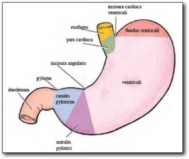
Fig. 1.1. SCHEMATIC Structure OF the stomach.
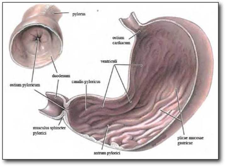
Fig. 1.2. STRUCTURE OF THE stomach.
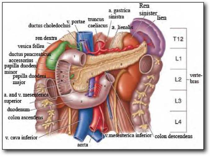
Fig. 1.3. Syntopy of the duodenum.
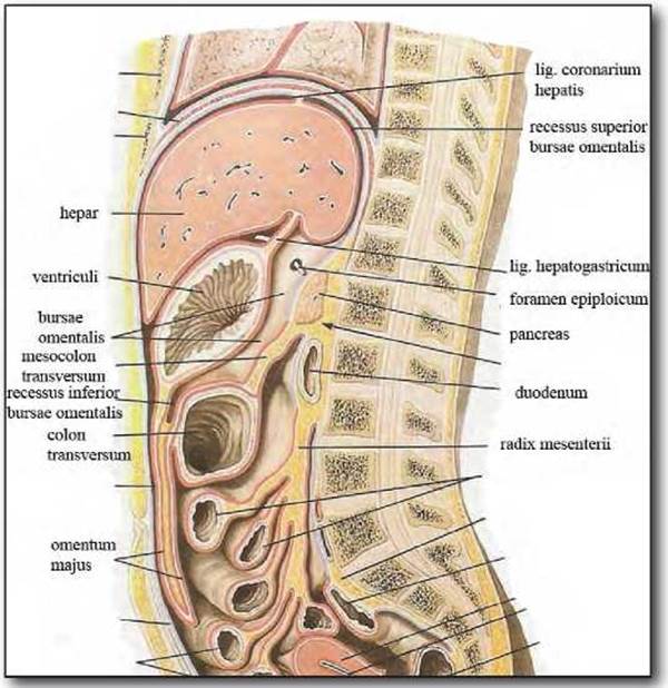
Fig. 1.4. Ligamentous apparatus of the stomach
The wall of the duodenum consists of three layers: the mucosa, muscularis, and serosa (tunica mucosa, tunica muscularis, tunica serosa).
On the medial wall of the descending part of the duodenum is the major duodenal papilla (papilla duodeni major), where the common Bile duct (ductus choledochus) and the pancreatic duct open. Proximal to the papilla duodeni major is a second, smaller papilla—the minor duodenal papilla (papilla duodeni minor), where the accessory pancreatic duct opens.
Ligamentous apparatus.
The ligaments surround the stomach in a continuous ring and play an important role in its fixation. Within the ligaments, between the peritoneal layers, lie adipose tissue, Blood and Lymphatic vessels, Lymph Nodes, and nerve branches. The following ligaments of the stomach are distinguished (Fig. 1.4).
The hepatogastric ligament, lig. hepatogastricum, is a peritoneal fold stretched between the porta hepatis and the lesser curvature of the stomach; on the left, it extends to the abdominal part of the esophagus, and on the right, it continues as the hepatoduodenal ligament. Together, these two ligaments form the lesser omentum, omentum minus.
Within the hepatogastric ligament, the pars tensa is distinguished, which lies closer to the cardiac part of the stomach. The ligament is trapezoidal in shape; its width near the lesser curvature is 10-19 cm, near the porta hepatis it is 5-10 cm, and its length from the porta hepatis to the gastric angle is 6-14 cm. Adipose tissue is located between the peritoneal layers of the hepatogastric ligament, with its layer thinning toward the Liver.
Sometimes, the adipose tissue between the layers of the hepatogastric ligament near the porta hepatis is almost completely absent. In such cases, the caudate lobe of The Liver and part of the body of the pancreas are visible through its translucent layers.
In the upper part of the ligament, the hepatic Branches of the anterior vagal trunk course. At the base of this ligament, the left gastric artery accompanied by its corresponding vein is located in some cases, though more often these vessels lie on the gastric wall along the lesser curvature. Additionally, an accessory hepatic artery arising from the left gastric artery is frequently (in 16.5% of cases) located in the pars tensa of the ligament. In some cases, the main trunk of the left gastric vein or its tributaries passes here.
During gastric mobilization along the lesser curvature near the porta hepatis (for Gastric Cancer), the potential presence of an accessory left hepatic artery must be considered, as dividing it can lead to Necrosis of the left hepatic lobe or a portion of it. On the right, near the Base of the hepatogastric ligament, the right gastric artery courses, accompanied by its corresponding vein.
The gastrophrenic ligament, lig. gastrophrenicum, is located to the left of the esophagus, between the fundus of the stomach and the Diaphragm. The ligament is shaped like a triangular plate and consists of a single peritoneal layer, at the base of which lies loose Connective Tissue. On the left, the ligament transitions into the Superficial layer of the gastrosplenic ligament, and on the right, onto the anterior circumference of the esophagus. Its length from the esophagus to the gastrosplenic ligament ranges from 2 to 7 cm, averaging 4 cm. The reflection of the Peritoneum from the gastrophrenic ligament onto the anterior esophageal wall and onto the hepatogastric ligament is termed the phrenicoesophageal ligament, lig. phrenicooesophageum.
The gastrosplenic ligament, lig. gastrolienale, is stretched between the fundus of the stomach, the upper part of the greater curvature, and the hilum of the Spleen, lying inferior to the gastrophrenic ligament. It consists of two peritoneal layers, between which course the short gastric Arteries accompanied by their corresponding Veins. The ligament is trapezoidal in shape; near the superior pole of the spleen, its width is 0.5-4 cm, and near the inferior pole, it is approximately three times wider. The width of the ligament between the superior and inferior poles of the spleen ranges from 6 to 12 cm. Continuing downward, it transitions into the gastrocolic ligament.
The gastrocolic ligament, lig. gastrocolicum, consists of two peritoneal layers. It represents the initial part of the greater omentum and is situated between the greater curvature of the stomach and the transverse colon. This is the widest ligament, extending as a band from the inferior pole of the spleen to the pylorus. In most cases, its width is 25-36 cm, and its length is 5-8 cm; in some instances, the length of the ligament reaches 12 cm. The ligament is loosely attached to the anterior circumference of the transverse colon, as well as to the tenia omentalis. The right and left gastro-omental arteries course within it. The left gastro-omental artery enters the ligament from beneath the pylorus and is therefore in close proximity to its wall. In the mid-region of the stomach, this artery lies 0.5-3 cm below the greater curvature. Near the anterior pole of the spleen, the left gastro-omental artery is located 3-10 cm from the gastric wall and approaches the greater curvature in an oblique direction. In the region of the pyloric part of the stomach, the gastrocolic ligament is often fused with the superior layer of the transverse mesocolon. In such cases, the right gastro-omental artery lies in close proximity to the middle colic artery. This anatomical feature must be taken into account during gastric resection, as mobilizing the pyloric part of the stomach and ligating the right gastro-omental artery may lead to accidental division of the middle colic artery, thereby compromising the arterial Blood supply to the transverse colon, which typically results in necrosis of its wall.
The gastropancreatic ligament, lig. gastropancreaticum, is located between the superior border of the pancreas, the cardiac part, and the fundus of the stomach. It is clearly visualized if the gastrocolic ligament is divided and the stomach is retracted anteriorly and superiorly. It can also be palpated through the hepatogastric ligament. The length of the ligament is variable, ranging from 2 to 5 cm. On the right, it consists of two peritoneal layers that form the gastropancreatic fold, plica gastropancreatica, while its left portion, consisting of a single peritoneal layer, reflects onto the posterior surface of the cardia and fundus of the stomach.
Within the free margin of the gastropancreatic ligament lie the initial segment of the left gastric artery and its accompanying vein, as well as Lymphatic vessels and gastropancreatic lymph nodes. Additionally, pancreaticosplenic lymph nodes are located at the base of the ligament along the superior border of the pancreas.
The pyloropancreatic ligament, lig. pyloropancreaticum, is a peritoneal fold located between the pylorus and the right part of the body of the pancreas. It is triangular in shape, with one side attached to the posterior surface of the pylorus and the other to the anteroinferior surface of the pancreatic body; the free margin of the ligament is directed to the left. Occasionally, this ligament is absent.
Small lymph nodes are concentrated within the pyloropancreatic ligament and may be involved in malignancies of the pyloric region of the stomach. Therefore, during gastric resection, this ligament must be completely excised along with these lymph nodes.
Between the gastropancreatic and pyloropancreatic ligaments lies the slit-like gastropancreatic opening. The shape and size of this opening depend on The Development of these ligaments. Sometimes, the ligaments are so well-developed that they overlap or fuse with each other, closing the gastropancreatic opening. Consequently, the cavity of the omental bursa is divided by the ligaments into two separate spaces. In such cases, if pathological contents (effusion, blood, gastric contents, etc.) accumulate in the omental bursa, they will be confined to one of these spaces.
The peritoneal layers of the ligaments surrounding the stomach reflect onto its anterior and posterior walls, making the stomach almost entirely covered by peritoneum. It lacks a peritoneal covering only along the lesser and greater curvatures, as well as on a portion of the posterior wall near the fundus and occasionally the cardia.
The bare area of the stomach along the lesser curvature extends from the right circumference of the abdominal esophagus to the pylorus. In the cardiac region, its width reaches 1.5–4 cm, gradually narrowing toward the pylorus to 0.3–0.5 cm.
The extraperitoneal area of the stomach along the greater curvature (1–2 cm wide) is bounded inferiorly by the layers of the gastrocolic ligament, and superiorly and to the left by the layers of the gastrosplenic ligament. Above the latter, it gradually widens (up to 3–4 cm) and extends onto the fundus of the stomach. Here, the extraperitoneal part of the stomach is bounded superiorly by the gastrophrenic ligament and inferiorly by the pancreatogastric ligament, extending to the esophagus and sometimes the cardia. Therefore, the dimensions of the extraperitoneal area on the posterior wall of the gastric fundus depend on the distance between the gastrophrenic and pancreatogastric ligaments. Sometimes these ligaments meet, forming a peritoneal fold up to 1–3 cm wide. In such cases, the extraperitoneal area in the fundus region is represented by a very narrow strip, only 0.3–0.5 cm wide.
The posterior wall of the cardiac part of the stomach and the posterior wall of the abdominal esophagus are covered by peritoneum only if the left part of the gastrophrenic ligament is adjacent to the peritoneum of the cardiac recess of the omental bursa. If these peritoneal layers do not meet, the posterior walls of the cardia and esophagus remain devoid of peritoneal covering.
Syntopy. The relations of the stomach to its surrounding Organs are highly variable. This is due to differences in the shape and size of the stomach, its degree of distension, and the varying shape and position of adjacent organs. The anterior wall of the stomach is in contact with the Inferior surface of the left lobe of the liver. If the stomach is small and located almost entirely in the left hypochondrium, the liver covers its entire anterior surface, or only a small part of the greater curvature projects from beneath it. In such cases, the majority of the anterior gastric surface lies adjacent to the transverse colon (especially when the latter is distended), or, less frequently, to the anterior abdominal wall, and sometimes to the loops of the Small Intestine that ascend into the upper compartment of the peritoneal cavity over the transverse colon and greater omentum. In rare cases, a highly distended sigmoid colon may lie adjacent to the anterior wall of the stomach.
The fundus of the stomach is adjacent to the diaphragm, the spleen, and the transverse colon. The pyloric part of the stomach may lie adjacent to the left, quadrate, or right lobes of the liver, as well as the Gallbladder. The pylorus usually contacts the quadrate lobe of the liver, less frequently only the left lobe, and rarely the right lobe. Superiorly and to the left, the anterior wall of the stomach is adjacent to the diaphragm and the transverse colon.
The posterior wall of the stomach is adjacent to the retroperitoneal organs and is separated from them by the omental bursa.
Throughout almost its entire extent, the posterior wall of the stomach is adjacent to the body and tail of the pancreas. In most cases, the omental tuber (tuber omentale) of the pancreatic body projects 2–4 cm above the lesser curvature and lies adjacent to the hepatogastric ligament. Slightly superior to the pancreas, the splenic vessels, the left Kidney, and the left suprarenal gland lie adjacent to the stomach. Only the superior pole of the left kidney contacts the gastric fundus. Slightly medial and superior to the left kidney, the left suprarenal gland lies adjacent to the cardiac part of the stomach. The pyloric part of the stomach is adjacent to the head of the pancreas. Near the greater curvature, the posterior wall of the stomach contacts the transverse mesocolon.
To the left, the stomach is adjacent to the gastric surface (facies gastrica) of the spleen, and inferiorly to the transverse colon and its mesocolon. Behind the stomach lie slit-like spaces: the pregastric bursa and the omental bursa.
The pregastric bursa is bounded anteriorly by the peritoneum lining the anterior abdominal wall and the diaphragm, posteriorly by the lesser omentum and the anterior wall of the stomach, and superiorly by the left lobe of the liver. To the right, it is separated from the hepatic bursa by the falciform ligament. The pregastric bursa is not closed and communicates freely with other compartments of the peritoneal cavity: inferiorly with the preomental space, to the left with the left paracolic gutter, and to the right with the hepatic bursa.
The omental bursa is bounded anteriorly by the lesser omentum, the posterior wall of the stomach, and the gastrocolic ligament; superiorly by the caudate lobe of the liver and the inferior surface of the diaphragm; posteriorly by the peritoneum covering the retroperitoneal organs: the pancreas, left suprarenal gland, superior pole of the left kidney, aorta, inferior vena cava, splenic artery and vein; and inferiorly by the transverse colon and its mesocolon. To the left, it extends to the spleen, where it is bounded by the gastrosplenic and splenophrenic ligaments, and to the right, it reaches the omental foramen. The width of the omental bursa cavity from the omental foramen to the splenic hilum ranges from 11 to 19 cm, averaging 16 cm, and its height is 8–14 cm, averaging 10 cm (Fig. 1.5).
The omental bursa is divided into several compartments. The portion of the bursa adjacent to the omental foramen is called the Vestibule of the omental bursa, vestibulum bursae omentalis. The superior compartment of the bursa is divided by the gastropancreatic fold, which runs obliquely upward, into two recesses: the superior recess, recessus superior omentalis, and the cardiac recess, recessus cardialis. The former is located to the right of the gastropancreatic fold, and the latter to its left. The superior recess extends deep upward, posterior to the caudate lobe of the liver, reaching the diaphragm; its right wall is formed by the peritoneum covering the inferior vena cava. The height of this recess corresponds to the length of the caudate lobe of the liver, which is 3–6 cm.
The cardiac recess is shallower; posterior to it lies the left suprarenal gland, anterior to it is the posterior wall of the stomach, and superiorly it reaches the cardia or the abdominal esophagus.
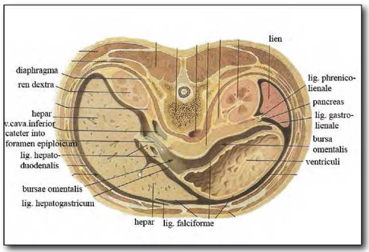
Fig. 1.5. Structure of the omental bursa.
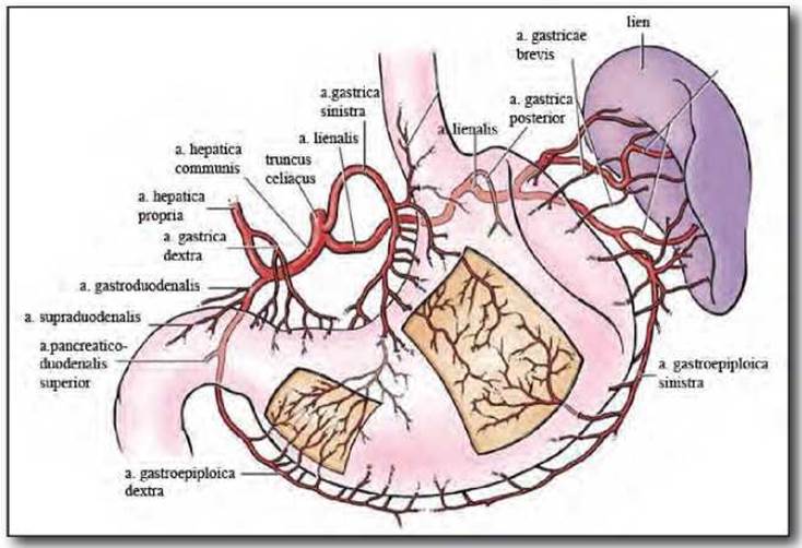
Fig. 1.6. Blood supply of the stomach.
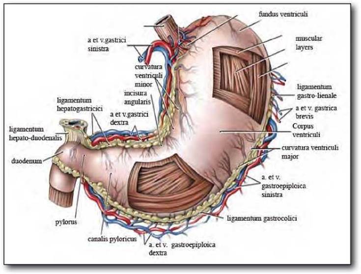
Fig. 1.7. Arterial Blood supply and venous drainage of the stomach.
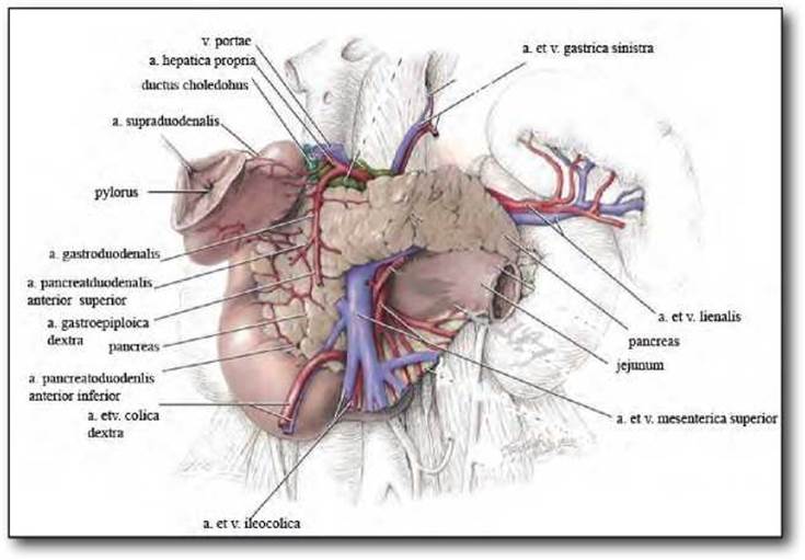
Fig. 1.8. Blood supply of the duodenum.
In the lower compartment of the omental bursa, above the transverse mesocolon, There are also two recesses: the inferior recess, recessus inferior omentalis, and the splenic recess, recessus lienalis. The former, recessus inferior omentalis, is bounded anteriorly by the posterior wall of the pyloric part of the stomach and the gastrocolic ligament, posteriorly by the parietal peritoneum covering the head of the pancreas, and inferiorly by the transverse mesocolon. The latter, recessus lienalis, is located near the inferior pole of the spleen; it is bounded by the gastrosplenic and phrenicosplenic ligaments, as well as the transverse mesocolon.
Inferiorly, the omental bursa communicates with a slit-like space located between the layers of the greater omentum (the cavity of the greater omentum). However, this space sometimes obliterates due to the fusion of the layers of the greater omentum.
To the right, the omental bursa communicates with the peritoneal cavity via the omental foramen, which is bounded anteriorly by the hepatoduodenal ligament, posteriorly by the hepatorenal ligament and the inferior vena cava running within it, superiorly by the caudate process of the liver, and inferiorly by the peritoneal reflection from the posterior surface of the hepatoduodenal ligament and the superior part of the duodenum onto the inferior vena cava.
Normally, the omental foramen easily admits 1-3 fingerbreadths. Occasionally (in 17%), inflammatory processes lead to its complete closure, resulting in the Isolation of the omental bursa. This circumstance must be taken into account in cases of perforated ulcers located on the posterior wall of the stomach, as the contents escaping through the gastric perforation will be confined solely to the omental bursa.
Blood supply. The blood supply to the stomach is provided by the left and right gastric arteries, the left and right gastro-omental arteries, and the short gastric arteries. All of these vessels belong to the System of the celiac trunk (Fig. 1.6, 1.7).
The celiac trunk, truncus coeliacus, arises from the aorta at the level of the XII thoracic to I lumbar vertebra and is a short arterial trunk 0.5-3 cm in length (averaging 1.7 cm) and 0.8-1.2 cm in diameter.
The celiac trunk divides into three branches: the left gastric, common hepatic, and splenic arteries. In some cases, other branches may arise from the truncus coeliacus: the inferior phrenic, superior mesenteric, accessory hepatic, or inferior pancreaticoduodenal artery.
All branches of the truncus coeliacus initially lie deep within the retroperitoneal space; then, coursing away from their origin, they branch out in different directions.
The left gastric artery, a. gastrica sinistra, is the largest artery of the stomach, with its diameter reaching 0.3-0.5 cm. It courses to the left from its origin, initially running within the gastropancreatic fold, and then reaches the lesser curvature approximately 3-4 cm below the gastroesophageal junction, running along it. Therefore, a distinction is made between the parietal, or ascending, part of the left gastric artery, which passes through the gastropancreatic fold, and its distal, or descending, part, which runs along the lesser curvature. This division of the artery has practical clinical significance, as the descending part of the a. gastricae sinistrae is ligated during a standard subtotal gastrectomy, whereas its ascending part is ligated during an extended resection or total gastrectomy. The length of the ascending part of the artery ranges from 2.5 to 4.5 cm, averaging 3 cm.
Not infrequently (in 19%), an accessory hepatic artery arises from the left gastric artery and courses to the liver within the lesser omentum.
Running along the lesser curvature, the left gastric artery gives off branches to the cardiac part of the stomach and then divides into two trunks (anterior and posterior). These trunks give off 4-5 branches to the respective walls of the stomach. Anastomosis between the left and right gastric arteries is most commonly formed by the posterior trunk, and occasionally by both trunks or the anterior trunk alone. Sometimes, the left and right gastric arteries do not anastomose. Figure 1.6 illustrates the branching patterns of the gastric arteries along the lesser curvature.
The common hepatic artery, a. hepatica communis, courses to the right from its origin and runs along the superior border of the pancreas, sometimes covered by it. At the level of the pylorus or slightly to its right, this artery divides into the hepatic artery proper and the gastroduodenal artery.
The hepatic artery proper runs within the hepatoduodenal ligament. The right gastric artery, a. gastrica dextra, most commonly (in 70%) arises from this artery or its left branch, coursing toward the lesser curvature from the pyloric side. In some cases, the right gastric artery may arise from the common hepatic artery or the gastroduodenal artery. Its diameter is 2-3 times smaller than that of the left gastric artery.
The gastroduodenal artery courses downward and crosses the posterior wall of the duodenal bulb near the pylorus; the posterior superior pancreaticoduodenal artery arises from its proximal portion. At the level of the inferior border of the pylorus, the gastroduodenal artery divides into the right gastro-omental and anterior superior pancreaticoduodenal arteries. The former, a. gastro-epiploica dextra, gives off branches to the greater omentum and the stomach, and anastomoses within the gastrocolic ligament with the left gastro-omental artery.
The splenic artery, a. lienalis, runs along the superior border of the pancreas. In the region of the pancreatic tail, it emerges from the gland and, near the splenic hilum, divides into 2-3 large branches (superior and inferior, or superior, middle, and inferior). The left gastro-omental artery, a. gastro-epiploica sinistra, arises from the main trunk of the a. lienalis or its primary branches. It runs in the lower part of the gastrosplenic ligament, gives off branches to the greater omentum, and reaches the greater curvature of the stomach on the left, 3-10 cm from the splenic hilum, subsequently coursing within the gastrocolic ligament.
Thus, along the greater curvature, the anastomosing left and right gastro-omental arteries form an arterial arcade from which 12-15 pairs of branches arise to supply the anterior and posterior walls of the stomach. This arcade is located within the gastrocolic ligament at a distance of 0.5-3 cm from the greater curvature of the stomach. Not infrequently, the left and right gastro-omental arteries do not anastomose with each other. In such cases, the left gastro-omental artery gives off 2-3 branches to the stomach wall, while the right gastro-omental artery branches out predominantly within the gastrocolic ligament.
The fundus of the stomach is supplied by the short gastric arteries, aa. gastricae breves. Their number is variable, ranging from one to six. The short gastric arteries arise from the splenic artery near the splenic hilum, from its main trunks, from the arterial branches entering the splenic parenchyma, or from the left gastro-omental artery.
Coursing toward the fundus of the stomach, they run within the gastrosplenic ligament, sometimes dividing into 2-3 branches within it.
In addition, the fundus of the stomach is supplied by the posterior gastric artery, which arises from the splenic artery 4-5 cm from its origin. It courses vertically upward behind the peritoneum covering the left suprarenal gland and reaches the fundus of the stomach in the left part of the gastropancreatic fold.
Occasionally, a branch of the left phrenic artery also participates in the blood supply to the stomach, coursing to the fundus within the phrenicogastric ligament.
In some cases, accessory arterial branches supply the cardia or the superior part of the lesser curvature of the stomach. They arise from the left branch of the hepatic artery or from an accessory hepatic artery and, coursing toward the stomach, run between the peritoneal layers of the hepatogastric ligament.
Thus, the blood supply to the stomach is provided by constant and accessory gastric arteries. The constant arteries include the left and right gastric arteries, the left and right gastro-omental arteries, the short gastric arteries, and the posterior gastric artery (a branch of the splenic artery); the accessory arteries include branches arising from the left hepatic, accessory hepatic, or left phrenic arteries.
The arterial Vessels of the stomach anastomose extensively with one another, forming a well-developed intramural arterial network.
The gastric veins belong to the portal Venous system. The left and right gastric veins, V. gastrica sinistra et dextra, run along the lesser curvature. The former accompanies the left gastric artery and its branches. Coursing downward, the left gastric vein runs within the gastropancreatic fold, where it lies anterior to or slightly below the corresponding artery; it then passes to the posterior surface of the pancreas, crossing either anteriorly or posteriorly to the common hepatic or, less frequently, the splenic artery, and most commonly drains into the portal or splenic vein, or less frequently, into the junction of the superior mesenteric and splenic veins. Superiorly, the left gastric vein anastomoses with the esophageal veins. This anastomosis connects the portal and SUPERIOR VENA CAVA systems and is of great clinical importance for collateral BLOOD FLOW IN portal Hypertension.
The right gastric vein drains into the portal vein superior to the pancreas within the hepatoduodenal ligament. Occasionally, it courses as a separate trunk directly into the liver parenchyma.
Along the greater curvature lie the right and left gastro-omental veins, v. gastro-epiploica dextra et sinistra, which accompany the corresponding arteries. The right gastro-omental vein joins the middle colic and anterior superior pancreaticoduodenal veins to form a common trunk, which drains into the superior mesenteric vein near the incisurae pancreatis; in some cases, the v. gastro-epiploica dextra forms a common trunk only with the anterior superior pancreaticoduodenal vein.
The left gastro-omental vein drains into the splenic vein or its tributaries near the splenic hilum.
The short gastric veins, vv. gastricae breves, which accompany the corresponding arteries, run within the gastrosplenic ligament and drain into the trunks of the splenic vein or the left gastro-omental vein.
Pyloric veins are located at the boundary between the stomach and the duodenum. The degree of development and number of these veins are variable. In some cases, There is a single well-developed pyloric vein lying in the pyloric sulcus, which drains superiorly into the portal vein and inferiorly into the right gastro-omental vein. In other cases, 3-5 poorly developed venous trunks run along the superior and inferior semicircles of the pylorus. Sometimes, pyloric veins are completely absent.
The gastric veins generally accompany the corresponding arteries; they anastomose extensively with each other, forming a continuous venous network that ensures blood drainage from the gastric walls in various directions.
Lymphatic system. The subserosal and submucosal layers of the gastric wall are extremely rich in lymphatic capillaries. It is through these channels that microscopic, subclinical spread and metastasis of tumors beyond their macroscopic boundaries occur. Therefore, when performing surgery for a malignant gastric ulcer, it is crucial to perform an intraoperative frozen section biopsy of the margins of the resected stomach to confirm the absence of microscopic tumor spread and ensure the radicality of the resection. Submucosal lymphatic plexuses and capillaries extend toward both the esophagus and the duodenum. Consequently, micrometastasis can occur in both proximal and distal directions toward these two organs.
The structure of the gastric lymphatic system is largely identical to the venous system. Lymphatic drainage from the antral part of the greater curvature of the stomach goes to the subpyloric and greater omentum lymph nodes. Lymphatic drainage from the proximal part of the greater curvature occurs via the pancreaticosplenic lymph nodes to the splenic and gastrocolic groups of lymph nodes. Lymphatic drainage from the upper part of the lesser curvature goes to the superior group of lymph nodes surrounding the left gastric artery, and partially to the left gastric, paracardial, and celiac trunk groups. Suprapyloric lymph nodes drain the antral region of the lesser curvature of the stomach.
Four lymphatic drainage basins of the stomach are distinguished. Basin I (inferior gastric): lymph drains into the subpyloric and omental lymph nodes. Basin II (splenic): lymph drains to the pancreaticosplenic lymph nodes. Basin III (superior gastric): lymph drains to the anterior gastric lymph nodes. Basin IV (hepatic): lymph drains to the suprapyloric lymph nodes (Fig. 1.9).
Lymph from all four lymphatic basins of the stomach flows to the celiac trunk lymph node group and subsequently drains into the Thoracic duct. Thus, the regional lymph nodes for the stomach are those located along the lesser (1,3,5) and greater (2,4a,4b,6) curvatures, along the left gastric (7), common hepatic (8), splenic (10,11) arteries, and truncus celiacus (9), as well as the lymph nodes of the hepatoduodenal ligament (12). Retropancreatic, mesenteric, and para-aortic lymph nodes are not regional lymphatic collectors (Fig. 1.10).
Based on the JRSGC (1998) studies, the lymph node groups described above form four consecutive stages of metastasis—from N1 to N4. The First stage (N1) includes perigastric lymph collectors located in the ligamentous apparatus of the stomach and along the a.gastrica sinistra. The Second Stage (N2) includes lymph nodes along the arterial trunks (common hepatic artery, splenic artery, splenic hilum, truncus celiacus). The Third Stage (N3) includes lymph nodes of the hepatoduodenal ligament, retropancreaticoduodenal nodes, and the ROOT of the mesentery of the transverse mesocolon. The Fourth Stage (N4) includes para-aortic LYMPH NODES AND nodes along the superior mesenteric artery.

Fig. 1.9. Lymphatic system of the stomach
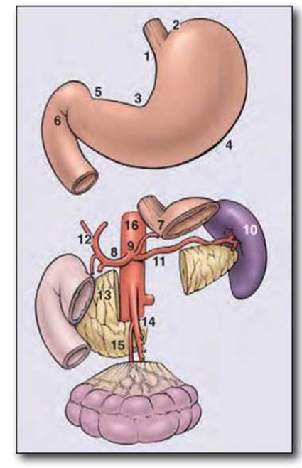
Fig. 1.10. Localization and anatomical Classification of gastric lymph node groups. Involvement of lymph nodes along the lesser or greater curvature of the stomach indicates N1 spread of the pathological process (groups 1-6), involvement of lymph nodes along the truncus celiacus and its three branches indicates N2 (7-11), N3 (12-14), N4 (15, 16). N1: 1, right paracardial; 2, left paracardial; 3, lesser curvature; 4, greater curvature of the stomach; 5, suprapyloric; 6, subpyloric. N2: 7, left gastric artery; 8, common hepatic artery; 9, truncus celiacus; 10, splenic hilum; 11, splenic artery. N3: 12, hepatic pedicle; 13, retropancreatic; 14, root of the mesentery. N4: 15, middle colic artery; 16, para-aortic.
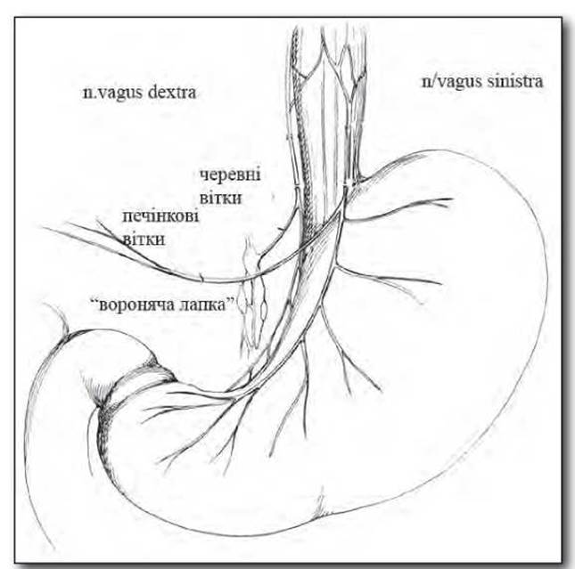
Fig. 1.11. Innervation of the stomach by the vagus nerves.
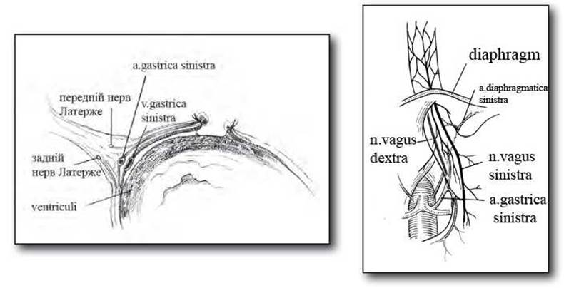
Fig. 1.12. Topography of the vagus nerves along the lesser curvature of the stomach.
Based on the above lymph node classification, the involvement of N1-2 lymph collectors in the oncological process is considered regional metastasis, whereas the involvement of N3-4 collectors is classified as distant metastasis (MILym).
Innervation. The stomach is innervated by sympathetic and parasympathetic nerves. Sympathetic nerves originate from the celiac plexus, plexus coeliacus, and its derivatives (plexus lienalis, plexus mesentericus superior). These nerves initially run along the greater and lesser curvatures, surrounding the arterial and venous vessels, and then enter the gastric wall. Parasympathetic innervation is provided by branches of the vagus nerves, which enter the abdominal cavity alongside the esophagus, usually as two trunks—truncus vagalis anterior et posterior—or less frequently, as separate branches.
The vagal trunks are located on the corresponding surfaces of the abdominal esophagus. The anterior vagal trunk is located anterior to the esophagus in 68% of cases, runs along its left wall in 30%, and is adjacent to the left part of the posterior esophageal wall in 2% of cases. In the vast majority of cases, the posterior trunk runs as a single trunk along the posterior wall of the esophagus on the right. A branch innervating the gastric fundus arises in the esophageal region. This branch can often be found in the region of the angle of His. In 1969, Grassi identified a separate branch of the posterior Vagus nerve that arises above the diaphragm and runs obliquely along the posterior wall of the esophagus. This is the so-called “criminal nerve of Grassi” (Fig. 1.11).
After giving off branches within the tissue of the lesser omentum, the vagal trunks run near (1-2 cm) the lesser curvature along its anterior and posterior surfaces. Here, they are often referred to as the nerves of Latarjet. The nerve of Latarjet terminates on the anterior surface of the gastric antrum, where 2-3 small branches penetrate its wall, innervating the pyloric and antral Regions of the stomach. This branching site of the nerve of Latarjet is commonly called the “crow's FOOT” of the nerve. The pyloric region of the stomach is additionally innervated by an accessory branch that runs within the hepatogastric ligament and accompanies the right gastric artery (Fig. 1.12).
From the anterior vagal trunk, the majority of fibers run to the cardia of the stomach and the lesser curvature. In addition, it gives off a hepatic branch that runs within the hepatogastric ligament to the left lobe of the liver.
The posterior vagal trunk runs near the left gastric artery and divides into right and left branches. The right branch gives off fibers to the celiac plexus, stomach, head of the pancreas, small intestine, and phrenic plexus. The left branch of the posterior vagal trunk sends fibers to the stomach, body of the pancreas, spleen, small intestine, and inferior mesenteric plexus. The posterior vagal trunk lies within the adipose tissue of the gastropancreatic fold along its entire length.
The vagal trunks anastomose with each other, as well as with branches arising from the celiac plexus.
The primary Functions of the stomach are the chemical and mechanical Processing of food, its storage, and its emptying into the intestine. The stomach also participates in Intermediary METABOLISM, hematopoiesis, Water-electrolyte balance, and the maintenance of acid-base balance (ABB).
The digestive function proper of the stomach is mediated by gastric juice secreted by the gastric glands. Gastric gland Cells secrete eight pepsinogen fractions, which form two immunologically heterogeneous groups. Group I pepsinogens are secreted by the fundic glands, while Group II pepsinogens are secreted by the antral glands. Gastric juice exhibits proteolytic activity over a wide pH range, with two distinct pH optima: at pH 1.5–2.0 and 3.2–3.5. At the first optimum, proteolysis is carried out by pepsins, and at the second, by gastricsin, which differs in its Amino Acid Composition, molecular weight, and several other properties. Under physiological conditions, The ratio of Pepsin to gastricsin ranges from 1:1.5 to 1:6. Pepsin and gastricsin account for 95% of the proteolytic activity of gastric juice. Gastric juice also exhibits minor lipolytic and amylolytic activities.
Chief Cells of the gastric glands are primarily stimulated by the vagus nerves via acetylcholine. Reflex stimulation of gastric enzyme secretion is also mediated by gastrin. Cholinergic stimulation increases the responsiveness of chief cells to gastrin. Histamine enhances enzyme secretion, though less potently than gastrin and the vagus nerves. Secretin, while inhibiting gastrin-stimulated Hydrochloric acid secretion, increases enzyme secretion. Cholecystokinin and pancreozymin exert a similar effect.
Parietal cells secrete hydrochloric acid, which participates in pepsinogen activation and the ESTABLISHMENT OF THE optimal pH for gastric enzyme activity. Under physiological conditions, parietal cells are stimulated by gastrin and histamine via cholinergic nerve fibers, and inhibited by secretin and cholecystokinin (pancreozymin). The secretory activity of the gastric glands is regulated by reflex and humoral mechanisms. Based on the mechanisms of gastric gland stimulation, a complex-reflex phase and a neurohumoral phase are distinguished. However, it is now established that there is no fundamental difference between them, as reflex stimulation is also mediated through humoral pathways (histamine, gastrin), and neural mechanisms modulate the sensitivity of the gastric glands to humoral agents. The vagus nerve is the primary mediator of central influence on the gastric glands. Gastrin is released from gastrin-producing cells of the antral mucosa both As a result of central influence (via the vagus nerves) and due to the action of Hydrolysis products of extractives (such as meat, ethanol, caffeine, etc.) on mechanoreceptors and chemoreceptors. When acidic solutions act on the antral mucosa, gastrin release is inhibited in proportion to the increase in acidity (at pH 1.0, gastrin release ceases completely). Following the passage of gastric chyme into the duodenum, Hormones are produced there that play a crucial role in regulating The activity of the hepatobiliary system, pancreas, stomach, and intestine. The phase of secretion regulated by the duodenum and small intestine is called the intestinal phase. Bypassing the transit of gastric contents through the duodenum increases the secretory response of the gastric glands to numerous stimulants. The primary inhibitors of gastric acid secretion are secretin and cholecystokinin (pancreozymin); however, several other Gastrointestinal Hormones also participate in this process.
Gastric motor activity ensures the storage of food, its mixing with gastric juice, and the portioned emptying of chyme into the duodenum. The reservoir function is primarily performed by the body and fundus of the stomach, whereas the emptying function is carried out by its pyloric region.
The REGULATION OF GASTRIC motility is mediated by neural and humoral mechanisms. Stimulation of the vagus nerves increases gastric motor activity, whereas stimulation of the sympathetic nerves decreases it (epinephrine has a similar effect). The enterogastric reflex plays a leading role in regulating gastric emptying: stimulation of the mechanoreceptors and chemoreceptors of the duodenum and small intestine inhibits gastric motility and emptying.
Thus, the secretory and motor functions of the stomach are closely interrelated, possessing a complex system of regulation and feedback-based self-regulation, thereby ensuring optimal conditions for the gastric phase of Digestion in close coordination with the neuro-reflex and intestinal phases.
The duodenum, in conjunction with the pancreas, liver, and biliary system, plays a leading role in the secretory, motor, and emptying functions of the digestive tract. Here, gastric chyme undergoes further mechanical and chemical processing. Pancreatic juice and bile are secreted into the intestinal lumen, altering the pH of its contents and, together with intestinal juice, enabling the further hydrolysis of nutrients by proteolytic, amylolytic, and lipolytic Enzymes. Normally, the pH in the duodenum ranges from 4.0 to 8.0.
Pancreatic secretion contains hydrolytic enzymes that ensure the digestion of Proteins, fats, and CARBOHYDRATES. Some enzymes are secreted in an active state (amylase, lipase, and Nucleases), while others (Proteolytic Enzymes, Trypsin, Chymotrypsin, and phospholipase A) are secreted in the form of zymogens. The latter account for about 70% of the total protein content in pancreatic secretion.
Trypsin activates the zymogens of almost all pancreatic enzymes. The activation of trypsin itself can occur either autocatalytically (under the action of trypsin itself) or under The Influence of enterokinase present in the duodenal contents. Activated trypsin breaks down proteins. Carbohydrates are cleaved by pancreatic amylase to maltose.
Lipase acts on emulsified fats and is capable of breaking down water-insoluble triglycerides. Pancreatic Ribonuclease and deoxyribonuclease break down Nucleic Acids into NUCLEOTIDES.
The secretion of hepatocytes, bile, is released into the lumen of the duodenum and facilitates the emulsification of fats. Bile acids promote their breakdown by activating Pancreatic and Intestinal lipases. Bile plays an important role in the absorption of Fatty acids, carotene, Vitamins D, F, A, E, K, Amino Acids, Cholesterol, and calcium salts. It increases the tone and enhances the peristalsis of the duodenum. Additionally, bile exerts a bacteriostatic effect and participates in membrane digestion.
The secretory activity of the duodenum is regulated by neural and humoral mechanisms. Stimulation of the vagus and sympathetic nerves enhances the secretion of Brunner's glands. Cholinomimetics and acetylcholinesterase inhibitors stimulate secretion, whereas anticholinergic and adrenomimetic agents inhibit it. Enterokinin is also produced in the duodenal mucosa, enhancing the secretion and sucrase activity of intestinal juice.
Thus, the functions of the duodenum, like those of the stomach, are complex and diverse. Its secretory and motor activities are regulated by numerous influences and are closely linked to the function of other Organs of the digestive tract based on the feedback principle. This must be taken into account when choosing a Surgical Treatment method for PEPTIC ULCER DISEASE, since the exclusion of the stomach or duodenal function, combined with insufficient compensatory capacity of the body, can lead to serious consequences, such as post-gastrectomy syndrome or Disorders of the operated stomach.
Last update: 11/08/2026
Editorial and Educational Adaptation: This material has been compiled based on the primary/original source text. The project team performed an editorial review, corrected technical inaccuracies, structured sections, and adapted the content for an educational format.
What was processed:
- elimination of formatting defects (OCR errors, structural breaks, corrupted characters);
- editorial organization of content;
- standardization of terminology in accordance with academic sources;
- verification of factual statements against the original source text.
All mentions of the author, publication year, and origin of the primary text have been preserved in accordance with the source.