BASICS OF MEDICAL BIOLOGY - 2012
Chromosomal diseases. Cytogenetic method of their study. Hereditary diseases and their classification
Depending on the interplay between heredity and the environment, all diseases can be divided into three groups: 1) Hereditary diseases; 2) Diseases with Hereditary predisposition (multifactorial); and 3) non-hereditary diseases. Hereditary diseases are conditions caused by Gene, chromosomal, or genomic Mutations. The manifestation of a mutation's pathological effect is practically independent of the environment, which can only alter symptom severity and the clinical course. Diseases with hereditary predisposition develop in individuals with a specific genotype under the Influence of Environmental factors. Non-hereditary diseases are caused by external environmental factors (such as trauma, Burns, or infectious diseases). However, even in these conditions, heredity can influence the course of the pathological process.
The term "hereditary diseases" should be distinguished from "congenital diseases" and "familial diseases," as they are not synonymous. Congenital conditions include all diseases present at birth, which may be caused by either hereditary or non-hereditary factors. Some hereditary diseases, such as hemophilia or Achondroplasia (shortening of the long tubular bones), manifest at birth and are therefore congenital. Other hereditary diseases, such as myopathies, progeria (premature Aging), which appear in adolescence, or Huntington's disease (with an average onset at 38–40 years of age), are not congenital in the full sense of the word. Familial diseases include all conditions (both hereditary and non-hereditary) that occur among members of the same family. Non-hereditary familial diseases arise from exposure to a shared harmful factor within that household (for example, occupational hazards).
Both genetic and clinical classifications of hereditary diseases are recognized. The genetic Classification is based on the etiological principle, namely the type of mutation and The Nature of the interaction with the environment. All hereditary pathology can be divided into 5 groups: 1) gene disorders; 2) Chromosomal Disorders; 3) diseases with hereditary predisposition (multifactorial); 4) somatic Cell Genetic Disorders; and 5) disorders of maternal-fetal genetic incompatibility. Strictly speaking, hereditary diseases are divided into two major groups: gene and chromosomal disorders.
Gene disorders are diseases caused by Gene Mutations that are transmitted across generations according to Gregor Mendel's Laws. Chromosomal disorders are caused by chromosomal and genomic mutations. Most chromosomal disorders resulting from aneuploidy are not inherited at all (having a lethal effect), whereas structural chromosomal rearrangements are transmitted with additional recombinations that occur during the Meiosis of the mutation carrier. Diseases with hereditary predisposition can be monogenic or polygenic. Their realization requires not only the appropriate genetic constitution of the individual, but also a factor or a complex of environmental factors that act as triggers in The Development of the pathology. Somatic cell genetic disorders are associated with The Emergence of specific Chromosomal aberrations in somatic Cells during oncological malignancies, leading to the activation of oncogenes. These include retinoblastoma and Wilms' Tumor (Kidney Cancer). Maternal-fetal genetic incompatibility disorders develop As a result of the mother's Immune Response against fetal Antigens (e.g., hemolytic disease of the newborn).
Hereditary pathology is so diverse that it is encountered in the practice of physicians across all specialties. Accordingly, a clinical classification of hereditary diseases exists, based on a systemic and organ-specific principle. It distinguishes: Hereditary diseases of The Nervous system; hereditary diseases of Internal Organs; hereditary Diseases of the Skin; and hereditary diseases of the eyes and other organs. There are very few hereditary diseases in which only a single system is selectively affected. Most hereditary disorders manifest as a complex of pathological signs—syndromes (such as Down syndrome or Klinefelter syndrome). The principles of diagnosing hereditary diseases are based on clinical Diagnosis data, which are refined using Genetic Methods during Genetic Counseling.
It is crucial to emphasize the core meaning of the term hereditary diseases: the defining factor is not necessarily the direct Inheritance of the disease (although this occurs in many cases), but rather that the ROOT cause lies in a defect within the hereditary (genetic) apparatus of the cells of one or both parents.
Class="center">Chromosomal diseases
Chromosomal diseases encompass Various Forms of pathologies clinically manifested by Multiple developmental malformations, the GENETIC BASIS OF which consists of chromosomal mutations (changes in Chromosome Structure) or genomic mutations (changes in chromosome number).
Most chromosomal disorders typically arise as de novo mutations and are not inherited across generations. The phenotypic basis of chromosomal diseases is determined by disruptions in early embryonic development; thus, pathological alterations originating in the prenatal period lead to the elimination of the embryo or fetus, or define the clinical picture in the newborn (with the exception of certain sexual development disorders that appear during Puberty).
Several frequently occurring chromosomal diseases were described long ago as clinical syndromes of developmental disorders, well before their connection to chromosomal aberrations was established. These include Down syndrome (1866), Klinefelter syndrome (1942), and Turner syndrome (1925, 1938). The Link Between these diseases and abnormal chromosome numbers was proven only in 1959. To date, more than 500 chromosomal disorders involving changes in chromosome number and structure have been identified.
The Role of chromosomal pathology is significant in prenatal Embryonic and Fetal mortality (accounting for about 40%), and approximately 6% of stillborn infants exhibit chromosomal abnormalities. Among live newborns, 3–4 per 1,000 have a chromosomal pathology, and approximately 40% of children with congenital birth defects harbor chromosomal aberrations.
The emergence of chromosomal diseases is associated with nondisjunction of Chromosomes during the First and Second anaphase divisions of meiosis, as well as with chromosomal aberrations (deletions, duplications, translocations, and other structural defects). Chromosomal diseases arise de novo due to mutations in the Gametes of one of the healthy parents or in the zygote during early Cleavage stages. If the mutation occurs in the gametes, it results in the full form of the disease; if it occurs during zygote cleavage, it leads to a mosaic form, in which some cells possess a normal karyotype while others harbor the mutation. Mosaic organisms may feature two, three, or more distinct cell clones. The pathology depends on the proportion of altered cells and the Nature of the mutation (whether involving autosomes, gonosomes, or partial monosomies/trisomies). In the full form, chromosomal changes are present in all Cells of the offspring. Unlike gene mutations, chromosomal mutations involve a much larger volume of genetic material and are characterized by multiple lesions resulting in lethality and Congenital Malformations. Patients with chromosomal diseases occupy nearly 25% of hospital beds worldwide. Cytogenetic analysis is employed to diagnose chromosomal diseases, as quantitative and structural chromosomal abnormalities are visible under a Microscope.
Genomic mutations associated with an increase or decrease in haploid chromosome sets are incompatible with human life. Only heteroploidies are observed clinically—trisomies, less commonly tetra- and pentasomies, and a single variant of monosomy; nullisomy is incompatible with life. Chromosomal diseases are divided into those caused by changes in autosome number and those associated with abnormalities in sex chromosomes. Examples of autosomal number disorders include Down syndrome, Edwards syndrome, and Patau syndrome.
Down syndrome (trisomy 21). The clinical picture of this syndrome was first described in 1866 by the English physician J.L. Down, who termed the condition "mongolian idiocy." In 1959, the French scientist J. Lejeune discovered an extra chromosome 21 in the karyotype of affected individuals. Their karyotypes are 47,XX,+21 or 47,XY,+21. The incidence is 1:1,100, and in some regions 1:700–1:800 newborns. The risk of having a child with Down syndrome increases with maternal age. Sex, race, geography, and population differences do not affect the frequency of its occurrence. The complex of congenital malformations characteristic of Down syndrome gives rise to the clinical impression that "all affected children look like they belong to the same family."
Clinical diagnostic features include short stature, varying degrees of intellectual disability, craniofacial anomalies (upslanted palpebral fissures, short neck, epicanthic folds, flat face, small short Nose, large Tongue, and small deformed ears, as shown in the figure). Muscle hypotonia, joint hypermobility, a single transverse palmar crease, and clinodactyly of the fifth finger are also characteristic. Congenital malformations of internal organs (such as The Heart) and reduced Immunity are frequently the cause of death in these children.
The cytogenetic Variants of the syndrome are diverse. The majority of cases (94%) comprise full trisomy 21 resulting from meiotic nondisjunction, with maternal nondisjunction accounting for 80% and paternal for 20% of cases. Approximately 4% of patients have a translocation form (where chromosome 21 is typically translocated onto chromosome 13 or 22), and 2% exhibit mosaicism due to mitotic nondisjunction, where one population of cells has a normal chromosome count (46) and another is aneuploid (47). The translocation form is independent of maternal age, carrying a high recurrence risk for affected children in the family.
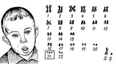
Fig. 33. Down syndrome (trisomy 21) and the patient's karyogram.
Patau syndrome (trisomy 13). Karyotype: 47,XX,+13 or 47,XY,+13. Incidence: 1:5,000 to 1:7,000 newborns. Clinical diagnostic features include cleft lip and palate, microcephaly, a sloped and low forehead, microphthalmia (abnormally small eyes), anophthalmia (absence of one or both eyes), a depressed nasal bridge, deformed auricles, polydactyly, and congenital Malformations of the heart and other internal organs. Cytogenetic testing is decisive for diagnosis. The prognosis for survival in Patau syndrome is poor; most infants die within the first weeks or months of life. The average life expectancy is 130 days, with 60% of affected infants dying within the first 3 months after birth, and only about 10% surviving past one year.
Edwards syndrome (trisomy 18). Karyotype: 47,XX,+18 or 47,XY,+18. Incidence: 1:5,000–1:7,000. The male-to-female ratio is 1:3. The reasons for the predominance of female patients remain unknown. Clinical diagnostic features include a dolichocephalic Skull (longitudinal HEAD diameter exceeding the transverse), a small Mouth and lower jaw, narrow palpebral fissures, deformed auricles, a flexed hand posture, and abnormal feet ("rocker-bottom feet"). The syndrome is characterized by congenital malformations of the heart, Skeletal System, Kidneys, and genitalia. Affected children typically die within the first 2 months. Diagnosis is established via cytogenetic testing.
Chromosomal diseases caused by changes in autosome structure: cri-du-chat syndrome.
Cri-du-chat syndrome (5p- syndrome — deletion of the short arm of chromosome 5). The incidence of this pathology among newborns is approximately 1:50,000, with a male-to-female sex ratio of 1:1.6. The primary phenotypic features include low birth weight (around 2,600 g), microcephaly, a round "moon-like" face in early childhood transitioning to a narrow face in older years, antimongoloid slant of the eyes, epicanthus, hypertelorism, strabismus, cataracts, areas of retinal depigmentation, optic atrophy, a depressed nasal bridge, a high palate (sometimes with a cleft), and microretrognathia. The auricles are malformed and positioned lower than normal, occasionally featuring preauricular pits. Musculoskeletal defects are frequently observed, such as clinodactyly of the fifth fingers, Syndactyly of the toes, Clubfoot, muscle hypotonia, diastasis recti, and umbilical or inguinal hernias. A pathognomonic symptom is a distinctive birth cry resembling the meow of a cat. Present During the first year of life, this cry is associated with both Central Nervous System impairment and laryngeal anomalies (a hypoplastic epiglottis, narrowed laryngeal opening, and mucosal edema). Patients with 5p- syndrome typically exhibit profound intellectual disability (imbecility and idiocy), speech deficits, pronounced physical and motor developmental delay, and limb paresis. Postmortem examinations reveal diffuse Atrophy of the Brain and Cerebellum, Hydrocephalus, and less commonly, malformations of the heart, kidneys, Lungs, or thymic Dysplasia.
The prognosis for survival in partial trisomies and monosomies of chromosome 5 depends on the severity of symptoms, with the majority of patients surviving into adolescence.
Chromosomal disorders caused by Changes in the number of sex chromosomes include Turner syndrome, Klinefelter syndrome, Triple-X syndrome, and Y-chromosome disomy syndrome.
Turner syndrome (-X monosomy). Karyotype 45, X0. Sex Chromatin bodies are absent in the cells. The incidence is 1:2000–1:5000. The syndrome was described by the Russian clinician M.A. Shereshevsky (1925) and H. Turner (1938). Clinical diagnostic features: the condition manifests in females and is characterized by short stature, a short webbed neck with excess skin folds (sphinx-like neck), a low posterior hairline, a shield-shaped chest with widely spaced nipples, and ovarian hypoplasia. Congenital heart defects are diagnosed in nearly a quarter of patients, most commonly aortic coarctation, pulmonary stenosis, ventricular septal defect, and patent ductus arteriosus. Renal anomalies are less frequently observed. During adolescence, short stature (150–153 cm) and a male-type physique are very common. The long-term prognosis for life is favorable. Patients are typically infertile, although rare cases have been documented of women with karyotypically confirmed Turner syndrome who gave birth to healthy children (Wray H.Z. et al., 1981).
Klinefelter syndrome. Karyotype 47, XXY. Incidence: 1:400. This syndrome is diagnosed exclusively in males, usually becoming apparent during puberty. Clinical diagnostic features: tall stature, long limbs, eunuchoidism, gynecomastia (enlargement of the Mammary Glands), lack of Spermatogenesis, and underdeveloped Gonads. Sex chromatin bodies are detected in 80% of cases. Occasionally, patients with Klinefelter syndrome present with 48 and 49 chromosomes (48, XXXY; 49, XXXXY). The greater the number of X chromosomes in the karyotype, the higher the likelihood of developing intellectual disability.
Triple-X syndrome (-X trisomy). Karyotype 47, XXX. The vast majority of these women have normal physical and mental development and are identified incidentally during medical examinations. Only a few experience reproductive dysfunction. Most women exhibit normal fertility, although there is an increased risk of spontaneous miscarriages and chromosomal aberrations in their offspring. Cells contain two sex chromatin bodies. As the number of X chromosomes increases, the degree of deviation from the norm becomes more pronounced. Women with tetra- and pentasomy are reported to have intellectual disability, craniofacial abnormalities, as well as dental, skeletal, and genital anomalies. Nevertheless, women even with X-tetrasomy have been known to have children.
Y-chromosome disomy syndrome. Karyotype 47, XYY. Incidence: 1:1000. This syndrome occurs in males. In terms of mental and physical development, these men do not differ from healthy individuals. No significant deviations in sexual or hormonal status have been identified. However, some clinicians have noted an increased tendency toward aggressiveness in certain individuals.
Cytogenetic Method
The primary method for diagnosing chromosomal disorders is the cytogenetic method, which includes: a) karyotyping (establishment and Analysis of the karyotype); b) differential chromosome banding; c) Determination of Sex chromatin (Y-chromatin, X-chromatin).
This is a human genetics method based on the microscopic study of Human chromosomes. It is used for: 1) studying karyotypes; 2) diagnosing human chromosomal disorders; 3) mapping chromosomes; 4) investigating mutation processes in human populations; 5) addressing various evolutionary questions.
Study of the Human Karyotype (karyotyping). To study the human karyotype, mitotic (metaphase) chromosomes are typically examined, and less frequently, meiotic (prophase and metaphase) chromosomes. Both Direct and Indirect methods are employed. In the direct method, fresh biopsy material is examined immediately after collection (Bone Marrow, tumors, embryonic Tissues, chorion, gonadal cells), whereas in the indirect method, the cells are first cultured in nutrient media. Currently, human karyotypes are most commonly analyzed using peripheral Blood leukocyte cultures, from which metaphase plate preparations are obtained.
The traditional method for identifying human chromosomes remains their systematic arrangement by cutting out individual chromosomes from a photomicrograph of a metaphase plate and mounting them on paper According to the Denver classification. Chromosomes are divided into 7 groups, within which homologous chromosome pairs are identified. This yields an idiogram (karyogram) of the chromosomes. The Denver system is based on chromosomal size characteristics and THE POSITION OF the primary constriction.
However, identifying chromosomes solely based on these parameters presents significant challenges. While it is usually possible to determine to which group a chromosome belongs, identifying its exact position and number within the group can be quite difficult. At the Paris Conference on Human Cytogenetic Standardization (1971), building upon the technique proposed by Swedish geneticist T. Caspersson (1969), differential chromosome banding was introduced. This significantly expanded diagnostic capabilities, enabling the precise identification of homologous chromosomes and structural chromosome abnormalities—chromosomal aberrations (using fluorescent staining methods with quinacrine mustard and its derivatives).
Determination of sex chromatin. There are X- and Y-sex chromatin types. X-sex chromatin (Barr body) is one of the two female X chromosomes that undergoes heterochromatinization in the Cytology/cytology/16.html">Early stages of Embryogenesis and transitions into a genetically inactive state, thereby achieving Gene Dosage Compensation. X-chromatin is most commonly examined in the cells of the oral mucosa, where it appears as a hemispherical mass of heterochromatin attached to the inner nuclear membrane. Sex chromatin can also be detected in neutrophil leukocytes as a drumstick-shaped appendage protruding from The surface of one of the nuclear segments (see figure). The number of sex chromatin bodies (or drumsticks) is one less than the number of X chromosomes in somatic cells, expressed by the formula: $a = n - 1$, where $a$ is the number of X-chromatin bodies and $n$ is the number of X chromosomes. In a healthy woman (XX), 60–70% of somatic cells contain a single Barr body; in X-trisomy (XXX), There are two Barr bodies; and in X-monosomy (X0), Barr bodies are absent. In most healthy men (XY), somatic cell nuclei lack Barr bodies entirely, although they may occasionally be found in up to 8% of cells in certain modern populations.
Y-chromatin (F-body) represents the Y chromosome in the somatic cell nuclei of males. To detect Y-chromatin, somatic cells are stained with fluorochromes and examined under a fluorescence microscope. The Y chromosome is distinguished from all other chromosomes by the intense green fluorescence of its long arm. The Nucleus of a somatic cell from a healthy male exhibits one fluorescent body, whereas in Y-polysomy (XYY), two are visible, and so forth.
The sex chromatin test is applied in the following scenarios: 1) determining genetic sex in cases of Hermaphroditism; 2) prenatal Sex Determination (via amniocentesis); 3) diagnosing hereditary disorders caused by sex chromosome imbalances; 4) evaluating hereditary pathology in children with intellectual disabilities; 5) investigating hereditary disorders associated with Primary and secondary Amenorrhea in women; 6) assessing male and Female Infertility; 7) in forensic practice (examining dried blood stains to determine the sex of the individual from whom the blood originated).
Significance: the determination of sex chromatin allows for rapid express Diagnostics of sex chromosome number anomalies without The Need for time-consuming complete karyotyping.
Relationship between the number of X chromosomes, the number of Barr bodies in oral mucosal somatic cells (A), and the number of "drumsticks" in leukocyte nuclei (B)
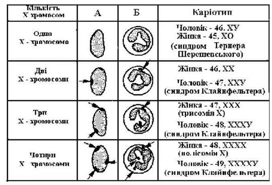
Fig. 34. Determination of X-sex chromatin.
The cytogenetic method allows for the determination of: a) the number of chromosomes in the karyotype; b) the genetic sex of the Organism; c) the presence and localization of chromosomal aberrations.
The application of the cytogenetic method in MEDICAL GENETIC COUNSELING makes it possible to promptly diagnose chromosomal pathologies and prevent the birth of a child with chromosomal syndromes.
Additionally, Biochemical Methods can be used to diagnose chromosomal disorders because these conditions exhibit a "gene dosage effect," which alters the concentration of certain Enzymes. For instance, in trisomy 21 (Down syndrome), The activity of the enzyme superoxide dismutase is increased by 1.5 times. The dosage effect has been established for 30 genes localized across various chromosomes.
Phenotypic analysis (such as facial gestalt diagnostics) and dermatoglyphics also play an important diagnostic role in identifying chromosomal pathologies.
Last update: 08/08/2026
Editorial and Educational Adaptation: This material has been compiled based on the primary/original source text. The project team performed an editorial review, corrected technical inaccuracies, structured sections, and adapted the content for an educational format.
What was processed:
- elimination of formatting defects (OCR errors, structural breaks, corrupted characters);
- editorial organization of content;
- standardization of terminology in accordance with academic sources;
- verification of factual statements against the original source text.
All mentions of the author, publication year, and origin of the primary text have been preserved in accordance with the source.