Vertebrate Zoology: A Study Guide - T. A. Dauda 2014
Multicellular Animals
Class Aves
The Class of birds encompasses over 8,000 species of highly specialized vertebrates adapted for flight. Representatives of this class are widely distributed across the globe and inhabit a great variety of environments.
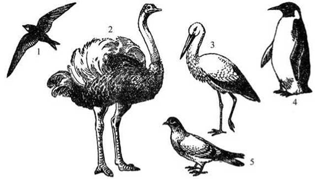
Fig. 29 Various body shapes in birds:
1 — swift; 2 — African ostrich; 3 — white stork; 4 — king penguin; 5 — wild rock dove.
Birds are closely related to reptiles and are regarded as their progressive evolutionary branch. The more advanced Organization of birds compared to reptiles is manifested in a higher level of development of The Nervous system and, consequently, in a more diverse and sophisticated adaptive behavior; in a developed thermoregulation capacity that determines a high body Temperature; in The ability to fly in the air while retaining the ability to move on land or climb; and in more advanced reproduction, which involves egg incubation and feeding of the nestlings.
Bird sizes vary widely: from the smallest (e.g., the hummingbird, the size of a bumblebee) to the largest (e.g., African ostriches up to 2.5 m tall). The body shape of birds is also diverse (Fig. 29), although their overall appearance and Structure share many common features. Externally, a bird is covered with feathers.
The HEAD of birds is small and lightweight. It bears the beak, eyes, and ear openings. The beak is formed by the elongated upper and lower jaws, which are covered with a horny sheath.
The upper part of the beak is called the upper Mandible (culmen), and the lower part is the lower mandible. The Base of the upper mandible in A number of birds (e.g., pigeons, diurnal birds of prey) is covered by a swollen whitish Skin called the cere, which Functions as an Organ of Touch. Paired external nostrils lie above the cere. In addition to the upper and lower eyelids, the eyes are equipped with a nictitating membrane. Below and behind each eye lies a rounded auditory opening leading to a shallow external auditory canal—the External ear. The head is connected to a flexible, usually long neck.
The body is compact and streamlined. The forelimbs are modified into Organs of flight—wings. The hindlimbs in many birds are small and pressed against the body during flight. The knee joint of the hindlimbs is concealed under the plumage. Their lower section is covered with horny scutes. The tips of the digits, which usually number four, bear claws. The tail is poorly developed.
Structural Features of birds
The skin of birds is thin and dry, as it is almost entirely devoid of glands. The only exception is the uropygial (preen) gland at the base of the tail, the secretion of which is used to lubricate the feathers and make the plumage waterproof. The preen gland is particularly well-developed in waterfowl. Conversely, in some birds inhabiting arid climates, the preen gland is absent (e.g., in ostriches and bustards). The surface layers of the epidermis keratinize. Horny derivatives of the epidermis include the beak, claws at the tips of the digits, horny scutes covering the digits, the tarsometatarsus (tarsus), and in some individuals, the Tibia as well.
The epidermis is followed by the Connective Tissue layer, which is subdivided into the dermis proper (containing Blood Vessels, feather quills, and bundles of smooth Muscle fibers) and the subcutaneous adipose tissue directly adjacent to the trunk musculature, where fat reserves are deposited.

Fig. 30 Structure of a bird feather:
1 — rachis (shaft); 2 — proximal secondary barbs; 3 — distal secondary barbs with hooklets; 4 — hooklets; 5 — calamus (quill); 6 — vane (vexillum).
Feathers are horny derivatives of the epidermis. They are distinguished by their STRUCTURE AND FUNCTIONS. Contour feathers are located on the exterior. They consist of a hollow shaft to which two lateral plates—the vanes—are attached (Fig. 30).
The lower part of the shaft, embedded in the skin, is the calamus (quill), while the upper part is the rachis. The vane consists of numerous primary barbs that bear secondary barbs. The latter are equipped with tiny hooklets that interlock the barbs, making the vane an elastic, resilient plate.
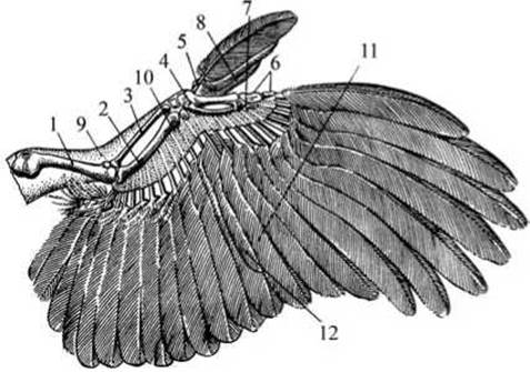
Fig. 31 Structure of a bird wing:
1 — humerus; 2 — ulna; 3 — radius; 4 — carpometacarpus; 5 — part of the carpus; 6, 7 — Phalanges of the digits; 8 — alula (bastard wing); 9 — patagium (wing membrane); 10 — bases of the remiges; 11 — primary remiges (flight feathers); 12 — secondary remiges.
Depending on their Location, contour feathers are divided into groups. For instance, the long feathers along the posterior margin of the forelimb that form the wing blade are called remiges (flight feathers). Long tail feathers are called rectrices (tail feathers), those covering the upper part of the wing are upper wing coverts, those covering the upper base of the tail are upper tail coverts, and so on.
Contour feathers are anchored in the skin within specific areas called pterylae, separated by apteria—regions where feathers do not grow. This shingle-like arrangement of overlapping feathers allows the entire body to be covered with a minimum number of feathers (Fig. 31). In flightless birds, feathers are distributed evenly across the body.
Beneath the contour feathers lie small down feathers. Their shaft is short, and secondary barbs are absent; consequently, the vane does not form a continuous plate. If the shaft of a down feather is so shortened that the barbs emerge in a single tuft, the feather is termed true down. Filoplumes, which are downy feathers lacking barbs, are interspersed among the down.
Feathers undergo regular replacement (molting). Many birds experience not one, but two or three molts per year. The pattern of molting varies among species. Birds of prey and insectivorous birds that catch their prey in flight molt gradually and do not lose their ability to fly. Other birds replace their plumage almost all at once, during which time many of them become flightless.
Due to the variety of their movements (such as flight, walking, and climbing), the musculature of birds is represented by a much larger number of Muscles than that of fish, amphibians, and reptiles. There are almost no muscles on the DORSAL SIDE OF the body; the bulk of them are located on the ventral side. The largest Muscles responsible for limb movement are situated on the trunk, while tendons extend to the limbs themselves. The pectoral muscles, which attach to the sternal keel and account for up to 20% of a bird's total body mass, are particularly well-developed. They are involved in the downward stroke of the wing. Lying beneath them are the subclavius muscles, which raise the wing and have a somewhat smaller mass. Birds are characterized by an accumulation of Myoglobin in their muscles, which provides a reserve supply of oxygen utilized during periods of intense exertion.
The avian Skeleton features a number of specific adaptations associated with flight and bipedal terrestrial locomotion (Fig. 32). These include unique modifications of the limbs and their girdles, as well as the lightweight yet robust Nature of the entire skeleton. Its lightness is ensured by the pneumaticity of most bones, while its strength is achieved through the fusion of many bones at Cytology/cytology/16.html">Early stages of embryonic development.
The Skull of birds consists of a large, thin-walled braincase, enormous orbits, and toothless jaws. A number of skull bones are pneumatic. In adult specimens, the skull bones are completely fused. These structural features ensure both durability and lightness of the skull capsule. The skull articulates with the first cervical vertebra via a single occipital condyle.
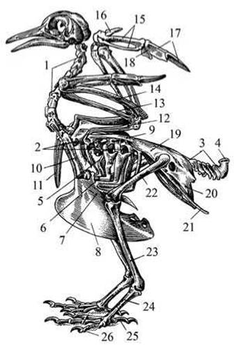
Fig. 32. Skeleton of a bird (pigeon):
1 — cervical vertebrae; 2 — thoracic vertebrae; 3 — caudal vertebrae; 4 — pygostyle; 5, 6 — Ribs; 7 — Sternum; 8 — keel; 9 — scapula; 10 — coracoid; 11 — clavicle (furcula); 12 — humerus; 13 — radius; 14 — ulna; 15 — metacarpus; 16-18 — phalanges of the digits; 19-21 — pelvic bones; 22 — Femur; 23 — tibiotarsus; 24 — tarsometatarsus; 25, 26 — phalanges of the digits.
The spinal Column in birds comprises cervical, thoracic, lumbar, sacral, and caudal regions.
The cervical region is long and highly flexible, consisting of 11–25 vertebrae. The first two cervical vertebrae—the atlas and axis—have a structure typical of amniotes (vertebrates with embryonic membranes).
The thoracic region includes 3–10 vertebrae fused with one another and with the compound sacrum. The thoracic vertebrae bear ribs, the lower ends of which attach to the sternum to form the ribcage. Each rib consists of two parts: a vertebral (dorsal) segment and a sternal segment. These parts are movably articulated, forming an angle with its apex directed backward.
Because of this rib structure, contraction of the corresponding muscles allows the sternum to move either toward or away from THE Vertebral Column. Consequently, the volume of the thoracic cavity changes, which plays an important role in Respiration. The sternum also exhibits a structure typical of birds: it is broad and slightly arched outward, with the thoracic ribs articulating along its margins. In the vast majority of modern birds, the sternum bears a bony keel to which the flight muscles attach on the left and right sides. Only a few species (such as ratites), which have lost the power of flight, lack a keel, and their sternum is slightly convex.
The lumbar vertebrae are completely fused with one another, with the iliac bones, and with the sacral vertebrae. A portion of the caudal vertebrae also fuses with the sacrum, resulting in The formation of a compound sacrum characteristic of birds. This structure is of great adaptive significance, as it Supports the body entirely on the hind limbs. The caudal region (6–9 vertebrae) terminates in a vertical bony plate known as the pygostyle, which serves as the anchoring base for the tail feathers.
Adapted for flight, the forelimbs of birds possess several distinctive features. The Pectoral Girdle consists of the scapula, coracoid, and clavicle. The scapula is elongated and rests upon the ribs, over which it can glide freely. The coracoid abuts the sternum at one end and fuses with the scapula and clavicle at the other, forming the shoulder joint socket. The left and right clavicles fuse to form the furcula (or wishbone), unique to birds, which imparts elasticity to the pectoral girdle. The wing skeleton comprises elements typical of a pentadactyl limb. The humerus and BONES OF THE forearm are largely unmodified, whereas the manus (hand) displays several specializations: the carpals are heavily reduced, and the metacarpals are fused into a single bone.
Pelvic strength is achieved through the fusion of the large iliac bones with the compound sacrum. The large ischial bones also fuse with the ilium, while the pubic bones are small and attach to the outer margin of the ischia. The femur is of typical structure. The lower leg (crus) consists of the tibiotarsus and a fused Fibula. During embryonic development, the proximal row of Tarsal Bones fuses with the tibia, forming the tibiotarsus; this fusion is so complete that no suture lines are visible in adult birds. The segment immediately following the lower leg is the tarsometatarsus (or shank). In adult birds, it consists of a single long bone embryonically formed by the fusion of the metatarsals and distal tarsals. The number of digits is most commonly 4, occasionally 3, and in only one case (the ostrich) reduced to 2.
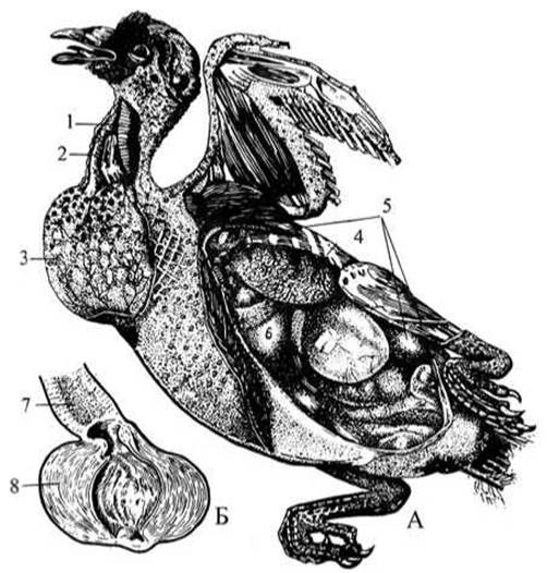
Fig. 33. Internal anatomy of a bird (pigeon):
A — dissected pigeon; B — section of the pigeon's Stomach; 1 — Trachea; 2 — Esophagus; 3 — crop; 4 — lung; 5 — air sacs; 6 — Heart; 7 — proventriculus (glandular stomach); 8 — gizzard (muscular stomach).
The Digestive System begins with the beak (Fig. 33), which lacks Teeth and bounds the Oral Cavity. A Tongue is attached to the floor of the Mouth. Salivary Glands vary in development among different species. The oral cavity leads into the esophagus, which in some birds forms an expanded pouch known as the crop. For instance, during the nestling-feeding period, the Cells of the crop wall in pigeons produce a fatty, curd-like substance referred to as "crop milk". The esophagus opens into the thin-walled proventriculus (glandular stomach), where food is acted upon by digestive secretions. The proventriculus is followed by the gizzard (muscular stomach), where food is mechanically ground down. After being pulverized and mixed with digestive Enzymes, the food passes into the Small Intestine, into which open the ducts of the well-developed Pancreas and the bilobed Liver. The intestine then continues as a short rectum that opens into the cloaca.
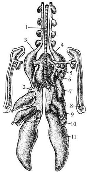
Fig. 34. Respiratory organs of a bird (pigeon):
1 — trachea; 2 — Lungs; 3–11 — air sacs.
The respiratory organs exhibit structural adaptations to an aerial lifestyle that are more pronounced than those of any other internal organ system.
Avian lungs are spongy structures attached to the dorsal wall of the thoracic cavity. Upon entering the lungs, the Bronchi branch out, permeate the lung tissue, and open into air sacs—thin-walled structures whose total volume vastly exceeds that of the lungs themselves (Fig. 34).
The air sacs are distributed among the Internal Organs, with diverticula extending between the muscles and into the bones. Modern research has demonstrated that avian respiration is a complex unidirectional process in which the air sacs play an exceptionally vital role.
When the posterior air sacs expand, oxygen-rich air quickly rushes into them through the Trachea and Bronchi. As these sacs decrease in volume, the air is forced forward into the lungs, where gas exchange takes place. From the lungs, the air passes into the anterior air sacs and is subsequently expelled outward. Volume Changes in the air sacs are driven primarily by Movements of the ribcage. Thus, airflow during respiration follows this pathway: trachea -> primary bronchi -> posterior air sacs -> lungs (gas exchange) -> anterior air sacs -> trachea -> exit to the exterior.
Consequently, during inhalation, air flows simultaneously into the lungs and air sacs; during exhalation, air from the posterior sacs is forced through the lungs, while air from the anterior sacs (which is already depleted of oxygen) is pushed out through the trachea. As a result, oxygenated air passes through the lungs in an almost continuous stream during both inhalation and exhalation—a mechanism known as double respiration—thereby heavily oxygenating the blood.
In addition to enhancing respiration, the air sacs protect the body from overheating during intense movement, as excess heat is continuously removed by the circulating air.
The main feature of avian Blood Circulation is the complete Separation of arterial and venous blood (see Fig. 35). The Heart is four-chambered, consisting of two atria and two ventricles.
Due to the separation of the arterial and venous blood streams, the organs are supplied exclusively with pure arterial blood. This factor, combined with rapid blood circulation and vigorous gas exchange, results in a high body temperature, averaging around 42°С.
Unlike reptiles, birds have completely separated SYSTEMIC AND PULMONARY circuits: venous and arterial blood streams never mix anywhere, and the right (venous) half of the heart is isolated from the left (arterial) one. The pulmonary circuit begins in the right ventricle, which receives venous blood from the entire body via the right atria.
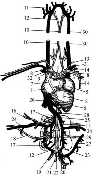
Fig. 35 Circulatory system OF a bird (pigeon):
1 — right atrium; 2 — right ventricle of the heart; 3 — left pulmonary artery; 4 — right pulmonary artery;
5 — left atrium; 6 — left ventricle of the heart; 7 — right aortic arch; 8, 9 — innominate Arteries; 10-12 — carotid arteries; 13 — Subclavian Artery; 14 — left thoracic artery; 15 — aorta; 16 — right femoral artery; 17 — renal artery; 18 — sciatic artery; 19 — iliac artery; 20 — posterior mesenteric artery; 21 — caudal artery; 22 — caudal vein; 23 — renal portal vein; 24 — femoral vein; 25 — iliac vein; 26 — posterior vena cava; 27 — intestinal vein; 28 — supra-intestinal vein; 29 — renal vein; 30 — jugular vein; 31 — subclavian vein; 32 — anterior vena cava.
The pulmonary artery extends from the right ventricle and divides into the right and left branches, through which venous blood enters the corresponding lung. The oxygenated arterial blood then flows through the right and left Pulmonary Veins into the left atrium.
The systemic circuit begins with the left ventricle, from which only a single vessel emerges — the right aortic arch (the left one is completely reduced in birds). The right aortic arch branches into two vessels: the right and left innominate arteries. It then arches sharply over the right bronchus and extends backward along the vertebral column as the dorsal aorta, which supplies arteries to all internal organs.
The Venous system of birds resembles that of reptiles, differing only in the partial reduction of the renal portal system and the reduction of the abdominal vein.
A characteristic feature of birds is the relatively large size of the heart (approximately 1% of body weight, and 1.5–2% in fast flyers). The heart works at a high intensity: during flight, birds weighing about 0.5 kg have a heart rate of 400–500 beats per minute (200–300 beats per minute at rest), while in small birds the flight heart rate can reach up to 1000 beats per minute. The total blood volume, erythrocyte count, and Hemoglobin content in birds are much higher than in reptiles and are quite comparable to those of mammals.
All Features of the AVIAN CIRCULATORY SYSTEM correspond to their High Metabolic Rate, ensuring the continuous and intensive supply of oxygen and nutrients to all organs and Tissues, as well as the removal of Metabolic waste products.
The Central Nervous System of birds is complex, which is associated with a high level of vital processes. The Brain is large: in flying birds, it accounts for up to 5–8% of body weight, driven by The Development of the Forebrain hemispheres and a strongly developed Cerebellum (see Fig. 36). This provides birds with a higher level of nervous activity and more complex forms of behavior compared to reptiles.
Sight and Hearing are well-developed in birds. Vision in most species is nearly monocular. The visual field of each eye is approximately 150 degrees. Visual acuity is high; for example, a peregrine falcon can spot a moving bird at a distance of up to 1100 m. In owls, vision is largely binocular, which has adaptive significance due to their nocturnal lifestyle.
All birds possess Color Vision, which is why bright color patches that serve as species-specific recognition marks are so common in avian plumage.
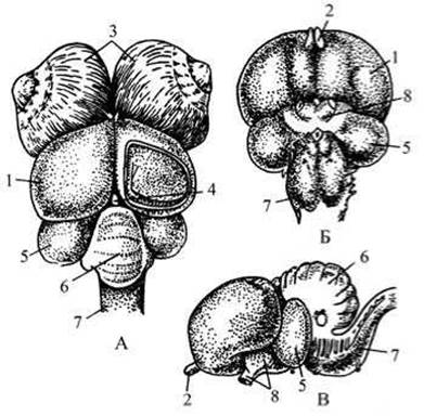
Fig. 36 Brain of a bird (pigeon):
A — dorsal view; Б — ventral view; В — lateral view; 1 — forebrain hemispheres; 2 — olfactory lobes;
3 — eyeballs; 4 — Pineal Gland; 5 — optic lobes; 6 — cerebellum; 7 — Medulla Oblongata; 8 — optic nerves.
The Organ of Hearing is anatomically similar to that of reptiles (especially crocodilians), but functionally differs little from the more complex mammalian ear. The avian ear is represented by the inner and Middle ear. Contour feathers covering the external auditory meatus serve not only for mechanical Protection of the ear canal but also act as a horn to channel sound waves. There are apparently no birds with poorly developed hearing. Some species are capable of perceiving ultrasounds up to 35–50 kHz present in their vocalizations. Many birds can determine THE POSITION OF a sound source with great precision (2–3 degrees); sound localization is especially precise (about 1 degree) in owls, which successfully capture prey "by ear" without seeing it. A few birds have been found to possess echolocation — the ability to capture reflected sound produced in the audible range rather than the ultrasonic range, unlike bats.
Acute hearing and the capacity for acoustic analysis are combined in birds with the ability to produce A wide variety of sounds. These convey rich and biologically important information, such as parent-chick communication calls, alarm signals, and sounds and postures that regulate territorial relations, partner interactions, etc.
It is generally believed that the SENSE OF SMELL is poorly developed in birds because the olfactory lobes of the forebrain are small. However, compared to reptiles, the surface area of the Nasal cavity in birds is increased due to the proliferation of nasal conchae. Furthermore, experimental data suggest that in certain birds (such as scavengers, shorebirds, ducks, and some passerines), the sense of smell is sufficiently well developed and used in foraging.
Birds also have well-developed sense of taste. Taste buds are located in the mucous membrane of the oral cavity, on the tongue, and at its base. Many species can distinguish sweet, salty, and bitter tastes.
The organs of touch are represented by clusters of sensory cells located on the skin, beak, oral cavity, and hind limbs. Some of these sensory cells function as thermodetectors, registering body temperature.
The excretory organs in adult birds are represented by pelvic metanephric Kidneys. Embryos initially develop trunk kidneys, which are subsequently replaced by pelvic ones. A Ureter extends from the ventral side of each Kidney and opens into the cloaca. Adult birds lack a Urinary Bladder. In birds, as in most reptiles, the end product of Protein METABOLISM is uric acid rather than urea; it readily precipitates out of solution as crystals, forming a white paste. This consistency of the excreted urine apparently accounts for the reduction of the urinary bladder.
The reproductive organs of birds include the Testes, Ovaries, and their respective ducts. The testes are two bean-shaped bodies located in the Abdominal cavity. Rudimentary epididymides attach to their inner margins, from which deferent ducts extend and open into the cloaca. Copulatory organs are present in only a few species. In most birds, internal Fertilization is achieved by bringing the cloacal openings close together. Females typically have only one (left) Ovary, which is an irregularly shaped, granular body lying anterior to the left kidney. An ovum released from the ovary passes through the unpaired oviduct, where it receives an albumen coating, and enters the wide Uterus, where it acquires a calcareous shell. The final section of the oviduct is the Vagina, from which the egg passes into the cloaca and then to the exterior.
Bird eggs are relatively large. Their main bulk consists of yolk and albumen (Fig. 37); however, the actual egg is strictly the yolk. The Contents of the yolk serve as a reserve of nutrients and Water required for the Development of the growing embryo. The embryo develops from the germinal disc located at one of the poles of the yolk (the animal pole).
The germinal disc consists of a Nucleus and Cytoplasm. The yolk is separated from the albumen by a thin vitelline membrane. The primary function of the albumen is to protect the yolk proper from mechanical damage and shocks, while also serving as a source of water for the embryo. Externally, the albumen is enclosed by two thin, parchment-like shell membranes that diverge slightly at the blunt end of the egg to form an air chamber, which accommodates changes in the egg's volume with temperature fluctuations. The yolk is suspended within the albumen by twisted, densified protein fibers known as chalazae, which extend from the vitelline membrane to the shell at the sharp and blunt poles of the egg, where they attach.
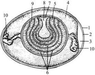
Fig. 37 Structure of a bird egg:
1 — chalazae; 2 — air chamber; 3 — inner shell membrane; 4 — outer shell membrane; 5 — shell; 6 — liquid albumen; 7 — dense albumen; 8 — dark yolk; 9 — light yolk; 10 — germinal disc.
The shell consists of 89–97% calcium carbonate and protects the egg from mechanical damage. Gas exchange is possible solely due to the porous nature of the shell. Beneath the outer shell membrane lies an extremely delicate cuticular film that prevents Bacteria from penetrating the egg.
Reproduction in all birds begins with courtship rituals, or display. Nest building takes place during the display period. Only a few birds do not build nests, laying their eggs directly on the ground (such as nightjars and certain waders).
Virtually all birds incubate their eggs. The majority of birds begin intensive incubation only after all eggs have been laid, ensuring that hatching occurs more or less simultaneously.
In birds of prey, owls, and certain other species, incubation begins after the first egg is laid, which stretches the hatching period over several days. The duration of incubation depends on the size of the egg and the bird, as well as the intensity of incubation.
Small passerines incubate for 11–14 days, crows for 17, mallards for 26, and swans for 35–40 days. The longest incubation period—about two months—is found in large penguins, albatrosses, and vultures.
Based on the physiological maturity of chicks at hatching, all birds can be divided into two groups (Fig. 38): precocial and altricial. Precocial chicks are downy and sighted; once dry, they can run well, and aquatic species can swim. Shortly after hatching, they leave the nest and wander about, feeding independently. Altricial chicks emerge from the egg helpless, blind, and naked; they remain in the nest for a long time, being fed and brooded by their parents.
A fairly large number of birds form an intermediate group. Chicks of loons and grebes hatch downy and sighted, capable of swimming and diving, but are fed by their parents until they fledge. In owls and diurnal raptors, chicks hatch downy and blind (though their eyes open quickly), and are likewise fed by their parents until fledging.
Approximately 80 species (mostly tropical) exhibit brood parasitism: they do not build nests, instead laying their eggs in the nests of other, often closely related, bird species, leaving both incubation and chick rearing to the hosts.
In the fauna of our country, representatives of this group include the common cuckoo and the Oriental cuckoo.
Bird behavior and significance will be discussed below in the descriptions of the various orders.
Birds are the most numerous class of terrestrial vertebrates. Within our country, approximately 750 species from 18 orders are found, accounting for about 8% of the world's avian fauna.
All extant bird orders belong to the subclass Neornithes (true birds).
This subclass is divided into several superorders (two of which are extinct).
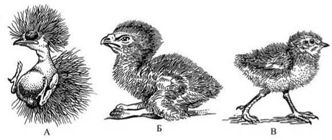
Fig. 38 Chicks of various birds at the same age:
A — altricial (pipit); B — semi-precocial (eagle); C — precocial (partridge).
We will examine the morphological and Biological features of orders belonging to three superorders: Impennes (penguins), Ratitae (flightless or ratite birds), and Carinatae (carinate birds).
Last update: 13/08/2026
Editorial and Educational Adaptation: This material has been compiled based on the primary/original source text. The project team performed an editorial review, corrected technical inaccuracies, structured sections, and adapted the content for an educational format.
What was processed:
- elimination of formatting defects (OCR errors, structural breaks, corrupted characters);
- editorial organization of content;
- standardization of terminology in accordance with academic sources;
- verification of factual statements against the original source text.
All mentions of the author, publication year, and origin of the primary text have been preserved in accordance with the source.