Human Anatomy, Part 2 - K. A. Diubenko, A. K. Kolomiitsev, Yu. B. Chaikovsky 2008
Specialized Part
Peripheral Nervous System, systema nervosum periphericum — General Overview
Cranial Nerves — Structure of Individual Cranial Nerves
1. The olfactory nerves, nervi olfactorii (I), are formed by the central processes of olfactory neurosensory Cells, which constitute 15–20 olfactory filaments, fila olfactoria. These filaments pass from the olfactory region, regio olfactoria, of the Nasal cavity through the cribriform plate, lamina cribrosa, to reach the olfactory bulb, bulbus olfactorius (for details, see "The Olfactory Organ").
Damage to the Components of the First and Second Neurons of the Olfactory nerve (specifically, the olfactory bulb and tract) results in ipsilateral loss of smell (anosmia). Ipsilateral involvement of the third neuron does not lead to olfactory impairment due to incomplete decussation. Irritation of the hippocampus in the temporal lobe of the cerebrum produces false olfactory perceptions (hallucinations).
2. The Optic nerve, nervus opticus (II) (see Fig. 219), consists of the processes of the retinal ganglion cells. Through the optic canal, canalis opticus, both nerves enter the cranial cavity, where they form an incomplete decussation in the region of the prechiasmatic sulcus, sulcus chiasmatis.
It should be noted that the optic nerve is enveloped by the pia mater, arachnoid mater, and dura mater, which extend onto it from the Brain. Intermeningeal spaces are preserved between these membranes, through which CEREBROSPINAL FLUID circulates. This explains the involvement of the optic nerve in meningitis.
Damage to the optic nerve causes blindness on the same side. Lesions of the optic chiasm result in heteronymous (contralateral) hemianopia (loss of half of the visual fields). Starting from the optic tract, tractus opticus, and extending to the visual cortex of the occipital lobe, homonymous (ipsilateral) hemianopia occurs on the opposite side.
3. The Oculomotor nerve, nervus oculomotorius (III) (Fig. 180), is formed by the axons of neurons located in the nerve nuclei within the Midbrain. The oculomotor nerve emerges on the basal surface of the brain near the medial margin of the cerebral peduncle, runs anteriorly along the lateral wall of the cavernous sinus, and then passes through the superior orbital fissure, fissura orbitalis superior, into the Orbit, where it divides into superior and inferior branches, rami superior et inferior. The superior branch innervates the levator palpebrae superioris Muscle, m. levator palpebrae superioris, and the superior rectus muscle of the Eyeball, m. rectus superior. The inferior branch innervates the medial and inferior rectus Muscles, m. rectus medialis and m. rectus inferior, and the inferior oblique muscle, m. obliquus inferior. A branch departs from the inferior branch to the ciliary ganglion, forming its parasympathetic ROOT, radix parasympathetica [oculomotoria], and runs toward the ciliary ganglion.
Class="center">
Fig. 180. Oculomotor nerve, n. oculomotorius; Trochlear nerve, n. trochlearis; Abducens nerve, n. abducens (Diagram based on I. Ye. Kefeli).
4. The trochlear nerve, nervus trochlearis (IV) (Fig. 180), is a motor nerve. It is formed by the axons of neurons of the motor Nucleus, nucleus n. trochlearis, in the midbrain. It emerges from the brain tissue in the region of the superior medullary velum, curves laterally around the cerebral peduncle, and appears on the basal surface of the brain lateral to the Pons. It then courses anteriorly in the wall of the cavernous sinus, sinus cavernosus, and further extends through the superior orbital fissure, fissura orbitalis superior, into the orbit, where it innervates the superior oblique muscle of the eyeball, m. obliquus superior.
Lesions of the trochlear nerve result in strabismus and double Vision (diplopia) exclusively when looking downward.
5. The Trigeminal nerve, nervus trigeminus (V) (Fig. 181), is a mixed nerve with a complex Structure. Its sensory fibers are represented by the processes of neurons of the trigeminal ganglion, ganglion trigeminale (Gasserian ganglion)*. The latter lies within the trigeminal impression, impressio trigemini, in the trigeminal cavity, cavum trigeminale, formed by a splitting of the dura mater (Meckel's cave). The ganglion is semilunar in shape and consists of sensory pseudounipolar Nerve Cells; its length is 15–30 mm and its height is 5–20 mm. The central processes (axons) of the ganglion cells form the sensory root, radix sensoria, and enter the Brainstem, reaching the three sensory nuclei of the nerve (nucl. mesencephalicus nervi trigemini, nucl. pontinus nervi trigeminali, nucl. spinalis nervi trigeminali). The peripheral processes (dendrites) run within the three Branches of the trigeminal nerve.
Motor fibers are represented by the axons of neurons of the motor Nucleus of the nerve, nucl. motorius n. trigemini. They form the motor root, radix motoria. On the basal surface of the brain, the sensory and motor roots are located at the border of the pons and the middle cerebellar peduncle, forming anatomical connections through which a certain number of fibers pass from one root to the other. The motor root passes beneath the trigeminal ganglion and becomes part of the mandibular nerve.
Thus, the first and second branches of the trigeminal nerve are sensory, while the third is mixed. In general, the sensory fibers of the trigeminal nerve are distributed in the scalp and mucous membranes such that the boundaries between the three branches correspond to the level of the lateral angle of THE EYE AND the angle of the Mouth. From THE ORIGIN OF each branch, a meningeal branch, ramus meningeus, departs toward the dura mater.
The first branch of the trigeminal nerve is the ophthalmic nerve, nervus ophthalmicus (Fig. 181), which runs forward from the ganglion within the wall of the cavernous sinus, sinus cavernosus, enters the superior orbital fissure, fissura orbitalis superior, and reaches the orbit, where it divides into three branches: the lacrimal, frontal, and nasociliary nerves.
1. The lacrimal nerve, nervus lacrimalis, runs along the lateral wall of the orbit, receives a communicating branch from the zygomatic nerve, ramus communicans cum n. zygomatico, and innervates the lacrimal gland, the Skin of the upper eyelid, and the lateral angle of the eye.
* Johann Laurentius Gasser (1727–1779) was a German physician and anatomist. He was awarded the degree of Doctor of Medicine without defending a dissertation. In 1765, the anatomist Hirsch (a student of Gasser) named the trigeminal ganglion (Gasserian ganglion) after him.

Fig. 181. Trigeminal nerve, n. trigeminus (Topography of the ophthalmic nerve, n. ophthalmicus, and maxillary nerve, n. maxillaris).

Fig. 181A. Trigeminal nerve, n. trigeminus (topography of the mandibular nerve, n. mandibularis).
2. The frontal nerve, n. frontalis, runs beneath the superior wall of the orbit and divides into the supraorbital nerve, n. supraorbitalis, which passes through the supraorbital notch, incisura supraorbitalis, to the skin of the forehead, and the supratrochlear nerve, n. supratrochlearis, which emerges from the orbit near its medial wall and innervates the skin of the upper eyelid, the medial angle of the eye, and the root of the Nose.
3. The nasociliary nerve, n. nasociliaris, lies along the medial wall of the orbit. It gives off long ciliary nerves, nn. ciliares longi, to the eyeball; the posterior ethmoidal nerve, n. ethmoidalis posterior, which passes through the posterior ethmoidal foramen to the mucous membrane of the sphenoidal sinus, sinus sphenoidalis, and the posterior ethmoidal air cells; and the anterior ethmoidal nerve, n. ethmoidalis anterior, to the mucous membrane of the frontal sinus and nasal cavity, as well as to the skin of the nasal tip and alae. In addition, a communicating branch to the ciliary ganglion, ramus communicans cum ganglio ciliari, arises from the nasociliary nerve. The nasociliary nerve terminates as the infratrochlear nerve, n. infratrochlearis, which emerges from the orbit and innervates the lacrimal sac, conjunctiva, and medial angle of the eye.
The ciliary ganglion, ganglion ciliare (Fig. 181B), lies posterior to the eyeball, lateral to the optic nerve. It contains parasympathetic multipolar neurons upon which the preganglionic parasympathetic fibers of the oculomotor nerve terminate. Transmission of nerve impulses to the ganglion's nerve cells occurs within the ciliary ganglion via sympathetic connections. Postganglionic fibers—axons of the ganglion cells—run within the short ciliary nerves, nn. ciliares breves. Sensory fibers from the nasociliary nerve pass through the ganglion and also join the short ciliary nerves. They are joined by a sympathetic branch from the cavernous plexus, consisting of postganglionic fibers from the cervical Sympathetic trunk ganglia. The short ciliary nerves innervate the tunics and Muscles of the eyeball, with their postganglionic sympathetic fibers innervating the Blood Vessels and the dilator pupillae muscle, m. dilator pupillae. Postganglionic parasympathetic fibers innervate the ciliary muscle, m. ciliaris, and the sphincter pupillae muscle, m. sphincter pupillae, thus participating in accommodation and adaptation processes of the visual system.
Thus, the first branch of the trigeminal nerve, n. ophthalmicus, innervates: the tentorium cerebelli, the skin of the forehead and parietal region, the upper eyelid, the dorsum and tip of the nose, the mucous membrane of the anterior nasal cavity and the frontal, sphenoid, and ethmoid sinuses, as well as the eyeball, conjunctiva, and Lacrimal Apparatus. The nervus ophthalmicus provides sensory proprioceptive innervation to the extraocular muscles via connections with cranial nerve pairs III, IV, and VI, and proprioceptive innervation to the facial expression muscles via connections with cranial nerve pair VII.
The second branch of the trigeminal nerve is the maxillary nerve, n. maxillaris (Fig. 181), which emerges from the cranial cavity into the pterygopalatine fossa through the foramen rotundum. It divides into the infraorbital nerve, zygomatic nerve, and ganglionic branches to the pterygopalatine ganglion.
1. The infraorbital nerve, n. infraorbitalis, emerges from the pterygopalatine fossa and enters the orbit through the inferior orbital fissure. Accompanied by Vessels of the same name, it runs along the infraorbital groove and canal (sulcus infraorbitalis and canalis infraorbitalis) to exit onto the face via the infraorbital foramen (foramen infraorbitale). Here, deep to the levator labii superioris muscle, it fans out into its terminal branches. The nerve gives off the posterior, middle, and superior anterior alveolar branches (gg. alveolares superiores posteriores, anteriores et medius), which form the superior dental plexus (plexus dentalis superior) within the alveolar process of the Maxilla. The plexus gives rise to the superior gingival branches (ramі gingivales superiores) supplying the mucosa of the alveolar process, gingiva, and tooth sockets, as well as the superior dental branches (rr. dentales superiores) supplying the upper Teeth. Specifically, the posterior branches innervate mainly the molars, their sockets, and the corresponding mucosa and gingiva, whereas the middle branch supplies the premolars along with their periodontium. Additionally, the posterior branches innervate the mucosa of the maxillary sinus, while the anterior branches supply the mucosa of the anterior nasal cavity.

Fig. 181 B. NERVES OF THE right orbit, superior view (after R. D. Sinelnikov)

Fig. 182 C. Diagram of the autonomic ganglia of the HEAD (after R. D. and Ya. R. Sinelnikov)
The terminal branches of the maxillary nerve include the inferior palpebral branches (rami palpebrales inferiores), which innervate the skin of the lower eyelids; the external nasal branches (ramі nasales externi), supplying the skin of the nasal ala; the internal nasal branches (ramі nasales interni), supplying the nasal mucosa; and the superior labial branches (ramі labiales superiores), which innervate the skin and mucosa of the upper lip.
2. The zygomatic nerve, n. zygomaticus, enters the orbit via the inferior orbital fissure (fissura orbitalis inferior), where its lateral wall features the zygomaticoorbitale foramen. Here it divides into two branches: the zygomaticofacial branch (ramus zygomaticofacialis), which emerges through the corresponding foramen to distribute to the skin of the cheek and the lateral angle of the eye; and the zygomaticotemporal branch (ramus zygomaticotemporalis), which exits through its own foramen, pierces the temporalis muscle and its fascia, and innervates the skin of the lateral temporal and frontal regions.
3. Ganglionic branches (rami ganglionares) connect the maxillary nerve to the pterygopalatine ganglion, supplying the latter with sensory fibers.
The pterygopalatine ganglion, ganglion pterygopalatinum (Figs. 181; 182 C), is located in the pterygopalatine fossa. It consists of parasympathetic neurons upon which the preganglionic fibers of the greater petrosal nerve terminate. Sympathetic (postganglionic) fibers also pass through the ganglion, reaching it as part of the deep petrosal nerve (n. petrosus profundus). Sympathetic, parasympathetic, and sensory fibers emerge from the ganglion within its branches. The orbital branches (ramі orbitales) pass through the inferior orbital fissure (fissura orbitalis inferior) into the orbit. Further along, as part of the posterior ethmoidal nerve, their fibers travel to the sphenoidal sinus and the posterior Cells of the ethmoidal labyrinth. The lateral and medial superior posterior nasal branches (ramі nasales posteriores superiores laterales et mediales) enter the nasal cavity via the sphenopalatine foramen. There, they supply the posterior PARTS OF THE superior and middle nasal conchae and meatuses, the superior surface of the choanae, and the pharyngeal opening of the auditory tube (lateral branches), as well as the upper part of the nasal septum (medial branches). One of the medial branches, the nasopalatine nerve (nervus nasopalatinus), runs forward beneath the mucosa of the nasal septum to reach the incisive canal (canalis incisivus). Through this canal, it enters the Oral Cavity to supply the mucosa of the anterior part of the hard palate (general and gustatory sensitivity). The greater palatine nerve (nervus palatinus major) passes through the greater palatine canal to the hard palate, innervating most of it, the mucosa of both the Hard and Soft palates, and the anterior gingiva (general and gustatory sensitivity). The inferior posterior nasal branches (ramі nasales posteriores inferiores) travel via the greater palatine canal to the Inferior nasal concha, the middle and inferior nasal meatuses, and the maxillary sinus. The lesser palatine nerves (nervi palatini minores) pass through the lesser palatine foramina to the soft palate and the region of the palatine tonsil. A portion of the parasympathetic fibers from the ganglion courses toward the trunk of the maxillary nerve, enters the zygomatic nerve, and via an anastomotic branch passes into the lacrimal nerve to provide secretomotor innervation to the lacrimal gland.
Thus, the second branch of the trigeminal nerve, n. maxillaris, innervates: the dura mater, the skin of the lower eyelid, upper lip, cheek, and nasal ala, as well as the mucosa of the posterior nasal cavity, maxillary sinus, palate, gingiva, and upper teeth. The nervus maxillaris also provides proprioceptive innervation to the mimic (facial) muscles through connections with the cranial nerve VII.
The third branch of the trigeminal nerve is the mandibular nerve, nervus mandibularis (Fig. 181 A), which exits the cranial cavity through the ovale foramen. It divides into the masseteric, deep temporal, lateral and medial pterygoid, buccal, auriculotemporal, lingual, and inferior alveolar nerves. It comprises both motor and sensory fibers.
Motor branches:
1. The masseteric nerve (nervus massetericus) frequently originates as a common trunk with other nerves supplying the masticatory muscles. It runs above the upper margin of the lateral pterygoid muscle and reaches the masseter muscle via the mandibular notch (incisura mandibulae). Before entering the muscle, it gives off a sensory branch to the temporomandibular joint.
2. The deep temporal nerves (nn. temporales profundi) loop around the infratemporal crest (crista infratemporalis) and innervate the temporalis muscle.
3. The lateral pterygoid nerve (n. pterygoideus lateralis) innervates the muscle of the same name.
4. The medial pterygoid nerve (n. pterygoideus medialis) innervates the corresponding muscle and also gives off branches to the tensor tympani muscle, the tensor veli palatini muscle, and the otic ganglion.
The otic ganglion, ganglion oticum, lies inferior to the foramen ovale, closely applied to the posteromedial surface of the mandibular nerve trunk (Fig. 182 C). It is composed of parasympathetic cells upon which the preganglionic fibers of the lesser petrosal nerve terminate. The ganglion also receives postganglionic sympathetic fibers from the plexus surrounding the middle meningeal artery (a. meningea media).
Sensory, parasympathetic, and sympathetic fibers leave the ganglion as part of its branches, the most important of which is the communicating branch with the auriculotemporal nerve (ramus communicans cum n. auriculotemporali). Its fibers provide secretomotor innervation to the Cytology/practical/97.html">Parotid salivary gland.
Sensory branches:
1. The meningeal branch (ramus meningeus [n. spinosus] LNA) enters the cranial cavity through the spinous foramen (foramen spinosum) alongside the middle meningeal artery (a. meningea media). The meningeal branch supplies the dura mater of the middle cranial fossa.
2. The buccal nerve (nervus buccalis) passes between the two heads of the lateral pterygoid muscle and then runs along the inner surface of the temporalis muscle, branching out over the outer surface of the buccinator muscle toward the angle of the mouth. It gives off fine filaments that pierce this muscle and innervate the buccal mucosa.
3. The auriculotemporal nerve (nervus auriculotemporalis) originates from two roots that encircle the middle meningeal artery (a. meningea media) before uniting into a single trunk. At the level of the neck of the mandibular condyle, it ascends, pierces the parotid gland, and enters the temporal region where it divides into its terminal superficial temporal branches (nervi temporales superficiales), which innervate the skin of this area. Along its course, the auriculotemporal nerve gives off parotid branches (rаmі parotidei), the nerve of the external acoustic meatus (n. meatus acustici externi), tympanic membrane branches (n. membranae tympani), and anterior auricular nerves (nervi auriculares anteriores) that supply the skin of the anterior auricle and the middle part of the temporal region.
4. The lingual nerve (n. lingualis) runs between the pterygoid muscles and joins the chorda tympani (a branch of the intermediate nerve). The nerve then courses between the Medial surface of the Mandible and the medial pterygoid muscle, superior to the Submandibular salivary gland, along the outer surface of the hyoglossus muscle to the Inferior surface of the Tongue. Between the hyoglossus and genioglossus muscles (m. hyoglossus and m. genioglossus), the nerve divides into its terminal branches, reaching the mucosa of the anterior two-thirds of the tongue as the lingual branches (ramі linguales). The fibers of the lingual nerve mediate general sensation (Touch, pain, Temperature)—their receptors are located within the filiform papillae. The sensory fibers of the chorda tympani, which travel within the lingual nerve to the tongue mucosa, mediate taste sensitivity (see "ORGAN OF TASTE").
Along its course, the lingual nerve gives off branches to the ISTHMUS OF THE fauces (rami isthmi faucium), the sublingual nerve (nervus sublingualis) supplying the mucosa of the floor of the mouth, gingiva, and sublingual salivary gland, and ganglionic branches (rami ganglionares) leading to the submandibular ganglion.
The submandibular ganglion, ganglion submandibulare, is located inferior to the trunk of the lingual nerve on the superior surface of the submandibular salivary gland (Fig. 182 C). The ganglion consists of multipolar nerve cells upon which the preganglionic fibers of the chorda tympani terminate. Sympathetic postganglionic fibers are supplied to the ganglion by the plexus surrounding the facial artery (a. facialis). Sensory, sympathetic, and parasympathetic fibers pass from the ganglion to the submandibular and sublingual Salivary Glands.
5. The inferior alveolar nerve, nervus alveolaris inferior, runs between the pterygoid muscles posterior to the lingual nerve, and then enters the mandibular canal, canalis mandibulae, where it gives off branches that anastomose with one another to form the inferior dental plexus, plexus dentalis inferior. The nerve exits the canal through the mental foramen, foramen mentale, becoming the mental nerve, nervus mentalis.
Before entering the mandibular canal, the inferior alveolar nerve gives off the mylohyoid nerve, n. mylohyoideus, which lies within the mylohyoid groove of the mandible and innervates the mylohyoid muscle and the anterior belly of the digastric muscle.
The inferior dental plexus gives rise to the inferior dental and inferior gingival branches, rаmі dentales et gingivales inferiores. It should be noted that the right and left plexuses cross the midline, extending to the level of the canines on the opposite side. This must be taken into account during the extraction of the lower incisors and canines. Novocain block must be performed bilaterally.
The mental nerve, n. mentalis, divides into mental branches to the skin of the chin, rаmі mentales, labial branches, rаmі labiales, and gingival branches, rаmі gingivales.
AUTONOMIC GANGLIA OF THE HEAD
Summary Table
|
Name of ganglion |
Roots |
Branches |
Innervated Organs |
|
1 |
2 |
3 |
4 |
|
Ciliary ganglion, gangl. ciliare |
Nasociliary (sensory) Oculomotor (parasymp.) Sympathetic root |
Short ciliary nerves |
Tunics of the eyeball, muscles of the eyeball |
|
Pterygopalatine ganglion, gangl. pterygopalatinum |
Ganglionic branches (sensory root) Greater petrosal nerve (parasympathetic) Deep petrosal nerve (sympathetic) |
Orbital branches Posterior superior nasal branches Palatine nerves |
Mucous membrane of the nasal cavity and Paranasal Sinuses, mucosal glands, palate |
|
Otic ganglion, gangl. oticum |
Sensory root (from the mandibular nerve) Lesser petrosal nerve (parasympathetic) Sympathetic root |
Communicating branch with the auriculotemporal nerve Communicating branch with the meningeal branch of the mandibular nerve Communicating branch with the chorda tympani |
Parotid gland, dura mater |
|
Submandibular ganglion, gangl. submandibulare |
Sensory root (from the lingual nerve) Parasympathetic root (from the chorda tympani) Sympathetic root |
Glandular branches Communicating branches with the lingual nerve |
Submandibular and sublingual salivary glands |
Thus, the third branch of the trigeminal nerve, n. mandibularis, is mixed. Its motor portion innervates the masticatory muscles, as well as the tensor veli palatini, tensor tympani, mylohyoid, and anterior belly of the digastric muscle.
The sensory portion of the nerve innervates: the dura mater, the skin of the lower lip, chin, cheek, temporal region, anterior auricle, external acoustic meatus, tympanic membrane, as well as the mucous membrane of the anterior two-thirds of the tongue (general sensation), the floor of the oral cavity, lower lip, gingiva, lower teeth, and salivary glands (general sensation). In addition, the n. mandibularis provides proprioceptive innervation to the temporomandibular joint, the masticatory muscles of the tongue via connections with the XII cranial nerve, and the Muscles of facial expression via connections with the VII cranial nerve.
Damage to the motor root of the trigeminal nerve results in peripheral paralysis of the masticatory muscles. Lesions of the sensory root lead to a loss of all modalities of facial sensation.
Innervation of the facial skin is provided by the branches of the trigeminal nerve. The skin of the forehead, parietal region, upper eyelid, bridge, and tip of the nose is innervated by the first branch of the trigeminal nerve. The skin of the lower eyelid, upper lip, cheek, and ala of the nose is innervated by the second branch of the trigeminal nerve. The skin of the lower lip, chin, cheek, temporal region, auricle, and external acoustic meatus is innervated by the third branch of the trigeminal nerve. The sites where the branches of the trigeminal nerve emerge subcutaneously (Valleix's points are tender in trigeminal neuralgia)
- supraorbital foramen - the site of subcutaneous emergence of the first branch of the trigeminal nerve;
- infraorbital foramen - the site of subcutaneous emergence of the second branch of the trigeminal nerve;
- mental foramen - the site of subcutaneous emergence of the third branch of the trigeminal nerve.
In the facial region, the Zander zones, zonae Zanderi, are also distinguished. Inflammatory processes in the sphenoid and ethmoid sinuses are accompanied by pain localized in the orbital, nasal, and infraorbital Regions of the face, which may radiate to the temporal and mastoid regions. Sometimes pain radiates to the occipital region, scapula, arm, forearm, and hand. Pain in these zones, the Zander zones, is accompanied by autonomic-vascular disorders (conjunctival hyperemia, increased lacrimation and salivation, nasal mucosal edema).
The Zander zones are also considered to be the areas of sensory loss resulting from lesions of the trigeminal nerve nuclei.
6. The abducens nerve, nervus abducens (VI) (Fig. 180), is a motor nerve formed by the axons of motor neurons originating from the abducens nucleus, nucleus n. abducentis. It emerges on the ventral surface of the brainstem between the posterior border of the pons and the pyramid. It passes through the cavernous sinus, sinus cavernosus, where it lies lateral to the Internal Carotid Artery, a. carotis interna. Entering the orbit via the superior orbital fissure, fissura orbitalis superior, it innervates the lateral rectus muscle of the eyeball. Damage to the abducens nerve causes paralysis of this muscle, leading to strabismus and diplopia, particularly when looking toward the affected muscle.
7. The Facial Nerve, nervus facialis (VII), [Intermediate-facial nerve], [n. intermediofacialis]. It consists of two nerves: the facial nerve, nervus facialis, and the intermediate nerve, n. intermedius, which form a common trunk (Fig. 183).
The facial nerve, nervus facialis, is formed by the axons of neurons from the motor nucleus, nucleus n. facialis. The root of the facial nerve emerges on the basal surface of the brain together with the root of the intermediate nerve, anterior to the Vestibulocochlear nerve, between the posterior part of the pons and the olive in the bulbopontine sulcus, sulcus bulbopontinus. The facial and intermediate nerves enter the facial canal via the internal acoustic meatus and canal; the facial nerve exits through the stylomastoid foramen, while the intermediate nerve gives off all its branches within the canal. Further on, the trunk of the facial nerve enters the parotid salivary gland, where, at a depth of 0.5–1 cm from its superficial surface, it divides into 2–5 primary branches. These subsequently branch into secondary branches, forming the intraparotid plexus, plexus intraparotideus.
Within the canal, the facial nerve gives off the stapedius nerve, n. stapedius, to the corresponding muscle. Immediately after exiting the canal, the facial nerve gives off the posterior auricular nerve, n. auricularis posterior, which courses superiorly and posteriorly along the anterior surface of the mastoid process and branches into several twigs. The occipital branch, ramus occipitalis, innervates the occipital belly of the epicranius muscle; the auricular branch, ramus auricularis, innervates the posterior auricular muscle; the digastric branch, ramus digastricus, innervates the posterior belly of the digastric muscle; the stylohyoid branch, ramus stylohyoideus, innervates the muscle of the same name.

Fig. 183. Facial nerve, n. facialis (right). Lateral view
The intraparotid plexus gives rise to the following branches (Fig. 183):
1. Temporal branches, rаmі temporales, innervate the upper part of the orbicularis oculi muscle, m. corrugator supercilii, the frontal belly of the epicranius muscle, and the superior and anterior auricular muscles;
2. Zygomatic branches, rami zygomatici, innervate the lateral and lower parts of the orbicularis oculi muscle and the zygomaticus muscle;
3. Buccal branches, rami buccales, innervate the mimic muscles of the nose and mouth;
4. Marginal mandibular branch, ramus marginalis mandibularis, innervates the mm. risorius, depressor labii inf., mentalis;
5. Cervical branch, ramus colli [cervicalis], innervates the subcutaneous muscle of the neck, m. platysma.
The intermediate nerve, nervus intermedius (Fig. 183 A), is mixed and formed by parasympathetic (secretory) fibers from the multipolar nerve cells of the superior salivatory nucleus, nucleus salivarius superior, and sensory (gustatory) fibers from the neurons of the geniculate ganglion, ganglion geniculi. The latter lies in the region of the geniculum of the facial canal and consists of pseudounipolar nerve cells. The central processes of its neurons extend to the sensory nucleus of the solitary tract, nucleus solitarius, forming the root of the intermediate nerve together with the parasympathetic fibers. The peripheral processes run alongside the facial nerve fibers through the facial canal, branching off just above the stylomastoid foramen, foramen stylomastoideum. A portion of the parasympathetic fibers travels with them. Thus, the chorda tympani is formed. It passes through the canaliculus chordae tympani into the tympanic cavity, where it lies beneath the mucous membrane between the long process of the incus and the handle of the malleus. Through the petrotympanic fissure, fisura petrotympanica, the chorda tympani emerges at the external cranial base and merges with the lingual nerve.

Fig. 183 A. Diagram of the facial nerve, n. facialis (after I. Ye. Kefeli)

Fig. 184. Diagram of the Glossopharyngeal nerve, n. glossopharyngeus (after I. Ye. Kefeli)
The second part of the parasympathetic fibers branches off from the common trunk of the facial and intermediate nerves in the area of the geniculate ganglion, forming the greater petrosal nerve, n. petrosus major. It emerges from the facial canal through the hiatus for the greater petrosal nerve, hiatus canalis n. petrosi majoris, and runs along the groove of the same name to the foramen lacerum, exiting through it onto the external cranial base. Here, the greater petrosal nerve joins the deep petrosal nerve, n. petrosus profundus, which is a branch from the plexus surrounding the internal carotid artery, meaning it consists of postganglionic sympathetic fibers from the cervical ganglia of the sympathetic trunk. The common trunk of both nerves is called the nerve of the pterygoid canal, n. canalis pterygoidei, or Vidian nerve (Vidius)*. It passes through the canal of the same name and enters the pterygopalatine fossa, fossa pterygopalatina.
Thus, the intermediate nerve has two branches: the chorda tympani and the greater petrosal nerve.
Lesions of the cranial nerve VII result in peripheral paralysis of the facial muscles and taste impairment due to damage to the chorda tympani.
8. Vestibulocochlear nerve, nervus vestibulocochlearis (VIII), is sensory and consists of two nerves: the cochlear nerve, nervus cochlearis, and the vestibular nerve, nervus vestibularis. The cochlear nerve is formed by the processes of bipolar neurons of the cochlear ganglion, while the vestibular nerve is formed by the processes of bipolar neurons of the vestibular ganglion. The peripheral processes of the cochlear ganglion cells (dendrites) transmit auditory impulses from the receptors of the Spiral organ of the cochlea, organum spirale. The cochlear ganglion, or Corti's ganglion, ganglion spirale [Cortii]**, is sensory and consists of bipolar nerve cells. The dendrites of the ganglion cells reach the receptors of the spiral organ, and the central processes (axons) pass through the modiolus, modiolus, forming the cochlear nerve. The cochlear nerve is a nerve of special sensitivity; it transmits auditory impulses from the cochlear organ, which is located in the cochlea and perceives sound stimuli. The central processes pass through the internal acoustic meatus and opening, meatus et porus acusticus internus, toward the inferior surface of the brain, forming the cochlear nerve, n. cochlearis, on its surface. The fibers enter the pons in the cerebellopontine angle region and reach the anterior and posterior cochlear nuclei, n. cochlearis anterior et n. cochlearis posterior (see "Vestibulocochlear Organ," p. 474).
* Vidius Vidius (1500–1569) was a professor of medicine in Paris. The pterygoid canal and the nerve passing through it were named after Vidius.
** Alfonso Corti (1822–1876) was an Italian anatomist. In 1851, he provided the first detailed Description of the STRUCTURE OF THE spiral (Corti's) organ.
The peripheral processes (dendrites) of the vestibular ganglion cells (vestibular ganglion is a sensory ganglion consisting of bipolar nerve cells) reach the sensory cells in the areas of the maculae of the utricle and saccule and the ampullary crests. The central processes (axons) of the vestibular ganglion reach the vestibular nuclei and form the vestibular nerve, nervus vestibularis (see p. 476). The vestibular nerve is a nerve of special sensitivity. It transmits impulses that provide information about the position and movement of the body in space. Within the internal acoustic meatus and on the inferior surface of the brain, the vestibulocochlear nerve lies in close proximity to the nervus intermediofacialis, which accounts for their combined involvement in pathological conditions of this region.
Damage to the cochlear nerve results in Hearing loss (anacusia). Damage to the vestibular nerve leads to vestibular ataxia, systemic vertigo, and nystagmus accompanied by vomiting and nausea.
IX. Glossopharyngeal nerve, nervus glossopharyngeus (IX) (Figs. 184, 185 B), is mixed, consisting of the axons of neurons from the ambiguous nucleus (motor), nucleus ambiguus, the inferior salivatory nucleus (autonomic, parasympathetic), nucleus salivarius inferior, and the peripheral processes of the superior and inferior ganglia (sensory). Both ganglia consist of pseudounipolar nerve cells. The superior ganglion, ganglion superius, lies within the jugular foramen, while the inferior ganglion, ganglion inferius, is located in the region of the petrosal fossa, fossula petrosa. The central processes of the cells of both ganglia run together with the motor and parasympathetic fibers as part of the nerve root, which emerges from the retroolivary sulcus, sulcus retroolivaris, of the Medulla Oblongata on the inferior surface of the brain. The central processes (axons) terminate primarily in the sensory solitary nucleus, nucleus solitarius, while the peripheral processes extend within the nerve trunk and its branches.
The glossopharyngeal nerve exits the cranial cavity along with the vagus and accessory nerves through the jugular foramen. In the cervical region, the trunk of the glossopharyngeal nerve initially lies between the internal carotid artery and the Internal jugular vein, then curves posteriorly and laterally around the stylopharyngeus muscle, passing medially to the hyoglossus muscle toward the root of the tongue, where it branches into its terminal branches.
In the region of the inferior ganglion, the tympanic nerve, n. tympanicus, branches off from the glossopharyngeal nerve. It enters the tympanic cavity through the canaliculus of the same name, where its sensory fibers, together with sympathetic fibers from the internal carotid plexus, plexus caroticus internus, form the tympanic plexus, plexus tympanicus. The latter innervates the mucous membrane of the tympanic cavity and the auditory tube. The parasympathetic fibers of the tympanic nerve exit the Middle ear cavity through the hiatus for the lesser petrosal nerve, hiatus canalis n. petrosi minoris, under the name of the lesser petrosal nerve, nervus petrosus minor. It runs through the groove and foramen lacerum of the same name, reaching the otic ganglion, ganglion oticum, where the fibers of the lesser petrosal nerve synapse. Postganglionic fibers then reach the parotid gland.

Fig. 185. Vagus and glossopharyngeal nerves and the cervical part of the sympathetic trunk
Among the branches of the glossopharyngeal nerve are the following:
1. rr. cardiaci inf. n. vagi;
2. n. laringeus inf.;
3. rr. cardiaci sup.;
4. pl. pharyngeus;
5. n. hypoglossus;
6. n. laryngeus sup.;
7. n. iingualis;
8. rr. pharyngei n. vagi;
9. n. glossopharyngeus;
10, 11. branches of n. accessorii;
12, 15, 17, 19, II, III, IV, V — cervical Spinal Nerves
13. g. cervicale sup.;
14, 16. n. vagus;
18. n. phrenicus;
20. g. cervicale med.;
21. pl. brachialis;
22. g. cervicale inf.;
23, 24, 26, 28. II, III, IV, V — thoracic ganglia of the sympathetic trunk (truncus sympathicus);
25. n. laryngeus recurrens;
27. pl. pulmonalis.
1) a branch to the stylopharyngeus muscle, ramus m. stylopharyngei, supplying the muscle of the same name and the pharyngeal mucosa; 2) the carotid sinus branch, ramus sinus carotici, carrying sensory fibers to the carotid sinus and glomus caroticus; 3) tonsillar branches, rami tonsillares, carrying sensory fibers to the palatine tonsil and arches; 4) pharyngeal branches, rami pharyngeales, which, together with the pharyngeal branches of the Vagus nerve and the sympathetic trunk, form the pharyngeal plexus, plexus pharyngeus, innervating the muscles and mucosa of the Pharynx; 5) lingual branches, rami linguales, the terminal branches of the nerve that provide general and taste sensitivity to the posterior third of the tongue; 6) a communicating branch (with the auricular branch of the vagus nerve), r. communicans (cum r. auriculari n. vagi), which joins one of the nerve branches of the vagus nerve—its auricular branch.
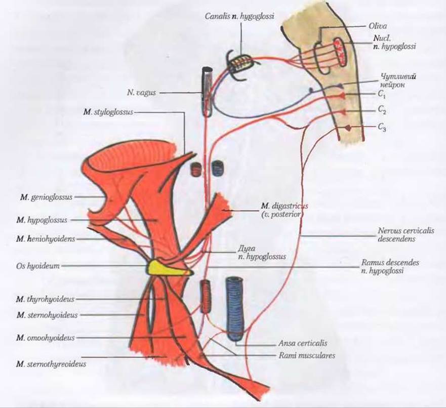
Fig. 186. Hypoglossal nerve, n. hypoglossus (diagram by I. Ye. Kefelii)
Loss of function of the glossopharyngeal nerve causes disorders of voice production, swallowing, and taste on the mucosa in the region of the tongue root and soft palate on one side, as well as general anesthesia of the mucosa of the upper half.
10. Vagus nerve, nervus vagus (X) (Fig. 185 A; B), is a mixed nerve consisting of axons of motor neurons from the ambiguous nucleus, nucleus ambiguus, parasympathetic neurons from the dorsal nucleus of the vagus nerve, nucleus dorsalis n. vagi, and peripheral processes of sensory neurons from the superior and inferior ganglia, gangll. superius et inferius. It is the longest of the Cranial Nerves and, due to its length, is called the vagus (wandering) nerve. It has numerous connections with the branches of the VIII, IX, XI, and XII cranial nerves and extends from the head region to the abdominal organs. Four parts are distinguished in it: cranial, cervical, thoracic, and abdominal. The central processes of the cells of both ganglia, together with motor and parasympathetic fibers, form the root of the nerve, which lies in the sulcus retroolivaris of the medulla oblongata, more caudal than the root of the glossopharyngeal nerve. The central processes terminate in the nucl. solitarius, while the peripheral processes, running within the trunk and branches of the nerve, terminate in receptors in the walls of The Heart, blood vessels, and Internal Organs.
Emerging from the cranial cavity through the jugular foramen, the vagus nerve enters the neurovascular bundle of the neck, lying between the internal carotid artery (and lower down, the common carotid artery) and the internal jugular vein. Through the superior thoracic aperture, the nerve enters the thoracic cavity. Here, the right vagus nerve passes between the subclavian vein (anteriorly) and the Subclavian Artery (posteriorly), descends along the Trachea (to the right of it), and crosses the right bronchus posteriorly. The left nerve passes anteriorly to the aortic arch and lower down posteriorly to the left bronchus. Further down, both nerves branch and anastomose with each other, forming the esophageal plexus, plexus oesophagealis. Near the esophageal opening of the Diaphragm, the anterior and posterior vagal trunks, truncus vagalis anterior et posterior, arise from the plexus and enter the Abdominal cavity through the esophageal hiatus of the diaphragm, hiatus oesophageus. The anterior trunk lies along the anterior surface of the lesser curvature of The Stomach, giving off branches to the stomach and Liver, while a small portion of its fibers passes to the plexus coeliacus. The posterior trunk lies along the posterior surface of the lesser curvature of the stomach, giving off branches to the stomach, while a larger portion of its fibers passes to the plexus coeliacus. From the latter, the fibers of the vagus nerves run as part of the periarterial plexuses to the abdominal organs (up to the border of the colon sigmoideum).
Along its course, the vagus nerve gives off numerous branches. Thus, from the superior ganglion arise:
I. Branches of the intracranial part of the vagus nerve: 1) meningeal branch, ramus meningeus, supplying the dura mater of the posterior cranial fossa, and 2) auricular branch, ramus auricularis, which passes through the mastoid canaliculus, canaliculus mastoideus, to the posterior wall of the external acoustic meatus and a small area of skin of the auricle.
II. Branches of the cervical part of the vagus nerve:
1. Pharyngeal branches, rami pharyngeales, originate from the inferior ganglion and take part in The formation of the pharyngeal plexus, plexus pharyngealis.
2. Superior laryngeal nerve, nervus laryngealis superior, arises from the inferior ganglion, descends downwards and forwards along the lateral wall of the pharynx, and at the level of the greater horn of the Hyoid bone divides into external and internal branches, ramus externus et internus. The first branch innervates the m. cricothyroideus and the inferior pharyngeal constrictor, while the second branch pierces the membrana thyrohyoidea and ramifies in the laryngeal mucosa above the rima glottidis and in the mucosa of the root of the tongue.
3. Superior cervical cardiac branches, rami cardiaci cervicales superiores, originate between the superior and recurrent laryngeal nerves and reach the cardiac plexus.
4. Recurrent laryngeal nerve, n. laryngealis recurrens, originates from the trunk of the vagus nerve immediately after the latter enters the thoracic cavity. The right one loops inferiorly and posteriorly around the right subclavian artery, and the left one loops around the aortic arch. Both nerves ascend in the grooves between the Esophagus and trachea, giving off branches to these organs. The terminal branch is called the inferior laryngeal nerve, nervus laryngealis inferior. Along its course, the recurrent laryngeal nerve gives off:
1. Tracheal branches, rami tracheales, which innervate the trachea (mucous membrane, glands, and smooth muscles).
2. Esophageal branches, rami oesophageales, pass towards the esophagus, innervating the mucous membrane, glands, and striated muscles.
3. Inferior cervical cardiac branches, rami cardiaci cervicales inferiores, originate from the trunk of the vagus nerve and from the recurrent laryngeal nerve, and reach the cardiac plexus.
The inferior laryngeal nerve innervates all muscles of the Larynx (except for the m. cricothyroideus) and the laryngeal mucosa below the vocal folds.
III. Branches of the thoracic part of the vagus nerve:
1. Thoracic cardiac branches, rami cardiaci thoracici, pass to the cardiac plexuses.
2. Bronchial branches, rami bronchiales, communicate with the branches of the sympathetic trunk and form the pulmonary plexus, plexus pulmonaris.
3. Esophageal plexus, plexus oesophagealis, is formed by the branches of the right and left trunks of the vagus nerve; its branches innervate the wall of the esophagus.
IV. Branches of the abdominal part of the vagus nerve:
1. Anterior gastric branches, rami gastrici anteriores, arise from the anterior trunk, truncus vagalis anterior, and form, together with sympathetic branches, the anterior gastric plexus along the lesser curvature of the stomach. The anterior gastric branches innervate the wall of the stomach (muscles, glands, and mucous membrane).
2. Posterior gastric branches, rami gastrici posteriores, arise from the posterior vagal trunk, truncus vagalis posterior, in the region of the pars cardiaca. The posterior gastric branches communicate with sympathetic branches and innervate the muscles, glands, and mucous membrane of the stomach.
3. Celiac branches, rami coeliaci, arise mainly from the posterior vagal trunk towards the plexus coeliacus. As part of the branches of the celiac plexus, fibers of the vagus nerves reach the abdominal organs (liver, Spleen, Pancreas, Kidney, small and Large Intestine up to the sigmoid colon), where they innervate the smooth musculature, glands, and mucous membranes.
4. Hepatic branches, rami hepatici, reach the hepatic plexus, plexus hepaticus.
5. Renal branches, rami renales, reach the renal plexus, plexus renalis.
Thus, the vagus nerve innervates the dura mater (posterior cranial fossa), the skin of the auricle and external acoustic meatus, the tympanic membrane, the muscles of the soft palate (except for the m. tensor veli palatini), the mucous membrane of the tongue (general and gustatory sensitivity), the muscles and mucous membrane of the pharynx (except for the m. stylopharyngeus), the muscles and mucous membrane of the larynx, the thyroid and Parathyroid glands, the Thymus, the Lymph Nodes of the neck, and the Organs of the thoracic and abdominal cavities.
When the vagus nerve is affected on one side, paralysis of the soft palate and vocal fold, absence of the pharyngeal reflex on the affected side, deviation of the uvula to the healthy side, as well as respiratory and cardiac disorders in bilateral lesions, are observed.
11. Accessory nerve, nervus accessorius (XI) (Fig. 185 A), motor, originates in two parts. The fibers of the cranial part arise from the motor ambiguous nucleus, nucl. ambiguus (shared with the IX and X cranial nerves) in the medulla oblongata, forming the cranial roots, radices craniales, which emerge in the sulcus dorsolateralis caudal to the rootlet of the vagus nerve. The fibers of the spinal part originate from the motor nucleus of the accessory nerve, nucl. spinalis nervi accessori, of the anterior horn, forming the spinal roots, radices spinales. The latter enter the cranial cavity through the foramen magnum and join the cranial part, thereby forming the trunk of the accessory nerve, truncus nervi accessorii. It divides into internal and external branches, ramus internus et externus. The first enters into the COMPOSITION OF THE vagus nerve (mainly into the laryngeal nerves), while the second exits the cranial cavity through the jugular foramen. Initially, it lies posterior to the posterior belly of the digastric muscle, and then medial to the m. sternocleidomastoideus. It pierces this muscle, descends downwards, and terminates in the m. trapezius. The muscular branches, rami musculares, of the external branch of the accessory nerve innervate two muscles: m. sternocleidomastoideus et m. trapezius. The accessory nerve communicates with the branches of the Cervical plexus.
When the accessory nerve is damaged, peripheral paralysis of the innervated muscles is observed.
12. Hypoglossal nerve, n. hypoglossus (XII), formed by axons of motor neurons. Its nucleus, nucleus nervi hypoglossi, is located in the medulla oblongata. The nerve emerges with 10–15 rootlets from the anterolateral sulcus, sulcus anterolateralis. After the rootlets join together into a common trunk, it exits the cranial cavity through the hypoglossal canal, canalis hypoglossi, into the neck. Initially, the nerve passes between the internal and external carotid Arteries, and then descends below the posterior belly of the digastric muscle across The surface of the hyoglossus muscle in an arch, bounding the lingual triangle superiorly. The terminal lingual branches, rami linguales, of the nerve innervate all muscles of the tongue.
It should be noted that fibers of the first and second cervical spinal nerves run within the trunk of the hypoglossal nerve. Part of them branches off approximately from the middle of the arch of the hypoglossal nerve and runs along the common carotid artery as the superior root of the cervical loop, radix superior ansae cervicalis. The second part departs slightly further and innervates the m. thyrohyoideus et geniohyoideus with independent branches. When the hypoglossal nerve is affected, peripheral paralysis of the tongue muscles occurs.
Cranial Nerves
Summary Table
|
Nerve and its functional characteristics |
Nuclei and their topography |
Brain exit or entry point |
Skull exit or entry point |
Innervation of organs and regions |
|
1 |
2 |
3 |
4 |
5 |
|
Olfactory nerves, nn. olfactorii (I) (sensory) |
Olfactory bulb |
Cribriform plate |
Mucous membrane of the superior nasal meatus (olfactory region) |
|
|
Optic nerve, n. opticus (II) (sensory) |
— |
Optic chiasm |
Optic canal |
Retina of the eyeball |
|
Oculomotor nerve, n. oculomotorius (III) (motor) |
nucl. n. oculomotorii (motor) located at the level of the superior colliculi of the midbrain; nucl. accessorius (parasympathetic) located medially to it |
Oculomotor sulcus of the cerebral peduncle |
Superior orbital fissure |
Superior, inferior, and medial rectus muscles of the eyeball |
|
Trochlear nerve n. trochlearis (IV) (motor) |
nucl. n. trochlearis (motor) located at the level of the inferior colliculi of the midbrain |
Lateral aspect of the cerebral peduncle (from the medullary velum) |
Superior orbital fissure |
Superior oblique muscle of the eyeball |
|
Trigeminal nerve n. trigeminus (V) (mixed) First branch n. ophthalmicus (sensory) |
nucl. motorius (motor), nucl. pontinus (sensory), nucl. spinalis (sensory), nucl. tractus mesencephali (sensory). Located in the tegmentum of the cerebral peduncle, lateral to the cerebral aqueduct |
Anterior to the middle cerebellar peduncle along the linea trigeminofacialis |
Superior orbital fissure, n. ophthalmicus (sensory) |
Tentorium cerebelli, lacrimal gland, lacrimal sac, conjunctiva, skin of the upper eyelid and forehead, mucosa of the ethmoid sinuses, anterior part of the nasal cavity, eyeball |
|
Second branch n. maxillaris (sensory) |
Foramen rotundum, n. maxillaris (sensory) |
Skin of the temporal region, lower eyelid, upper lip, External Nose, zygomatic and buccal regions |
||
|
Dura mater in the middle cranial fossa, gingiva, maxillary teeth, nasal mucosa, palate, paranasal sinuses, nasal glands. |
||||
|
Third branch, n. mandibularis (mixed) 1. Motor part 2. Sensory part |
Foramen ovale, n. mandibularis (mixed) |
1. Muscles: masticatory, mylohyoid, anterior belly of digastric, tensor veli palatini, and tensor tympani. 2. Dura mater, anterior 2/3 of the tongue, oral mucosa, palatine Tonsils, skin of the external acoustic meatus, auricle and temporal region, chin, lower lip, parotid gland. |
||
|
Abducens nerve, n. abducens (VI) (motor) |
Located in the dorsal part of the pons, facial colliculus area (internal genu of facial nerve) |
In the sulcus between the pons and the pyramid |
Superior orbital fissure |
Lateral rectus muscle of the eyeball |
|
Facial nerve, n. facialis (VII) (mixed) |
nucl. n. facialis (motor) located in the dorsal part of the pons, reticular formation |
Posterior border of the pons, posterior to the middle cerebellar peduncle |
Internal acoustic meatus, facial canal, stylomastoid foramen |
Muscles of facial expression, posterior belly of the digastric muscle, stylohyoid and stapedius muscles |
|
Intermediate nerve, n. intermedius (VII) (sensory and Parasympathetic part of the facial nerve) |
nucl. solitarius (sensory), nucl. salivatorius superior (secretomotor, parasympathetic). In the reticular Formation of the pons, dorsal to the facial nucleus |
Dorsal part of the pons |
Exits the tympanic cavity through the petrotympanic fissure. On the anterior surface of the petrous part of the Temporal bone through the opening of the canal for the greater petrosal nerve. |
Mucous membrane of the anterior 2/3 of the tongue and soft palate (gustatory) |
|
Vestibulocochlear nerve, n. vestibulocochlearis (VIII) (sensory) |
Pars cochlearis - nucl. cochleares ventralis et dorsalis (sensory) located in the Rhomboid fossa. Pars vestibularis - nuclei vestibulares - nuclei vestibulares medialis, lateralis, superior et inferior, lateral angle of the rhomboid fossa (area vestibularis inferior) |
Lateral to the olive, posterior border of the pons |
Internal acoustic meatus |
Spiral organ of the Inner ear. Vestibular apparatus of the inner ear. |
|
Glossopharyngeal nerve, n. glossopharyngeus (IX) (mixed) |
nucl. solitarius (sensory), located in the medulla oblongata (trigonum n. vagi area); nucl. ambiguus (motor), located in the reticular formation of the medulla oblongata; nucl. salivatorius inferior (secretomotor) parasympathetic, located in the medulla oblongata between the ambiguous nucleus and the olivary nucleus. |
Medulla oblongata, upper part of the dorsolateral sulcus |
Jugular foramen Anterior surface of the petrous part of the temporal bone, tympanic canaliculus |
Mucous membrane of the pharynx, posterior third of the tongue, carotid sinus Stylopharyngeus muscle Mucous membrane of the tympanic cavity and auditory tube, parotid gland |
|
Vagus nerve, n. vagus (X) (mixed) |
nucl. solitarius (sensory) in the trigonum n. vagi area of the medulla oblongata; nucl. ambiguus (motor), located in the reticular formation of the medulla oblongata; nucl. dorsalis n. vagi (parasympathetic) |
Medulla oblongata, posterior to the olive in the dorsolateral sulcus, inferior to the glossopharyngeal nerve |
Jugular foramen |
Respiratory organs, digestive tract down to the sigmoid colon. Heart. Dura mater of the posterior cranial fossa. Auricle, external acoustic meatus, muscles of the pharynx, soft palate, and larynx. Parasympathetic innervation of the heart, respiratory organs, digestive tract down to the sigmoid colon, liver, pancreas, Kidneys |
|
Accessory nerve, n. accessorius (XI) (motor) |
nucl. ambiguus (motor) in the medulla oblongata, being a continuation of the corresponding nuclei of the IX and X pairs; spinal motor nucleus located in the Gray matter of the Spinal Cord between the anterior and posterior horns. |
From the posterolateral sulcus of the medulla oblongata |
Jugular foramen |
Trapezius and sternocleidomastoid muscles |
|
Hypoglossal nerve, n. hypoglossus (XII) (motor) |
nucl. n. hypoglossi medulla oblongata trigonum n. hypoglossi of the rhomboid fossa |
Between the pyramid and the olive |
Hypoglossal canal |
Muscles of the tongue and muscles located inferior to the hyoid bone (via branches of the radix superior). |
Last update: 08/08/2026
Editorial and Educational Adaptation: This material has been compiled based on the primary/original source text. The project team performed an editorial review, corrected technical inaccuracies, structured sections, and adapted the content for an educational format.
What was processed:
- elimination of formatting defects (OCR errors, structural breaks, corrupted characters);
- editorial organization of content;
- standardization of terminology in accordance with academic sources;
- verification of factual statements against the original source text.
All mentions of the author, publication year, and origin of the primary text have been preserved in accordance with the source.